. 2002 Jan 1;22(1):10-20.
doi: 10.1523/JNEUROSCI.22-01-00010.2002.

Rapid tyrosine phosphorylation of neuronal proteins including tau and focal adhesion kinase in response to amyloid-beta peptide exposure: involvement of Src family protein kinases

Affiliations

Rapid tyrosine phosphorylation of neuronal proteins including tau and focal adhesion kinase in response to amyloid-beta peptide exposure: involvement of Src family protein kinases

Ritchie Williamson et al. J Neurosci. .

Abstract

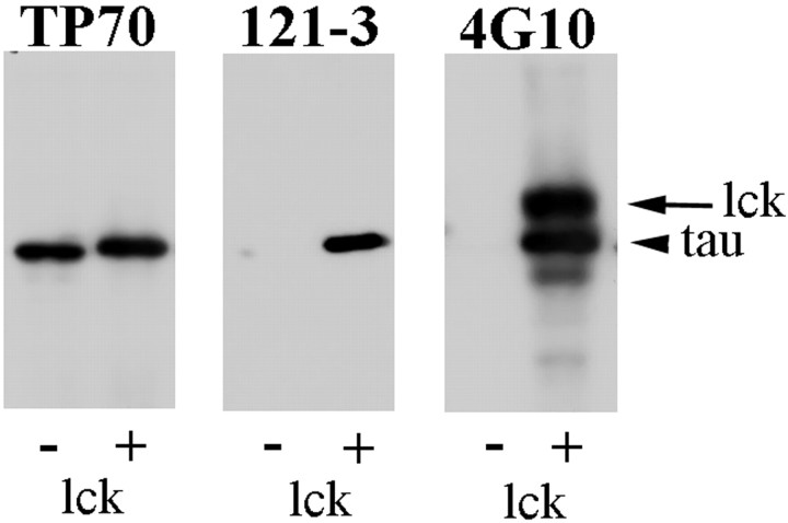
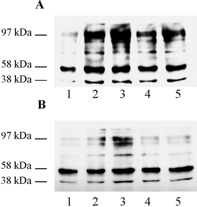
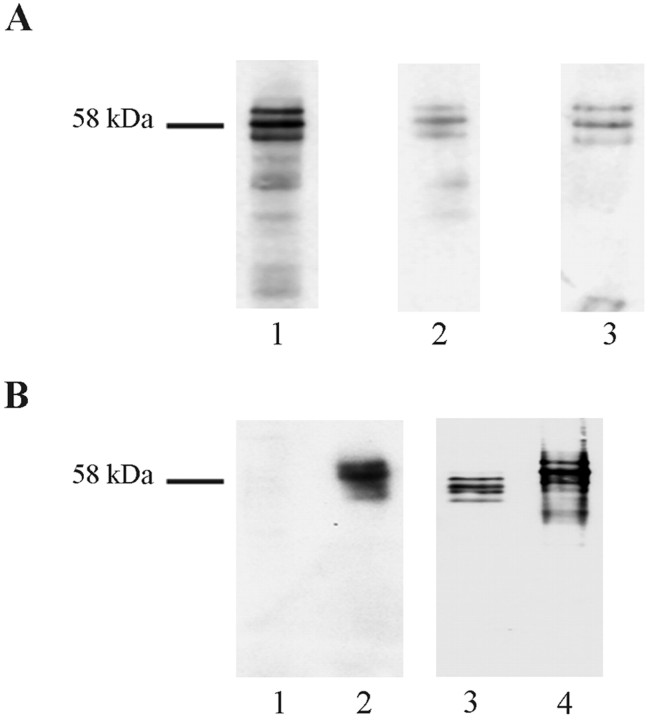
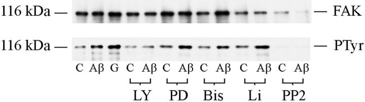
The increased production of amyloid beta-peptide (Abeta) in Alzheimer's disease is acknowledged to be a key pathogenic event. In this study, we examined the response of primary human and rat brain cortical cultures to Abeta administration and found a marked increase in the tyrosine phosphorylation content of numerous neuronal proteins, including tau and putative microtubule-associated protein 2c (MAP2c). We also found that paired helical filaments of aggregated and hyperphosphorylated tau are tyrosine phosphorylated, indicating that changes in the phosphotyrosine content of cytoplasmic proteins in response to Abeta are potentially an important process. Increased tyrosine phosphorylation of cytoskeletal and other neuronal proteins was specific to fibrillar Abeta(25-35) and Abeta(1-42). The tyrosine phosphorylation was blocked by addition of the Src family tyrosine kinase inhibitor 4-amino-5-(4-chlorophenyl)-7(t-butyl)pyrazol(3,4-d)pyramide (PP2) and the phosphatidylinositol 3-kinase inhibitor LY 294002. Tyrosine phosphorylation of tau and MAP2c was concomitant with an increase in the tyrosine phosphorylation and subsequent putative activation of the non-receptor kinase, focal adhesion kinase (FAK). Immunoprecipitation of Fyn, a member of the Src family, from Abeta(25-35)-treated neurons showed an increased association of Fyn with FAK. Abeta treatment of cells also stimulated the sustained activation of extracellular regulated kinase-2, which was blocked by addition of PP2 and LY 294002, suggesting that FAK/Fyn/PI3-kinase association is upstream of mitogen-activated protein (MAP) kinase signaling in Abeta-treated neurons. This cascade of signaling events contains the earliest biochemical changes in neurons to be described in response to Abeta exposure and may be critical for subsequent neurodegenerative changes.


Figures

Fig. 1.
Fig. 1.
Characterization of tyrosine-phosphorylated tau-specific antibody 121–3. Western blots of 2N4R recombinant tau that was either unphosphorylated (−) or phosphorylated (+) by lck were probed with the antibodies TP70, 121–3, and 4G10 as indicated.Arrowhead indicates the tau band; arrowindicates the lck band.
Fig. 2.
Fig. 2.
Aβ-induced neuronal death of primary rat cortical cultures. A, B, Hoechst nuclear labeling and immunolabeling (C, D) of activated caspases in untreated control cultures (A,C) and Aβ25–35-treated cultures (10 μm, 48 hr) (B, D) of 7-d-old primary rat cortical cultures. Scale bars, 50 μm.E, LDH release from control (■) cultures and cultures treated (▪) with Aβ25–35 (10 μm;24 hr, 48 hr, 72 hr,96 hr), expressed as fold increase in LDH release over control LDH release after 24 hr, normalized to 1. Error bars indicate SDs based on three independent experiments, 15 readings per experiment. *p < 0.0001; Mann–Whitney Utest.
Fig. 3.
Fig. 3.
Aβ-induced tyrosine phosphorylation of neuronal proteins. Rat primary cortical neurons (A) (7 d in culture) and human primary cortical neurons (B) (14 d in culture) were treated with 10 μm25–35 as follows: lane 1, control (no Aβ25–35); lane 2, 1 min; lane 3, 2 min; lane 4, 5 min;lane 5, 10 min. Whole-cell lysates were prepared, and Western blots were probed with anti-phosphotyrosine antibody 4G10. Apparent molecular mass (kDa) markers are indicated to the left.
Fig. 4.
Fig. 4.
Full-length Aβ induces tyrosine phosphorylation of neuronal proteins. Rat primary cortical neurons (7 d in culture) were treated with 10 μm1–42 as follows:lane 1, control (no Aβ1–42); lane 2, 1 min; lane 3, 2 min; lane 4, 5 min; lane 5, 10 min. A, Whole-cell lysates were prepared, and the Western blot was probed with anti-phosphotyrosine antibody 4G10. B, An identically loaded gel was blotted and subsequently treated with SHP-1 protein tyrosine phosphatase before probing with anti-phosphotyrosine antibody 4G10. C, Blot from B was stripped and reprobed with mAb PHF-1. D, An identically loaded Coomassie-stained gel. Apparent molecular mass (kDa) markers are indicated to the left.
Fig. 5.
Fig. 5.
Aβ-induced tyrosine phosphorylation of cytoskeletal proteins. A, Heat-stable extracts of 7-d-old rat and 14-d-old human primary neuronal cultures were prepared after treatments as described below and probed with 4G10 (lanes 1–5). Lanes 1–3, Rat primary neuronal cultures (7 d in culture) were either untreated (lane 1, untreated control) or treated for 5 min with 10 μm25–35 (lane 2) or with 10 μm35–25 (lane 3, reverse-sequence control). Lanes 4–5, Human primary cultures (14 d in culture) were either untreated (lane 4) or treated for 5 min with 10 μm25–35 (lane 5). Untreated control rat lysates were also probed with the anti-MAP2 antibody HM-2 (lane 6) or the anti-tau antibody TP70, which also reacts with MAP2c as well as with tau (lane 7).B, C, Western blots of total cell lysates from 14-d-old human primary cortical cultures treated with 10 μm25–35 as follows: lane 1, untreated control; lane 2, 1 min; lane 3, 2 min; lane 4, 5 min; lane 5, 10 min. B was probed with the polyclonal antibody 121–3 specific to tau in which tyrosine 29 is phosphorylated;C was probed with the polyclonal antibody TP70 to total tau. D, Western blot with 4G10 of heat-stable preparations from control 7-d-old primary rat brain cortical cultures (lane 1) or cultures treated with Aβ25–35alone for 5 min (lane 2), Aβ25–35 for 5 min in culture pretreated with the Src-family kinase inhibitor PP2 (lane 3), Aβ25–35 for 5 min in culture pretreated with the protein kinase C inhibitor bis-indolylmaleimide (lane 4), and Aβ25–35 for 5 min in culture pretreated with the PI3-kinase inhibitor wortmannin for 15 min (lane 5). Apparent molecular mass (kDa) markers are indicated to the left. Bands labeled MAP2c and tau in A had an interpolated molecular mass of 77 and 58 kDa, respectively.
Fig. 6.
Fig. 6.
PHF-tau is tyrosine phosphorylated.A, Western blots of partially purified PHF-tau isolated from AD brain probed with TP70 (lane 1), PHF-1 (lane 2), 4G10 (lane 3).B, Western blot of tau extracted from a control (non-AD brain; lanes 1 and 3) and an AD brain (lanes 2 and 4) probed with polyclonal antibody 121–3 (lanes 1 and2) and with TP70 (lane 3 and4). Apparent molecular mass (kDa) marker is indicated to the left.
Fig. 7.
Fig. 7.
Aβ induces FAK/Fyn association with increased tyrosine phosphorylation of FAK. Double immunolabeling of 7-d-old primary rat brain cortical cultures with the polyclonal antibody FAK (A) and the monoclonal antibody to Fyn (B). Scale bars, 50 μm.C–E, Western blots of protein lysates immunoprecipitated with anti-Fyn polyclonal antibody from control 7-d-old primary rat cortical cultures (lane 1) and cultures treated with Aβ25–35 (10 μm) for 1 min (lane 2), 2 min (lane 3), 5 min (lane 4), 10 min (lane 5), or treated with Aβ35–25 (10 μm) for 5 min (lane 6). C was probed with the polyclonal antibody Fyn. D was probed with the polyclonal antibody FAK. E was probed with the monoclonal antibody 4G10 to phosphotyrosine. Apparent molecular mass (kDa) is indicated on the left.
Fig. 8.
Fig. 8.
The rate of Aβ-induced FAK activation is similar in primary rat brain cortical cultures and primary human brain cortical cultures. A, B, Western blots with 4G10 of protein immunoprecipitated with anti-FAK polyclonal antibody from cell lysates of 7-d-old primary rat cortical cultures (A) and of 14-d-old human cortical cultures (B). Shown are untreated cultures (lane 1) and cultures treated with Aβ25–35 (10 μm) for 1 min (lane 2), 2 min (lane 3), 5 min (lane 4), and 10 min (lane 5). C, Western blot with 4G10 of protein immunoprecipitated with anti-FAK polyclonal antibody from cell lysates of control 7-d-old primary rat cortical cultures left untreated or treated for 5 min with 10 μm25–35 or 10 μm1–42 as indicated. Apparent molecular mass (kDa) is indicated on theleft.
Fig. 9.
Fig. 9.
PI3-kinase and Fyn are involved in Aβ-induced FAK activation. Shown are Western blots with 4G10 (bottom panel) and polyclonal antibody to FAK (top panel) of protein immunoprecipitated with anti-FAK polyclonal antibody from cell lysates of 7-d-old primary rat brain cortical cultures. Cultures that had been pretreated with LY 294002 (LY), PD 98059 (PD), bis-indolylmaleimide (Bis), lithium (Li), or PP2 or left untreated as outlined in Materials and Methods were either harvested without further treatment (C) or treated with Aβ25–35 (10 μm) for 5 min (Aβ); a further culture was treated with glutamate (1 mm) alone for 1 hr (G). Apparent molecular mass (kDa) is indicated on the left.
Fig. 10.
Fig. 10.
Aβ-induced ERK phosphorylation is downstream of FAK activation. A, Western blots of RIPA buffer lysates from control 7-d-old primary rat cortical cultures (lane 1) and cultures treated with Aβ (10 μm) and harvested after 5 min (lane 2), 10 min (lane 3), 30 min (lane 4), 60 min (lane 5), with PP2 alone (30 μm) for 18 hr (lane 6), Aβ25–35 for 5 min in culture pretreated with PP2 (lane 7), PD 98059 alone (lane 8), Aβ25–35 for 5 min in culture pretreated with PD 98059 (lane 9), LY 294002 alone (lane 10), and Aβ25–35 for 5 min in culture pretreated with LY 294002 (lane 11). Top panel blot was probed with monoclonal antibody MK12 to ERK;bottom panel blot was probed with a monoclonal antibody E10 to phosphorylated ERK1 (p44) and ERK2 (p42). B, Quantification of ERK kinase phosphorylation of the experiments shown in A. The activity, as indicated numerically beneath the abscissa, is expressed relative to the control (0 min) that was normalized to 1.

References

    1. Abe K, Saito H. Amyloid beta neurotoxicity not mediated by the mitogen-activated protein kinase cascade in cultured rat hippocampal and cortical neurons. Neurosci Lett. 2000;292:1–4. - PubMed
    1. Alvarez A, Toro R, Caceres A, Maccioni RB. Inhibition of tau phosphorylating protein kinase cdk5 prevents beta-amyloid-induced neuronal death. FEBS Lett. 1999;459:421–426. - PubMed
    1. Alvarez G, Munoz-Montano JR, Satrustegui J, Avila J, Bogonez E, Diaz-Nido J. Lithium protects cultured neurons against beta-amyloid-induced neurodegeneration. FEBS Lett. 1999;453:260–264. - PubMed
    1. Behl C, Davis JB, Lesley R, Schubert D. Hydrogen peroxide mediates amyloid beta protein toxicity. Cell. 1994;77:817–827. - PubMed
    1. Bradford MM. A rapid and sensitive method for the quantitation of microgram quantities of protein utilizing the principle of protein-dye binding. Anal Biochem. 1976;72:248–254. - PubMed

Publication types

MeSH terms

Substances

LinkOut - more resources