. 2002 Jan 1;22(1):21-8.
doi: 10.1523/JNEUROSCI.22-01-00021.2002.

Assessing the role of calcium-induced calcium release in short-term presynaptic plasticity at excitatory central synapses

Affiliations

Assessing the role of calcium-induced calcium release in short-term presynaptic plasticity at excitatory central synapses

Adam G Carter et al. J Neurosci. .

Abstract

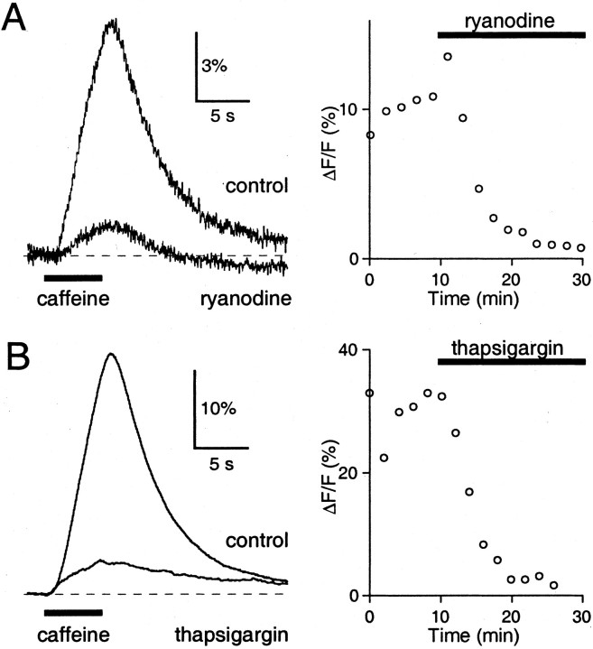
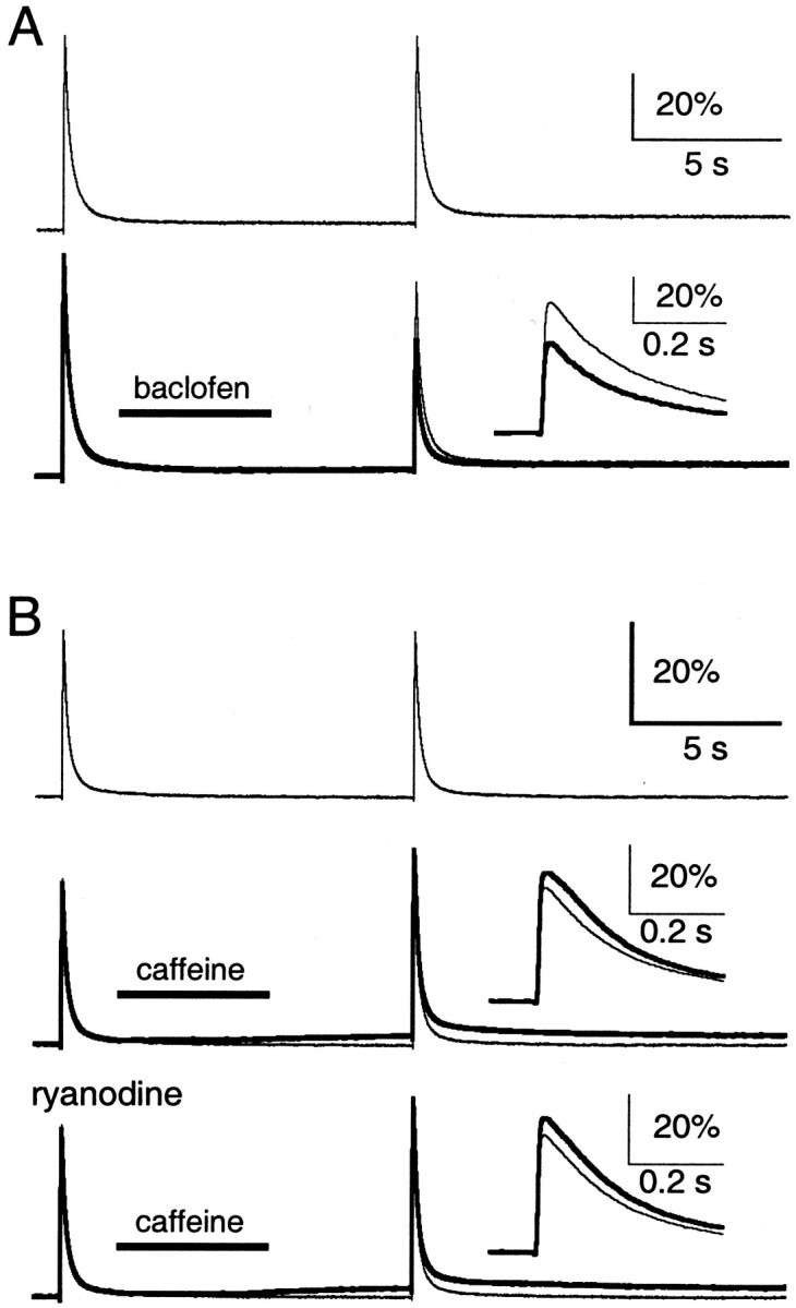
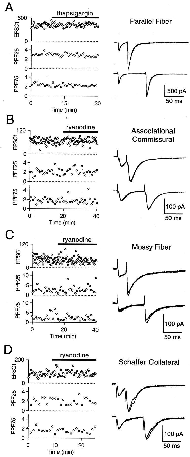
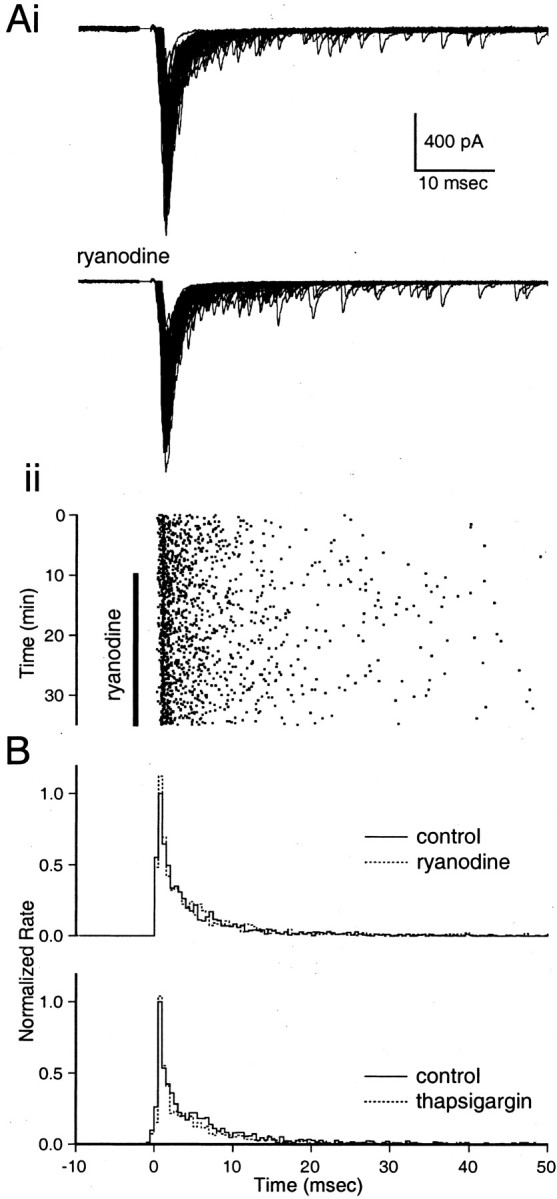
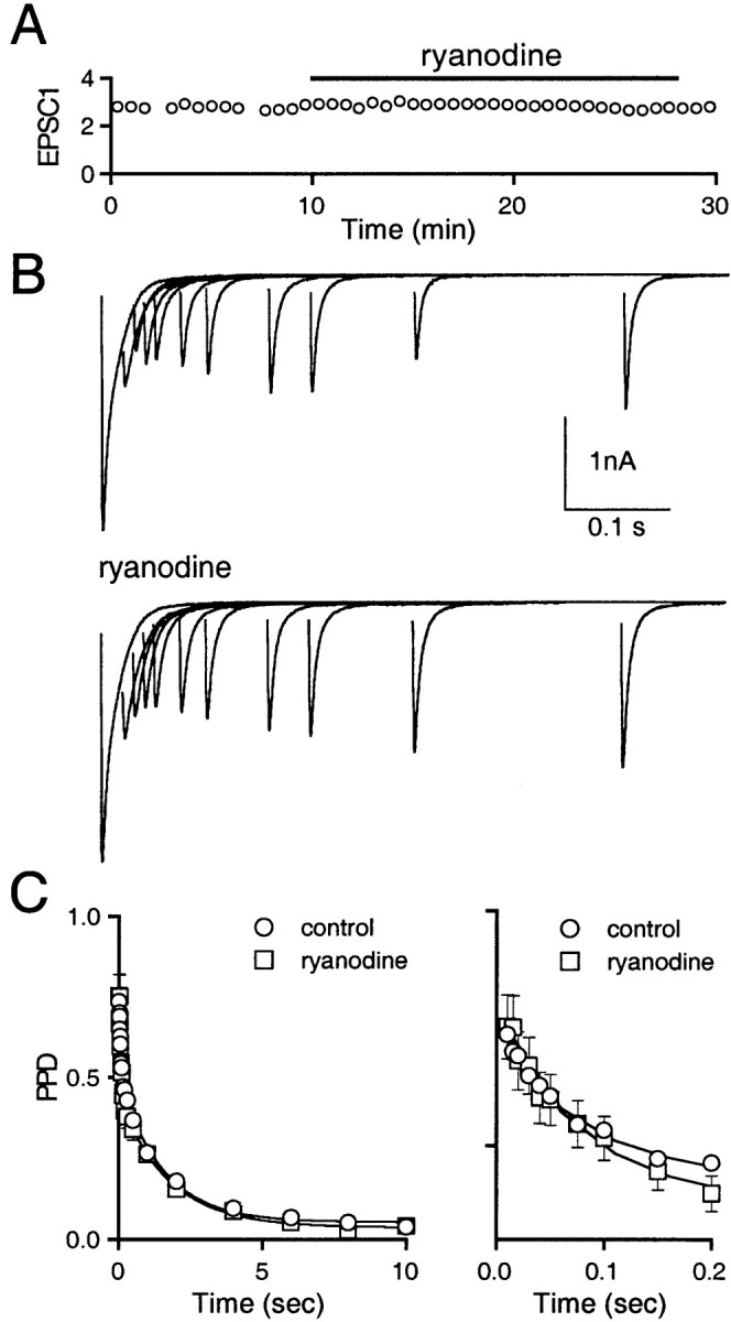
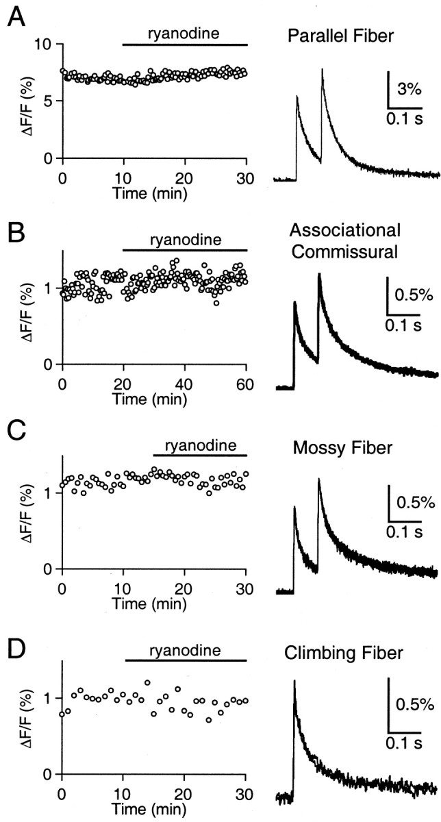
Recent evidence suggests that internal calcium stores and calcium-induced calcium release (CICR) provide an important source of calcium that drives short-term presynaptic plasticity at central synapses. Here we tested for the involvement of CICR in short-term presynaptic plasticity at six excitatory synapses in acute rat hippocampal and cerebellar brain slices. Depletion of internal calcium stores with thapsigargin and prevention of CICR with ryanodine have no effect on paired-pulse facilitation, delayed release of neurotransmitter, or calcium-dependent recovery from depression. Fluorometric calcium measurements also show that these drugs have no effect on the residual calcium signal that underlies these forms of short-term presynaptic plasticity. Finally, although caffeine causes CICR in Purkinje cell bodies and dendrites, it does not elicit CICR in parallel fiber inputs to these cells. Taken together, these results indicate that for the excitatory synapses studied here, internal calcium stores and CICR do not contribute to short-term presynaptic plasticity on the milliseconds-to-seconds time scale. Instead, this plasticity is driven by the residual calcium signal arising from calcium entry through voltage-gated calcium channels.


Figures

Fig. 1.
Fig. 1.
Ryanodine and thapsigargin abolish caffeine-evoked CICR in Purkinje cells. A Purkinje cell was filled with 200 μm Oregon Green 488 BAPTA-1 via a whole-cell recording pipette, and 40 mm caffeine was applied using a 5 sec pressure puff from a nearby micropipette. Fluorescence measurements were restricted to a small area that included the Purkinje cell soma and proximal dendrites. A, In control conditions, a ΔF/F signal was recorded (left) in response to caffeine application (solid bar). Bath application of 10 μm ryanodine abolished this ΔF/F signal. The time course is shown on the right, with the solid bar indicating ryanodine application. B, Similar results were found for bath application of 10 μmthapsigargin. Representative traces are averages of four or five trials.
Fig. 2.
Fig. 2.
Lack of caffeine-evoked CICR at the parallel fibers. Parallel fibers were filled with Oregon Green 488 BAPTA-1 AM. In each trial, parallel fibers were stimulated with a control and test stimulus separated by 10 sec. A, ΔF/F signals in the absence of drug application (top, bottom, light traces) and with puff application of 500 μm baclofen (bottom, bold trace).Inset, Test ΔF/F signals with baclofen (bold trace) or without (light trace) on an expanded time scale. B, ΔF/F signals in the absence of drug application (top, middle, bottom, light trace) and with puff application of 40 mm caffeine, in the absence (middle, bold trace) and presence (bottom, bold trace) of 10 μm ryanodine. Insets, Test ΔF/F signals with caffeine (bold trace) or without (light trace) on an expanded time scale. Representative traces are averages of three to five trials. For insets in B, the slow ΔF/F signal has been subtracted.
Fig. 3.
Fig. 3.
Disrupting CICR has no effect on paired-pulse facilitation at four excitatory synapses. A, At the PF→PC synapse, peak EPSC (EPSC1, picoamperes), PPF25, and PPF75 remain unchanged after bath application of 10 μm thapsigargin (solid bar). Representative traces (right) are superimposed averages of five trials before and after thapsigargin application. Similar results were found using 10 μmryanodine at the AC (B) and MF (C) synapses, and 100 μm ryanodine at the SC synapse (D).
Fig. 4.
Fig. 4.
Disrupting CICR has no effect on delayed release at the parallel fiber to stellate cell synapse. Ai, Seventy consecutive traces before (top) and after (bottom) bath application of 10 μm ryanodine. Aii, Raster plot of quantal events, with the vertical bar indicating the time of ryanodine application. B, Average PSTH plots of quantal events before (solid line) and after (dashed line) 10 μm ryanodine (B, top;n = 4) or 10 μm thapsigargin (B, bottom; n = 4). PSTH plots for each experiment were made for 10 min periods in both control conditions and after 10 min drug application. These PSTH plots were then normalized with respect to the peak rate in control conditions. Normalized PSTH plots from the different experiments were then averaged.
Fig. 5.
Fig. 5.
Disrupting CICR has no effect on calcium-dependent recovery from depression at the climbing fiber to Purkinje cell synapse. A, Initial EPSC amplitude (EPSC1, nanoamperes) remains unchanged after bath application of 100 μm ryanodine (solid bar). B, Representative tracesbefore (top) and after (bottom) bath application of ryanodine. C, PPD curves showing recovery from depression over 10 sec, with the first 200 msec expanded on theright. Representative traces inB are averages of two trials. PPD curves are averages ± SEM from 17 (control) or 5 (ryanodine) experiments.
Fig. 6.
Fig. 6.
Disrupting CICR has no effect on the residual calcium signal at four excitatory synapses. A, At the PF synapse, peak ΔF/F signal (left) remains unchanged after bath application of 10 μm ryanodine (solid bar). Representativetraces (right) are superimposed averages of five trials before and after ryanodine application. Similar results were found using 10 μm ryanodine at the AC synapse (B), and 100 μm ryanodine at the MF (C) and CF (D) synapses.

Similar articles

Cited by

References

    1. Alford S, Frenguelli BG, Schofield JG, Collingridge GL. Characterization of Ca2+ signals induced in hippocampal CA1 neurones by the synaptic activation of NMDA receptors. J Physiol (Lond) 1993;469:693–716. - PMC - PubMed
    1. Araque A, Sanzgiri RP, Parpura V, Haydon PG. Calcium elevation in astrocytes causes an NMDA receptor-dependent increase in the frequency of miniature synaptic currents in cultured hippocampal neurons. J Neurosci. 1998;18:6822–6829. - PMC - PubMed
    1. Araque A, Carmignoto G, Haydon PG. Dynamic signaling between astrocytes and neurons. Annu Rev Physiol. 2001;63:795–813. - PubMed
    1. Atluri PP, Regehr WG. Determinants of the time course of facilitation at the granule cell to Purkinje cell synapse. J Neurosci. 1996;16:5661–5671. - PMC - PubMed
    1. Atluri PP, Regehr WG. Delayed release of neurotransmitter from cerebellar granule cells. J Neurosci. 1998;18:8214–8227. - PMC - PubMed

Publication types

MeSH terms

LinkOut - more resources