. 2002 Jan 1;22(1):103-13.
doi: 10.1523/JNEUROSCI.22-01-00103.2002.

Inhibition of the c-Jun N-terminal kinase signaling pathway by the mixed lineage kinase inhibitor CEP-1347 (KT7515) preserves metabolism and growth of trophic factor-deprived neurons

Affiliations

Inhibition of the c-Jun N-terminal kinase signaling pathway by the mixed lineage kinase inhibitor CEP-1347 (KT7515) preserves metabolism and growth of trophic factor-deprived neurons

Charles A Harris et al. J Neurosci. .

Abstract

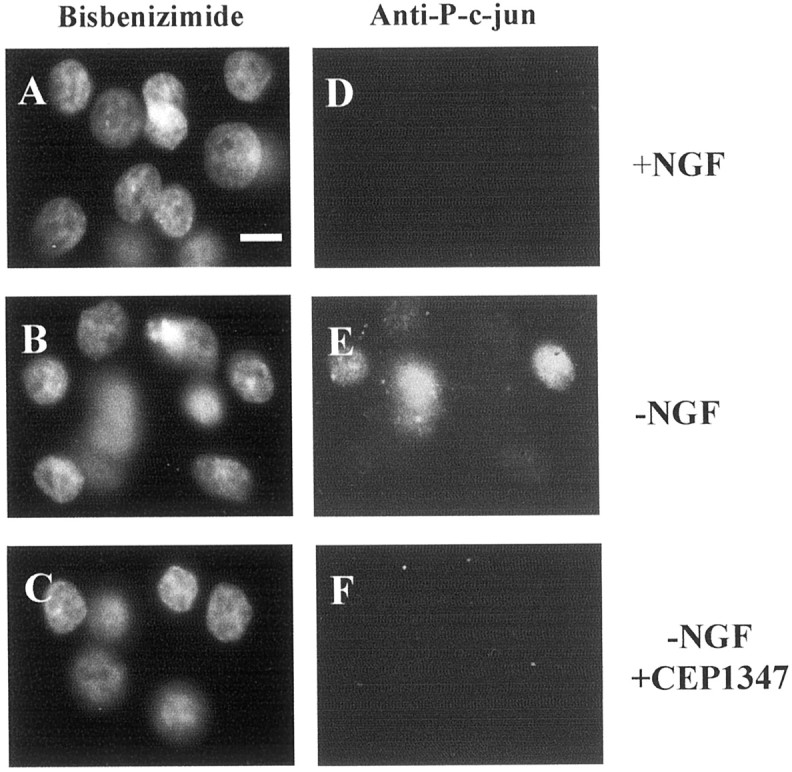
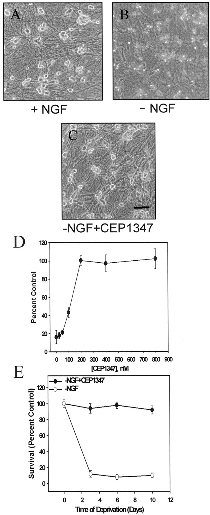
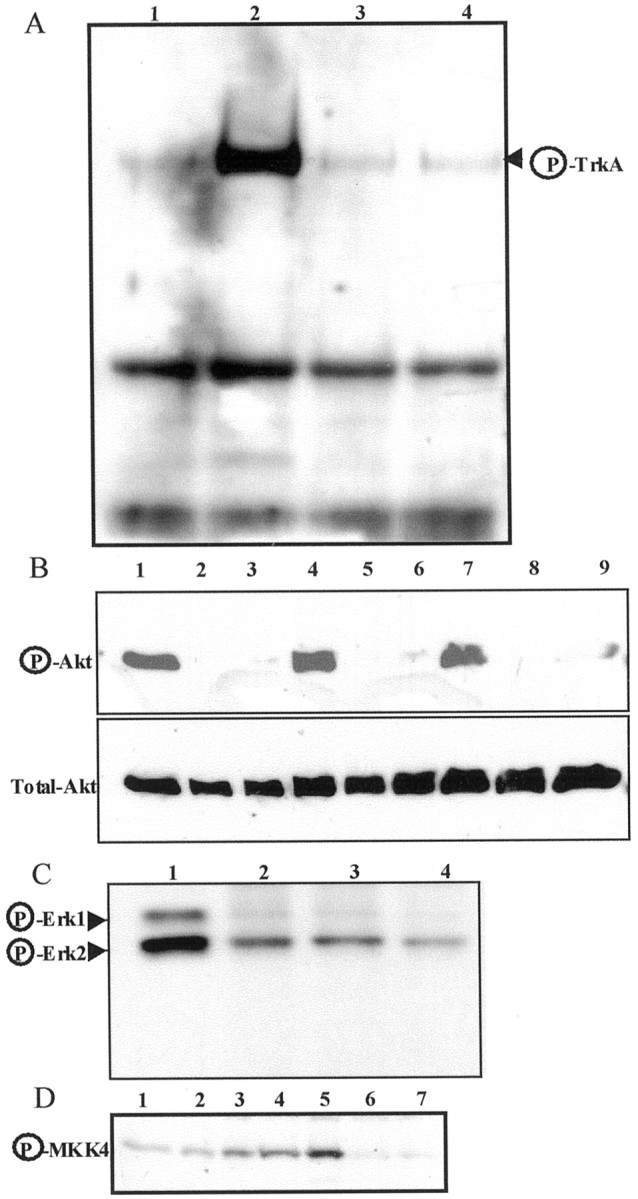
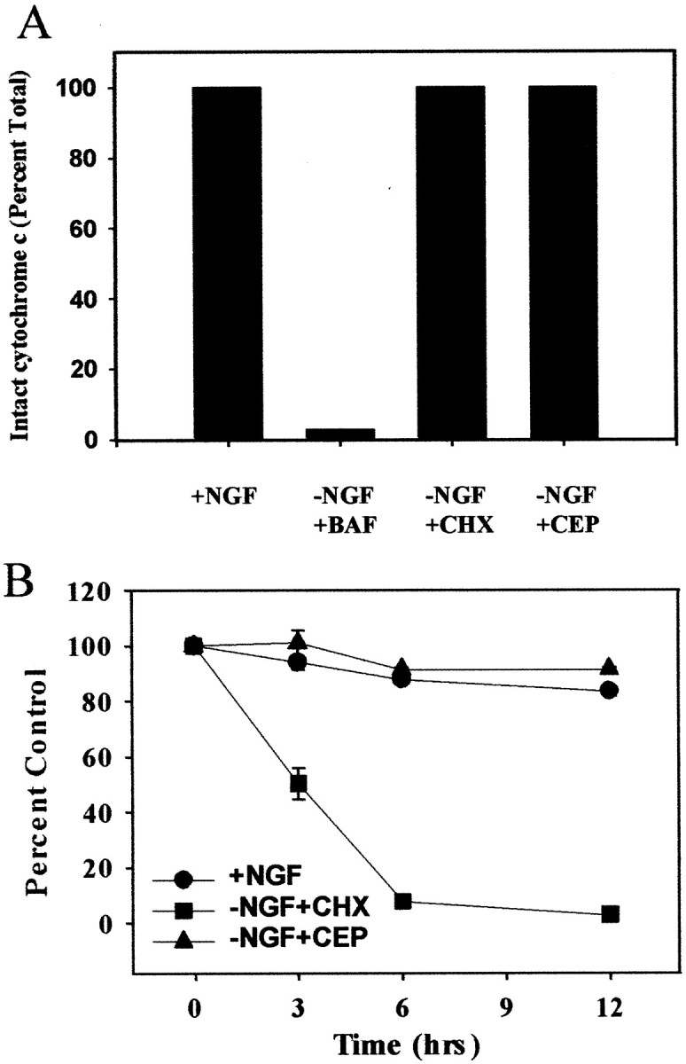
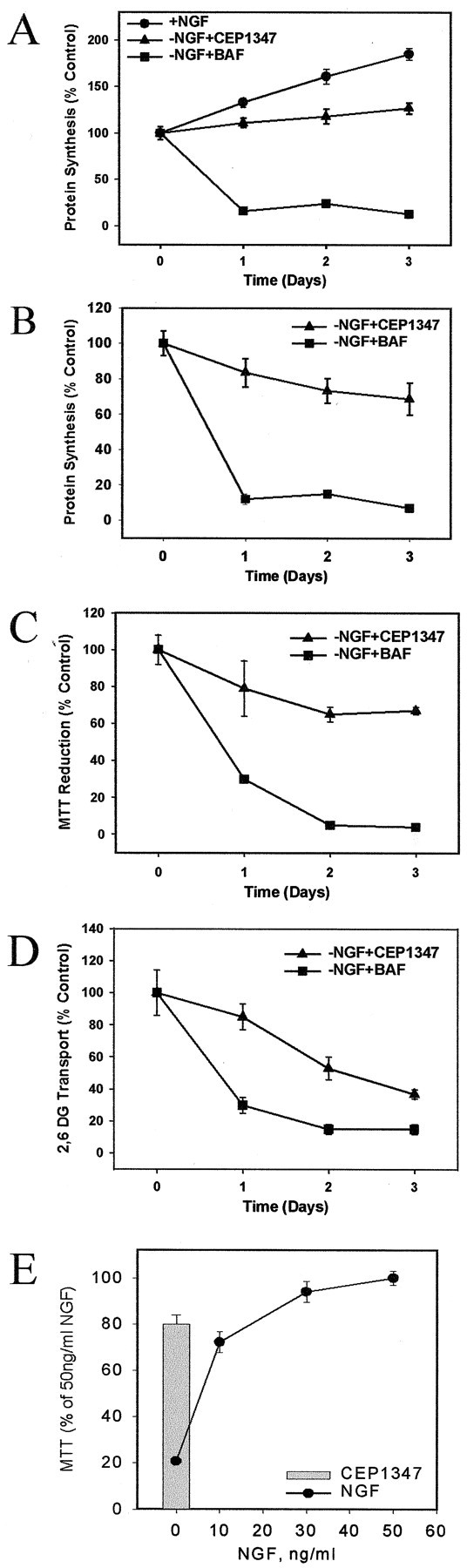
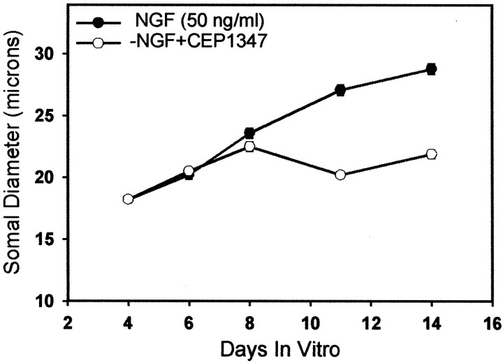
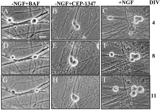
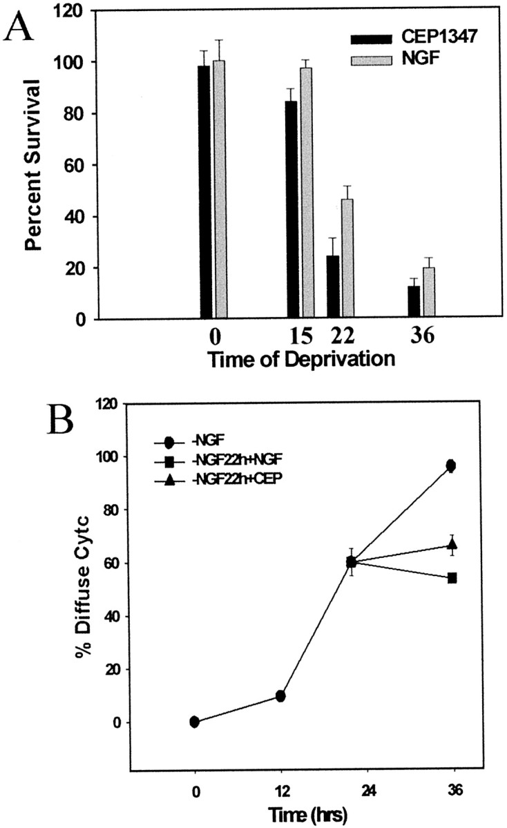
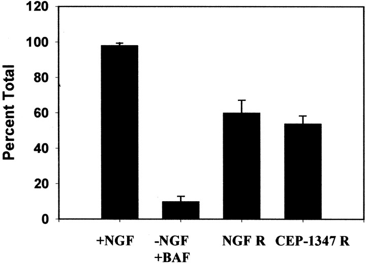
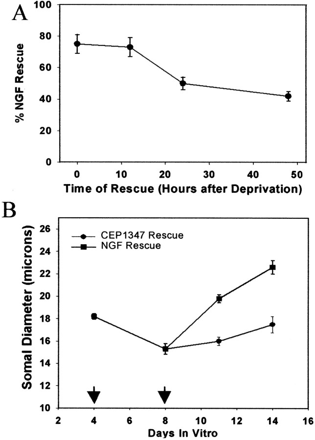
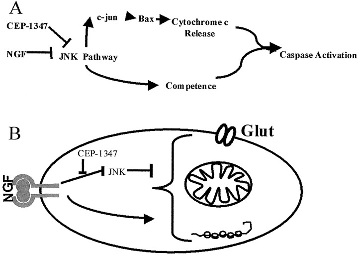
Nerve growth factor (NGF) deprivation triggers metabolic changes in sympathetic neurons that precede cell death. Here, we investigate the role of the c-Jun N-terminal kinase (JNK) pathway in downregulating neuronal metabolism. We show that, in the presence of CEP-1347 (KT7515), a small molecule known to block cell death upstream of JNK, cellular metabolism is preserved in neurons deprived of NGF. Biochemical data that are presented are consistent with the mechanism of action of CEP-1347 being the inhibition of the mixed lineage kinases (MLKs), known activators of JNK signaling. We demonstrate that CEP-1347-saved neurons continue to grow even in the absence of NGF, indicating that inhibition of the JNK pathway is permissive for neuronal growth in the absence of trophic support. These trophic effects are seen despite the fact that CEP-1347 does not stimulate several known survival kinase pathways. In addition to blocking Bax-dependent cytochrome c release, the inhibition of the JNK signaling pathway with CEP-1347 also blocks the development of competence-to-die in response to cytosolic cytochrome c. Therefore, inhibition of the JNK signaling pathway with the MLK inhibitor CEP-1347 inhibits both limbs of the apoptotic pathway. Finally, we demonstrate that neurons that have been NGF-deprived long-term but that have been kept alive by caspase inhibitors can be rescued metabolically by CEP-1347 as assessed by soma size, cytochrome c localization, and protein synthesis rates. Therefore, we conclude that, in addition to converting extracellular signals into decisions of life and death, the JNK pathway can modulate cellular metabolism directly and thereby maintain not only survival but the "quality of life" of neurons.


Figures

Fig. 1.
Fig. 1.
CEP-1347 blocks c-Jun phosphorylation after NGF deprivation. Neurons were maintained in NGF (A, D), deprived of NGF (B, E), or deprived of NGF in the presence of CEP-1347 (C, F) for 8 hr. Nuclei were stained with Hoechst 33258 (A–C), and immunohistochemistry was performed in parallel with an antibody specific for c-Jun that was phosphorylated at Serine residue 63 (D–F). Robust staining was seen in cultures deprived of NGF, but not in NGF-maintained cultures or NGF-deprived CEP-1347-treated cultures. Scale bar, 10 μm.
Fig. 2.
Fig. 2.
CEP-1347 is a potent long-term neuroprotectant and maintains both somal and neuritic morphology in sympathetic neurons. Neurons were grown in the presence of NGF for 5 d and then switched to media containing NGF (A), no NGF (B), or no NGF plus 500 nm CEP-1347 (C). Pictures were taken 3 d later.D, Sympathetic neurons were cultured in NGF for 5 d and then switched to serum-free medium containing 0, 25, 50, 100, 200, 400, or 800 nm CEP-1347. Neuronal viability was assessed 3 d later; p < 0.01 for CEP-1347 >50 nmversus no CEP-1347. E, CEP-1347 promotes long-term survival of NGF-deprived sympathetic neurons. Neurons were grown in the presence of NGF for 5 d and then deprived of NGF or deprived of NGF in the presence of 500 nm CEP-1347 for the indicated time periods; p < 0.001 for −NGF versus −NGF + CEP-1347 for all time points. Scale bar, 75 μm.
Fig. 3.
Fig. 3.
CEP-1347 blocks MKK4 phosphorylation and does not stimulate known survival kinase pathways. A, CEP-1347 does not stimulate TrkA. Neuronal lysates were subjected to immunoprecipitation with Trk antibody, followed by immunoblotting with phosphotyrosine antibody. Lane 1, Neurons deprived of NGF for 1 hr; lane 2, neurons maintained in NGF;lane 3, neurons deprived of NGF in the presence of CEP-1347 for 1 hr; lane 4, neurons deprived of NGF in the presence of CEP-1347 for 24 hr. B, CEP-1347 does not activate PI3-kinase activity. Lysates were prepared from NGF-maintained (lanes 1, 4, 7), NGF-deprived (lanes 2, 5, 8), or NGF-deprived CEP-1347-saved neurons (lanes 3, 6, 9) that were treated for 12 (lanes 1–3), 18 (lanes 4–6), or 24 hr (lanes 7–9). Lysates were immunoblotted with phospho-Akt (Serine-473) antibodies. C, CEP-1347 does not activate MAP Erk kinase activity. Lysates were blotted with antibodies that recognize phospho-Erks. Lane 1, NGF-maintained neurons;lane 2, neurons deprived of NGF for 1 hr; lane 3, neurons deprived of NGF but treated with CEP-1347 for 1 hr;lane 4, neurons deprived of NGF but treated with CEP-1347 for 24 hr. D, CEP-1347 blocks MKK4 phosphorylation induced by NGF deprivation. Neuronal lysates were immunoblotted with antibody specific to phospho-MKK4. Lane 1, NGF-maintained neurons; lane 2, neurons deprived of NGF for 45 min; lane 3, neurons deprived of NGF for 2 hr; lane 4, neurons deprived of NGF for 4 hr;lane 5, neurons deprived of NGF for 8 hr; lane 6, neurons deprived of NGF for 8 hr in the presence of CEP-1347; lane 7, NGF-maintained neurons in the presence of CEP-1347.
Fig. 4.
Fig. 4.
CEP-1347 blocks the release of cytochromec and the development of competence. A, Neurons were maintained in NGF for 5 d and then either refed with NGF (+NGF) or deprived of NGF in the presence of the pan-caspase inhibitor BAF (−NGF+BAF), cycloheximide (−NGF+CHX), or CEP-1347 (−NGF+CEP) for 48 hr. Cells were fixed and processed for immunohistochemistry to stain for cytochrome c as described in Materials and Methods. B, Rat sympathetic neurons (5 DIV) were maintained in NGF (+NGF), were deprived of NGF in the presence of cycloheximide (−NGF+CHX), or were deprived of NGF in the presence of 400 nm CEP-1347 (−NGF+CEP) for 36 hr. Then the cells were microinjected with cytochromec (5 mg/ml) along with rhodamine dextran. The viability of microinjected cells was determined at various times after the injections; p < 0.01 for −NGF + CHX versus −NGF + CEP-1347 at all time points.
Fig. 5.
Fig. 5.
CEP-1347 maintains the metabolism of NGF-deprived neurons. A, Neurons were maintained in NGF for 5 d, at which time they were switched to serum-free media containing NGF (50 ng/ml), BAF (50 μm), or CEP-1347 (500 nm) for the specified times. At each time point the protein synthesis rates were measured as described in Materials and Methods, and the value of 5 DIV NGF-maintained cultures was taken as 100%. The absolute protein synthesis rates of CEP-1347-treated cultures actually increase over time, albeit to a lesser extent than NGF-maintained cultures. B, Data plotted relative to NGF-maintained cultures of the same age.C, Neurons were treated identically as inB but assayed for mitochondrial activity by measuring MTT reduction as described in Materials and Methods. D, Neurons were treated as in B, except that the cultures were assayed for 2,6-deoxyglucose transport as described in Materials and Methods. E, CEP-1347 treatment is equivalent to ∼15 ng/ml NGF. Neurons were grown in 50 ng/ml for 5 d and then switched to various amounts of NGF for 36 hr (filled circles). MTT reduction was measured. Vertical bar, CEP-1347 treatment with no NGF. A–D,p < 0.05 for −NGF + BAF versus −NGF + CEP-1347 for all time points.
Fig. 6.
Fig. 6.
CEP-1347 promotes the growth of neurons in the absence of NGF. At 5 DIV sister cultures either were continued in 50 ng/ml NGF or were deprived of NGF in the presence of 500 nmCEP-1347. Somal diameters were measured as described in Materials and Methods.
Fig. 7.
Fig. 7.
CEP-1347 prevents neuronal atrophy that occurs after NGF deprivation. Neurons (4 DIV) were maintained in 50 ng/ml NGF (C, F, I), NGF-free medium plus caspase inhibitor BAF (A, D, G), or NGF-free medium plus CEP-1347 (B, E, H). Photomicrographs were taken at 4 DIV (A–C), 8 DIV (D–F), and 11 DIV (G–I). Scale bar, 20 μm.
Fig. 8.
Fig. 8.
CEP-1347 can abort the death process: commitment point and blockade of cytochrome c release.A, Neurons were maintained in NGF for 5 d and then deprived of NGF for the specified times. Then sister cultures were rescued with either NGF or CEP-1347 for 3 d, and viability was assessed. B, CEP-1347 can block cytochromec release acutely. Neurons were deprived of NGF for 12, 22, or 36 hr. Sister cultures were deprived of NGF for 22 hr and then rescued with either NGF or CEP-1347 for an additional 14 hr. At 36 hr,p < 0.01 for −NGF versus NGF rescue, −NGF versus CEP-1347 rescue.
Fig. 9.
Fig. 9.
CEP-1347 allows atrophic neurons once again to sequester cytochrome c mitochondrially. Neurons were grown in NGF for 5 d, at which time sister parallel cultures were deprived of NGF in the presence of BAF for 48 hr (−NGF+BAF). Then the deprived cultures were rescued with either NGF (NGF R) or CEP-1347 (CEP-1347 R). Both NGF and CEP-1347 treatments resulted in resequestration of mitochondrial cytochrome c;p < 0.01 for −NGF + GAF versus NGF R, −NGF + BAF versus CEP-1347 R.
Fig. 10.
Fig. 10.
CEP-1347 can reverse somal atrophy and dysfunction. A, Neurons were deprived of NGF for 12, 24, or 48 hr in the presence of BAF. At each time point sister cultures were rescued with either NGF or CEP-1347 for 2–3 d. The ability of CEP-1347 to rescue protein synthesis is plotted as a function of the time of deprivation. The protein synthesis rates acquired by NGF-rescued neurons were set at 100%, whereas the rate for neurons receiving no rescue (treatment with BAF alone) was set to 0%.B, CEP-1347 can promote the growth of atrophic neurons. Neurons were deprived of NGF in the presence of BAF for 4 d, at which time sister cultures were rescued with either NGF or CEP-1347. Subsequently, somal diameter was determined; p < 0.05 fort = 14 d CEP-1347 rescue versus 8 d.
Fig. 11.
Fig. 11.
CEP-1347 blocks cell death, and JNK acts upstream of the divergence of cell death pathways. A, CEP-1347 blocks the development of competence-to-die by cytochromec and the Bax-dependent release of cytochromec, which cooperate to activate caspases.B, NGF regulates metabolism via both activating and inhibiting signaling pathways. Stimulation of metabolism appears to result from the inhibition of JNK signaling because NGF-deprived neurons display active metabolic rates in the presence of the JNK inhibitor CEP-1347. A second pathway independent of JNK activation would account for the difference between NGF-maintained cultures and NGF-deprived CEP-1347-maintained neurons. Here metabolism is represented by polysomes (right bottom, protein synthesis), mitochondrial activity (right center), and glucose uptake (right top, Glut, a glucose transporter).

Similar articles

Cited by

References

    1. Borasio GD, Horstmann S, Anneser JM, Neff NT, Glicksman MA. CEP-1347/KT7515, a JNK pathway inhibitor, supports the in vitro survival of chick embryonic neurons. NeuroReport. 1998;9:1435–1439. - PubMed
    1. Deckwerth TL, Johnson EM., Jr Temporal analysis of events associated with programmed cell death (apoptosis) of sympathetic neurons deprived of nerve growth factor. J Cell Biol. 1993;123:1207–1222. - PMC - PubMed
    1. Deckwerth TL, Easton RM, Knudson CM, Korsmeyer SJ, Johnson EM., Jr Placement of the BCL-2 family member BAX in the death pathway of sympathetic neurons activated by trophic factor deprivation. Exp Neurol. 1998;152:150–162. - PubMed
    1. Deshmukh M, Johnson EM., Jr Evidence of a novel event during neuronal death: development of competence-to-die in response to cytoplasmic cytochrome c. Neuron. 1998;21:695–705. - PubMed
    1. Deshmukh M, Vasilakos J, Deckwerth TL, Lampe PA, Shivers BD, Johnson EM., Jr Genetic and metabolic status of NGF-deprived sympathetic neurons saved by an inhibitor of ICE family proteases. J Cell Biol. 1996;135:1341–1354. - PMC - PubMed

Publication types

MeSH terms