. 2002 Jan 1;22(1):142-55.
doi: 10.1523/JNEUROSCI.22-01-00142.2002.

Identification of the differentiation-associated Na+/PI transporter as a novel vesicular glutamate transporter expressed in a distinct set of glutamatergic synapses

Affiliations

Identification of the differentiation-associated Na+/PI transporter as a novel vesicular glutamate transporter expressed in a distinct set of glutamatergic synapses

Helene Varoqui et al. J Neurosci. .

Abstract

Glutamate transport into synaptic vesicles is a prerequisite for its regulated neurosecretion. Here we functionally identify a second isoform of the vesicular glutamate transporter (VGLUT2) that was previously identified as a plasma membrane Na+-dependent inorganic phosphate transporter (differentiation-associated Na+/P(I) transporter). Studies using intracellular vesicles from transiently transfected PC12 cells indicate that uptake by VGLUT2 is highly selective for glutamate, is H+ dependent, and requires Cl- ion. Both the vesicular membrane potential (Deltapsi) and the proton gradient (DeltapH) are important driving forces for vesicular glutamate accumulation under physiological Cl- concentrations. Using an antibody specific for VGLUT2, we also find that this protein is enriched on synaptic vesicles and selective for a distinct class of glutamatergic nerve terminals. The pathway-specific, complementary expression of two different vesicular glutamate transporters suggests functional diversity in the regulation of vesicular release at excitatory synapses. Together, the two isoforms may account for the uptake of glutamate by synaptic vesicles from all central glutamatergic neurons.


Figures
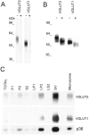
Fig. 1.
Fig. 1.
Antibody specificity and subcellular localization of VGLUT1 and VGLUT2 isoforms. A, Western blot strips of purified rat brain synaptic vesicles (Hell et al., 1988) were prepared and probed with rabbit polyclonal antisera raised against a GST fusion protein containing the C-terminal tails of rat VGLUT2 (DNPI) or VGLUT1 (BNPI). Broad bands are observed for VGLUT2 and VGLUT1 (−lanes) unless 10 μg/ml of the cognate fusion proteins are added (+ lanes). B, VGLUT2 and VGLUT1 are N-glycosylated. Rat brain synaptic vesicles (10 μg) were incubated for 1 hr at 37°C in the absence (−) or presence (+) of peptide-N-glycosidase F. A faster migrating species is observed after digestion. C, Fractionation of rat brain synaptosomes. The following rat brain fractions were analyzed for the presence of VGLUT2, VGLUT1, and the synaptic vesicle marker synaptophysin (p38): brain homogenate (TOTAL), low-speed pellet (P1), washed synaptosomes (P2), supernatant of the synaptosomal fraction (S2), low-speed pellet of the lysed synaptosomes (LP1), high-speed pellet of the lysed synaptosomes (LP2), synaptosomal cytosol (LS2), synaptic vesicles (SV) purified from LP2 by continuous sucrose density gradient fractionation, and the microsomal fraction (Microsome) of lysed synaptosomes.
Fig. 2.
Fig. 2.
Characterization of vesicular glutamate transport by VGLUT2 in transiently transfected PC12 cells. A, Transfection efficiency close to 100% and high expression levels are obtained as illustrated by the strong labeling visible after 30 min incubation with X-gal at 37°C in β-galactosidase-expressing PC12 cells.B, VGLUT2 is detected in transfected but not in mock-transfected PC12 cell homogenates. C, Time course of glutamate uptake in VGLUT2-transfected (●) and mock-transfected (○) PC12 cells. D, Saturation analysis of glutamate uptake by VGLUT2. VGLUT2-specific (mock-subtracted) uptake velocity reaches a plateau at ∼2 mm substrate.Inset, Lineweaver-Burk analysis of VGLUT2 initial velocity determines a Km of 0.8 mm and a Vmax of 190 pmol · min−1 · mg−1protein. E, VGLUT2 is highly specific for glutamate. Transport of glutamate (50 μm) was measured in 4 mm Cl-containing medium in the absence (100%) or presence of various amino acids (10 mm), K2PO4 (20 mm), or trypan blue (TB) (1 μm). F, VGLUT2 activity is H+ dependent. H+-ATPase inhibitors and dissipaters of the H+ electrochemical gradient abolish transport. Vesicle preparations were preincubated for 2 min with 1 μm bafilomycin A1 (bafilo), 200 μmN-ethyl maleimide (NEM), 50 μm carbonyl cyanidep-trifluoromethoxyphenylhydrazone (FCCP), or 20 mm KSCN (SCN). G, Anion specificity of VGLUT2 stimulation. Vesicle preparations were incubated in the absence or presence of the potassium salts of various inorganic anions (4 mm), chloride (Cl), bromide (Br), iodide (I), phosphate (PO4), thiocyanate (SCN), and in the presence of Cl plus 1 μmDIDS or 4 mm SCN.H, Transport is dependent on Δψ but can be driven by ΔpH. Vesicle preparations were incubated in the absence (control) or presence of 1 μmnigericin (+Nig), 1 μm valinomycin (+Val), or both (+Nig,+Val), in a medium containing 4 or 40 mm Cl and 5 mm Mg-ATP. Nonspecific transport (corresponding mock values) was subtracted.
Fig. 3.
Fig. 3.
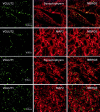
Confocal laser scanning double-immunofluorescence microscopy for VGLUT2 or VGLUT1 and the synaptic vesicle nerve terminal marker synaptophysin or the dendritic marker MAP-2. False color micrographs of confocal images from double immunofluorescence for VGLUT2 or VGLUT1 (green) and synaptophysin and microtubule-associated protein-2 (MAP2) (red) in the spinal dorsal horn. VGLUT2 and VGLUT1 staining coincide with staining for synaptophysin and are absent from neuronal dendrites. Note the presence of VGLUT2 staining in perisomatic and peridendritic synapses. Scale bar, 10 μm.
Fig. 4.
Fig. 4.
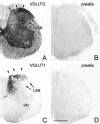
The distribution and assessment of the specificity of VGLUT2 and VGLUT1-ir in lumbar spinal cord. Immunoreactions inA and C are fully preabsorbed with the homologous recombinant fusion protein (B,D). Punctate immunostaining for VGLUT2 is present in the superficial dorsal horn (A, arrowheads mark lamina 1 and substantia gelatinosa) where VGLUT1-ir is minimal (C,arrowheads). Note accumulation of strongly positive punctate VGLUT1-ir in the deep dorsal horn where VGLUT2-ir is comparatively low. VGLUT2-ir is present in the lateral spinal nucleus (LSN) (A, arrows) where VGLUT1-ir is minimal (C, arrows). VGLUT1 immunostaining is low to moderate in the lateral ventral horn and sparse in the medial ventral horn. Fine punctate VGLUT2 staining is dense and abundant in the ventral horn (VH). Scale bar, 500 μm.
Fig. 5.
Fig. 5.
The distribution of VGLUT2 and VGLUT1 in the forebrain. A, H, Punctate VGLUT2-ir is relatively abundant throughout the hypothalamus and thalamus where VGLUT1 is restricted to the hypothalamic ventral premammillary nucleus (PMV) and to parts of the thalamic nuclei including the lateral posterior thalamic nucleus (LP), dorsal lateral geniculate nucleus (DLG), and ventral posteromedial thalamic nucleus (VPM).OPT, Olivary pretectal nucleus; APTD, dorsal anterior pretectal nucleus; PrC, precommissural nucleus. VGLUT2 staining is moderate in a band of the neocortex comprising lamina IV, weak in a neocortical band comprising lamina VI, and minimal in the other neocortical layers. Intense punctate VGLUT1-ir is abundant throughout the cortex including the piriform cortex (Pir) and somewhat less strong in the neocortical band of lamina IV where moderate VGLUT2 staining accumulates. Note mutual exclusion of VGLUT1 and VGLUT2 staining in the layers of the retrosplenial granular cortex (A,H, RSG) shown at high magnification inE and L. High magnifications (G, N) from lamina IV in F andM demonstrate different densities of VGLUT1- and VGLUT2-ir puncta between immunonegative neuronal cell bodies and processes. Sparse VGLUT2-ir puncta are mostly confined to the granular layer (g) of the dentate gyrus (DG) and pyramidal layer (p) of the fields CA1, CA2, and CA3 of the hippocampus. Dense VGLUT1-ir puncta are abundant throughout the hippocampus with the exception of the granule (g) and pyramidal (p) cell layers. Areas inrectangles in A and corresponding areas on adjacent section in H are shown at high magnification in BD andIK, respectively. Note differential distribution and density of VGLUT1-ir and VGLUT2-ir in the oriens layer (o), pyramidal layer (p), stratum radiatum (r), and stratum lacunosum moleculare (l) of CA1 (B,I) and CA3 (C,J) and in the molecular (m), granular (g), and polymorphic (p) layer of the dentate gyrus (DG) (D, K). Note some overlap but differential density and intensity of immunostaining for VGLUT1-ir and VGLUT2-ir puncta in the posterior basomedial amygdaloid nucleus (BMP), in the lateral amygdaloid nucleus (La), and in the cortical amygdaloid nucleus (Co) as well as in the adjacent dorsal endopiriform nucleus (DEn). Note that white matter and fiber tracts are VGLUT1 and VGLUT2 negative. pc, Posterior commissure; f, fornix; fr, fasciculus retroflexus; mt, mammillothalamic tract. Scale bars:A, E, 1.5 mm;BD, 50 μm;FH, 50 μm; I,J, 100 μm; K, L, 50 μm; M, N, 12.5 μm.
Fig. 6.
Fig. 6.
The distribution of VGLUT2 and VGLUT1 in the forebrain. Marked differences are apparent in the distribution, density, and intensity of punctate VGLUT2-ir (AD) and VGLUT1-ir (E–H) in the neocortex (laminaIV, VI), caudate putamen (CPu), globus pallidum (GP), piriform cortex (Pir), nucleus accumbens core (AcC), nucleus accumbens shell (AcSh), ventral pallidum (VP), olfactory tubercle (Tu), islands of Calleja (ICj), ventral diagonal band (VDB), and lateral septum (LS). In the CPu, VGLUT2 is somewhat less abundant than VGLUT1. VGLUT2 is present in the globus pallidum (GP) (B), where VGLUT1 is virtually absent (F). In piriform cortex (Pir) and islands of Calleja (ICj), punctate VGLUT1-ir is stronger and denser than VGLUT2-ir. Note accumulation of low to moderate VGLUT2-ir in the pyramidal cell layer in C where VGLUT1-ir is scarce (G). Note also some overlap and reciprocity in staining for VGLUT1 and VGLUT2 in the ICj (D, H). Note the absence of VGLUT1-ir and VGLUT2-ir from commissural fiber tracts.cc, Corpus callosum; ac, anterior commissure. Scale bars: A, C, 1 mm;B, D, 500 μm;EH, 200 μm.
Fig. 7.
Fig. 7.
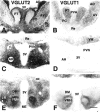
Nucleus-specific and differential abundance of VGLUT2 and VGLUT1 in thalamic and hypothalamic nuclei. Note high abundance of VGLUT2-ir (A) in the paraventricular thalamic nucleus (PVA), reuniens thalamic nucleus (Re), reticular thalamic nucleus (Rt), paracentral thalamic nucleus (PC), and anterodorsal thalamic nucleus (AD). Here, VGLUT1-ir (B) is almost absent or at low abundance. VGLUT1 (B) is moderately abundant in the posterior thalamic nucleus (PT) where VGLUT1 (A) is sparse. VGLUT1 is virtually absent from the stria medullaris (sm) where VGLUT2-ir is sparse. Adjacent frontal sections (C, D) of the diencephalon demonstrate the abundance of VGLUT2 (C) in the anterior hypothalamic nucleus (AH) but scarcity in the paraventricular nucleus (PVN) and extreme scarcity of VGLUT1-ir in the anterior hypothalamic nucleus (AH) and absence from the PVN. Note presence of VGLUT1 (D) but absence of VGLUT2 (C) in the ventromedial thalamic nucleus (VM). Adjacent frontal sections of the hypothalamus (E, F) demonstrate abundance of VGLUT2 in the lateral hypothalamic nucleus (LH), ventromedial hypothalamic nucleus (VMH), and dorsomedial hypothalamic nucleus (DM) and sparsity of VGLUT1 in the core of the VMH but moderate abundance in its shell. Note that VGLUT2 is less abundant in the shell and more abundant in the core of the VMH. Note some faint staining for VGLUT2 in the median eminence (ME). VGLUT1 and VGLUT2 are absent from fiber tracts of the fornix (f) and the mammillothalamic tract (mt). 3V, Third ventricle. Scale bar, 500 μm.
Fig. 8.
Fig. 8.
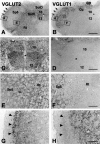
The distribution of VGLUT2 and VGLUT1 in the epithalamus. High-power micrographs from adjacent sections alternately stained for VGLUT2 (A) and VGLUT1 (B) demonstrating abundance of VGLUT2 in both the medial habenular nucleus (MHb) and the lateral habenular nucleus (LHb). Note that VGLUT1-ir in the MHb is less dense than VGLUT2-ir. VGLU1 is virtually absent from LHb. Scale bar, 100 μm.
Fig. 9.
Fig. 9.
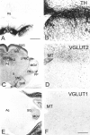
The distribution of VGLUT2, VGLUT1, and tyrosine hydroxylase (TH) in the mesencephalon and metathalamus. VGLUT2-ir puncta are concentrated in the tectum, with highest levels in the superficial gray layer of the superior colliculus (SuG) and lower levels in the intermediate gray layer of the superior colliculus (InG) and scarcity in the optic nucleus layer of the superior colliculus (Op). VGLUT2-ir puncta are present throughout the tegmentum, including the nucleus ruber (R) and the TH-positive pars compacta of the substantia nigra (SNC) and are enriched in the dorsal periaqueductal gray (PAG) and, in particular, in the medial terminal nucleus of the accessory optic tract (MT) as well as in the mediocaudal part of the lateral posterior nucleus (LPMC), in the posterior intralaminar thalamic nucleus (PIL), in the peripeduncular nucleus (PP), and in the suprageniculate thalamic nucleus (SG). Here, VGLUT1-ir is minimal. VGLUT1 staining is present at moderate levels in the ventral medial geniculate nucleus (MGV) where VGLUT2 is minimal. VGLUT1 is minimal throughout the tectum, periaqueductal gray, and tegmentum and virtually absent from the substantia nigra pars compacta (SNC) and pars reticularis (SNR). Weak VGLUT2 staining is present in neuronal perikarya and puncta in the SNR (D) high-power micrograph from area inrectangle in C where VGLUT1 is virtually absent (corresponding area in F).Aq, Mesencephalic aqueduct. Scale bars:A, C, E, 1 mm;B, F, D, 200 μm.
Fig. 10.
Fig. 10.
The distribution of VGLUT2 and VGLUT1 in the pontomedullary brainstem. High abundance of punctate VGLUT2-ir (B, D) in the medial superior olive (MSO) is observed where VGLUT1-ir is low (G, I). VGLUT2-ir is relatively low in the nucleus of the trapezoid body (TZ) (B, C) where VGLUT1-ir is at high abundance (G, H). Note that strongly VGLUT1-positive confluent large puncta encircle immunonegative neuronal cell bodies in the TZ (H). Strongly positive VGLUT-ir puncta are present in the principal sensory nucleus of the trigeminal nerve (Pr5) where VGLUT2-ir is very low. Moderately positive VGLUT1 puncta are present in the motor trigeminal nucleus (Mo5) where VGLUT-ir is low. VGLUT-ir and VGLUT2-ir are present at low abundance in the lateral and medial parabrachial nucleus (LPB,MPB). Moderate VGLUT2-ir accumulates in the locus coeruleus (LC), where VGLUT1-ir is very scarce. VGLUT1-ir and VGLUT2-ir are absent from the pyramidal tract (pyr). Adjacent sections (E,J) alternately stained for VGLUT2 (E) and VGLUT1 (J) reveal abundance of VGLUT1-ir in the anterior ventral cochlear nucleus (VCA) where VGLUT2 is virtually absent. High-power micrograph (inset) from J demonstrates strongly positive VGLUT1-ir puncta encircling immunonegative neuronal cell bodies and processes. VGLUT1-ir puncta outnumber VGLUT-2-positive puncta in the dorsal cochlear nucleus (DC), particularly in the superficial layer where VGLUT2 is absent (E,J). Scale bars: A,E, 500 μm; B, F,I, J, 200 μm; C,D, G, H, 25 μm.
Fig. 11.
Fig. 11.
The distribution of VGLUT2 and VGLUT1 in the lower brainstem. Moderate abundance of fine punctate VGLUT2-ir in superficial spinal trigeminal nucleus (Sp5) marked byarrowheads (A, G) is observed where VGLUT1-ir is virtually absent (B,H, arrowheads). VGLUT2 is moderately abundant in the dorsal motor nucleus of the vagus (10), hypoglossal nucleus (12), and reticular formation (Rt), and in the ventral part of the solitary tract (SolV) (A, C,E) where VGLUT1 is virtually absent (B,D, F). VGLUT2-ir is very low in the dorsal solitary tract (SolD). Note preponderance of VGLUT1 in deep Sp5 (B, F,H), in the cuneate (Cu), and in gracile nucleus (GR) where VGLUT2-ir is low.Asterisks mark the central canal. Scale bars:A, B, 500 μm; C,D, 200 μm; E, F, 100 μm; G, H, 100 μm.
Fig. 12.
Fig. 12.
The distribution of VGLUT1 and VGLUT2 in the cerebellum. A, B, Extreme density of intensely stained VGLUT1-positive puncta in the molecular layer (m), very sparse VGLUT1 puncta around somata of Purkinje cells in the Purkinje cell layer (p), and dense glomerula-like accumulation of strongly stained confluent VGLUT1 puncta in the granular layer (g) is observed. C, D, VGLUT2-ir puncta are much less dense in the molecular layer where they are arranged in a strand-like manner. VGLUT2-ir puncta forming glomerula-like structures in the glomerular layer (g) are less dense than those staining for VGLUT1. Scale bars: A,C, 500 μm; B, D, 100 μm.

Similar articles

Cited by

References

    1. Aicher SA, Milner TA, Pickel VM, Reis DJ. Anatomical substrates for baroreflex sympatho-inhibition in the rat. Brain Res Bull. 2000;51:107–110. - PubMed
    1. Aihara Y, Mashima H, Onda H, Hisano S, Kasuya H, Hori T, Yamada S, Tomura H, Yamada Y, Inoue I, Kojima I, Takeda J. Molecular cloning of a novel brain-type Na(+)-dependent inorganic phosphate cotransporter. J Neurochem. 2000;74:2622–2625. - PubMed
    1. al-Awqati Q. Chloride channels of intracellular organelles. Curr Opin Cell Biol. 1995;7:504–508. - PubMed
    1. Bellocchio EE, Hu H, Pohorille A, Chan J, Pickel VM, Edwards RH. The localization of the brain-specific inorganic phosphate transporter suggests a specific presynaptic role in glutamatergic neurotransmission. J Neurosci. 1998;18:8638–8659. - PMC - PubMed
    1. Bellocchio EE, Reimer RJ, Fremeau RT, Jr, Edwards RH. Uptake of glutamate into synaptic vesicles by an inorganic phosphate transporter. Science. 2000;289:957–960. - PubMed

Publication types

MeSH terms