. 2002 Jan 1;22(1):218-25.
doi: 10.1523/JNEUROSCI.22-01-00218.2002.

Zic2 controls cerebellar development in cooperation with Zic1

Affiliations

Zic2 controls cerebellar development in cooperation with Zic1

Jun Aruga et al. J Neurosci. .

Abstract

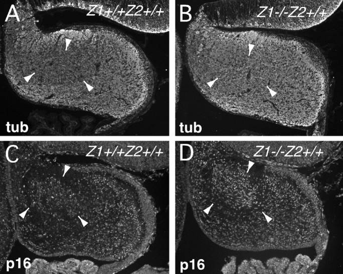
Mouse Zic genes encode zinc finger proteins and are expressed in the developing and mature CNS. Reduced expression of Zic2 in mice results in spina bifida and holoprosencephaly. However, the disruption of Zic1, a strong homolog of Zic2 that has an overlapping expression pattern, results in cerebellar malformation with no apparent abnormalities in the forebrain or in posterior neuropore closure. Here we revealed that Zic2 and Zic1 cooperatively control cerebellar development by regulating neuronal differentiation. Both Zic1 and Zic2 are expressed in the precursor cells of the granule neuron and the neurons in cerebellar nuclei. Mice carrying one mutated Zic1 allele together with one mutated Zic2 allele (Zic1(+/-)Zic2(+/kd)) showed a marked cerebellar folial abnormality similar to, but distinct from that found in mice homozygous for the Zic1 mutation (Zic1(-/-)). The Zic1(+/-)Zic2(+/kd) cerebellum is missing a lobule in the anterior vermis and has a truncation of the most posterior lobule. Expression of transverse zonal markers is shifted anteriorly in the developing cerebellum, indicating that the anterior part of the cerebellum is poorly developed. Abnormalities in the developing Zic1(+/-)Zic2(+/kd) cerebellum share the following features with those of the Zic1(-/-) cerebellum: a preceding reduction of cell proliferation in the anterior external germinal layer, a reduction in cyclin D1 expression, and enhanced expression of the mitosis inhibitors p27 and p16, and enhancement of Wnt7a expression. These results indicate that Zic1 and Zic2 may have very similar functions in the regulation of cerebellar development.


Figures

Fig. 1.
Fig. 1.
The expression of Zic1 andZic2 in the developing cerebellum. In situ hybridization was performed on coronal (A–H), horizontal (I, J, M, N), and sagittal (K, L, O, P) sections of E16 (A, B, E, F, I, K, L, M, O, P) and E18 (C, D, G, H, J, N) wild-type cerebella with Zic2(A–D, I–L) and Zic1 (E–H, M–P) probes. B, D,F, and H are higher magnifications ofA, C, E, andG, respectively. Zic1 andZic2 are expressed in a similar pattern. A significant difference between Zic1 and Zic2expression was found in the vermal (medial) intermediate region (I, J, M, N). Asterisks inK and O indicate midsagittal expression in I and M, respectively.EGL, External germinal cell layer; MS, medial septum; PN, pontine nuclei; PO, preoptic nuclei, TN, thalamic nuclei.
Fig. 2.
Fig. 2.
Cerebellar phenotype in Zic2knock-down mice. Coronal (A, C) and sagittal (B, D) hematoxylin and eosin-stained sections were prepared from E16 wild-type (Zic2+/+) (A, B) and Zic2kd/kd(C, D) embryos. The distorted shape suggests oppression, which is probably caused by disturbed CSF circulation. It is difficult to judge whether Zic2 has a primary role in the cerebellar development, from Zic2kd/kdphenotypes. CN, Cerebellar nuclei forming cells;EGL, external germinal cell layer.
Fig. 3.
Fig. 3.
Macroscopic observation of theZic1+/−Zic2+/kdcerebella. Cerebella from wild-type (Zic1+/+Zic2+/+) (A) andZic1+/−Zic2+/kd(B, C) at P17 are seen from dorsoposterior (A, B) or posterior (C) direction. Folial patterns are markedly altered in theZic1+/−Zic2+/kd, different from that ofZic1−/−. Vermis lobules are indicated by V–IX. a, Anterior colliculus; p, posterior colliculus; CI, CrusI lobule; CII, CrusII lobule; S, simplex lobule; P, paramedian lobule. Scale bar, 5 mm.
Fig. 4.
Fig. 4.
Comparison of the cerebellar folial patterns in sections. Sagittal sections through cerebellar vermis (left) and hemisphere (right) fromZic1+/+Zic2+/+(A),Zic1+/−Zic2+/+(B),Zic1+/+Zic2+/kd(C),Zic1+/−Zic2+/kd(D), andZic1−/−Zic2+/+(E), at P16 (A–D) or 3 weeks (E) mice. Vermis lobules are indicated byIII–X. A, Anterior lobe;CI, CrusI lobule; CII, CrusII lobule;S, simplex lobule; PM, paramedian lobule;P, pyramidis. Asterisks indicate dorsal (*) and ventral (**) paraflocculus lobules. Circlesindicate abnormally located lobules, which are not assigned at this point.
Fig. 5.
Fig. 5.
Morphometric analyses of the mutant cerebella. The areas of the granular cell layer (GCL) and the molecular layer (ML) in midsagittal (vermis) and parasagittal sections through hemisphere (hemisphere) were measured. The cerebella were derived from P16 single litter. The areas are indicated in square millimeters. The error bars indicate SD.White bar,Zic1+/+Zic2+/+(+/+); black bar,Zic1+/−Zic2+/kd(Z1/Z2); hatched bar,Zic1+/−Zic2+/+(Z1/+); stippled bar,Zic1+/+Zic2+/kd(+/Z2); *p < 0.05; **p < 0.01.
Fig. 6.
Fig. 6.
Anterior to posterior patterning abnormality found in theZic1+/−Zic2+/kdcerebella. Sagittal sections from E18 (A–F) and E16 (G, H) cerebella ofZic1+/+Zic2+/+(A, C, E, G),Zic1−/−Zic2+/+(B), andZic1+/−Zic2+/kd(D, F, H). A, C,E, and G are the littermates ofB, D, F, andH, respectively. A–D, Hematoxylin and eosin staining; E, F, immunohistochemical staining using anti-calbindin antibody (immature Purkinje cells).Arrowheads indicate the corresponding posterior and anterior boundaries of the clusters of Purkinje cells.G, H, Anti-EphA3 antibody staining, which stains a transversal zone corresponding to prospective lobule VII.Arrowheads indicate the strongly stained zone in each panel. I, J, Coronal sections of E18Zic1+/+Zic2+/+(I) andZic1+/−Zic2+/kd(J) embryos. K, L, En2expression in the E17 horizontal section ofZic1+/+Zic2+/+(K) andZic1+/−Zic2+/kd(L) are shown by in situhybridization. Note that the anterior region is reduced, as indicated by the folial and the marker expression patterns, whereas there are no apparent abnormalities in the medial to lateral patterning.
Fig. 7.
Fig. 7.
Proliferating cell numbers in theZic1/Zic2 cerebellum. Cells stained with anti-phospho-histone H3 antibodies (mitotic cells) were counted in the indicated regions of the E16 (A) and E17 (B) cerebellar sagittal sections. The ordinate indicates the mean number of the labeled cells per section. InC, mean numbers of the labeled cells in 1 mm of the EGL are indicated. The error bars indicate SD. ant EGL, Anterior EGL; post EGL, posterior EGL;VL, ventricular layer; int(intermed), intermediate zone that includes the area in the cerebellar primorium except EGL and VL; +/+,Zic1+/+Zic2+/+; Z1/Z2,Zic1+/−Zic2+/kd; **p < 0.01
Fig. 8.
Fig. 8.
Genes controlling cerebellar development are deregulated in theZic1−/− andZic1+/−Zic2+/kdcerebella. RT-PCR analysis was performed on cDNAs prepared from cerebella of E17.5Zic1−/− andZic1+/−Zic2+/kdand their littermate wild-type controls. G3PDH is a ubiquitously expressed gene. In the absence of reverse transcriptase [G3PDH (-RT)], no bands are detected, ensuring the absence of DNA. Amounts of cyclin D1, cyclin D2, p27, and Wnt7a transcripts were compared.
Fig. 9.
Fig. 9.
Premature neuronal differentiation occurs in theZic1−/− cerebella. Immunohistochemical staining showing localization of βIII tubulin (A, B) and p16 (C, D) proteins were examined in the in E16 cerebella ofZic1−/−. Wild-type littermates (A, C) are shown as controls forB and D, respectively. The area surrounded by the arrowheads indicates the corresponding region between the mutants and wild-type controls, which showed significant differences.

Similar articles

Cited by

References

    1. Altman J, Bayer SA. Development of the cerebellar system. CRC; New York: 1997. Comparative anatomy of the cerebellum, an evolutionally perspective. pp. 3–20.
    1. Aruga J, Yokota N, Hashimoto M, Furuichi T, Fukuda M, Mikoshiba K. A novel zinc finger protein, Zic, is involved in neurogenesis, especially in the cell lineage of cerebellar granule cells. J Neurochem. 1994;63:1880–1890. - PubMed
    1. Aruga J, Nagai T, Tokuyama T, Hayashizaki Y, Okazaki Y, Chapman VM, Mikoshiba K. The mouse Zic gene family: homologues of Drosophila pair-rule gene odd-paired. J Biol Chem. 1996;271:1043–1047. - PubMed
    1. Aruga J, Minowa O, Yaginuma H, Kuno J, Nagai T, Noda T, Mikoshiba K. Mouse Zic1 is involved in cerebellar development. J Neurosci. 1998;18:284–293. - PMC - PubMed
    1. Aruga J, Mizugishi K, Koseki H, Imai K, Balling R, Noda T, Mikoshiba K. Zic1 regulates the patterning of vertebral arches in cooperation with Gli3. Mech Dev. 1999;89:141–150. - PubMed

Publication types

MeSH terms