. 2002 Jan 1;22(1):226-38.
doi: 10.1523/JNEUROSCI.22-01-00226.2002.

Interaction between metabotropic and NMDA subtypes of glutamate receptors in sprout suppression at young synapses

Affiliations

Interaction between metabotropic and NMDA subtypes of glutamate receptors in sprout suppression at young synapses

Frank Miskevich et al. J Neurosci. .

Abstract

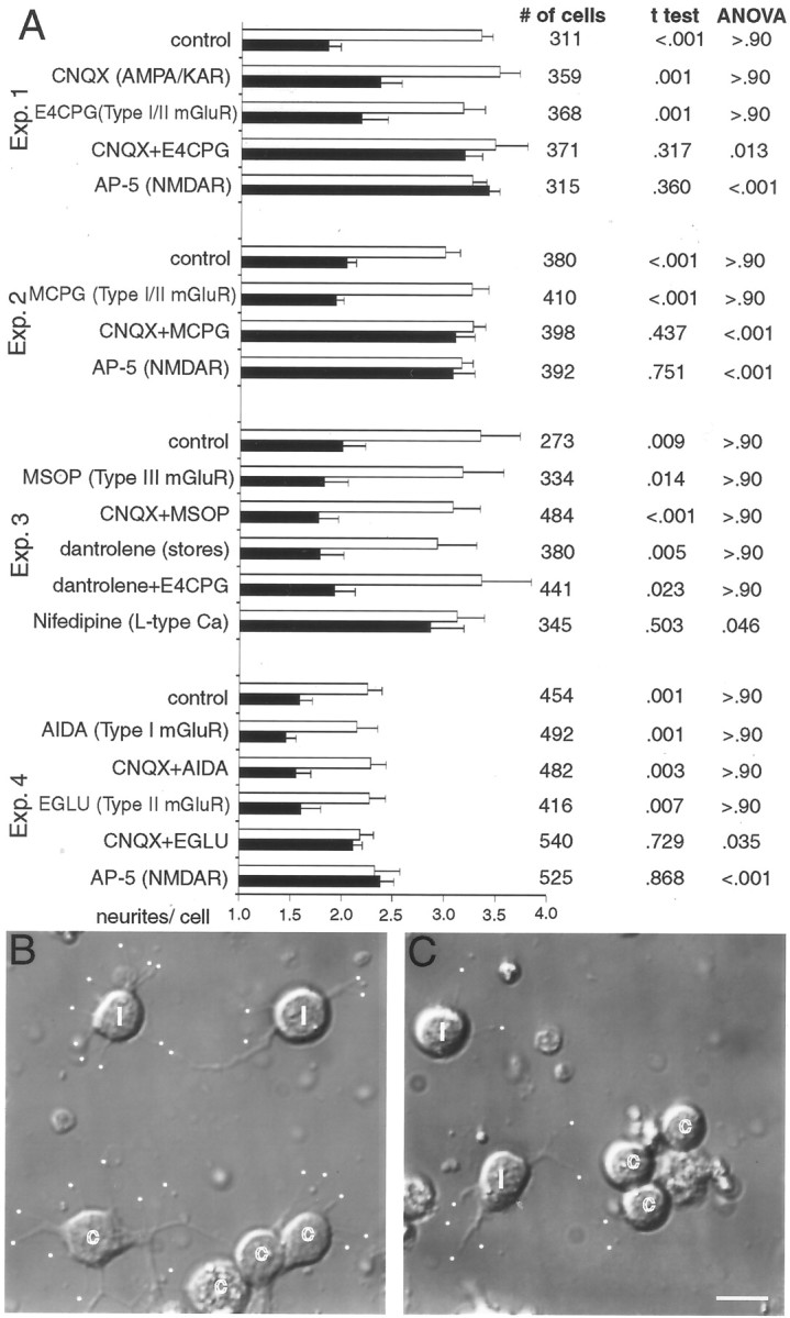
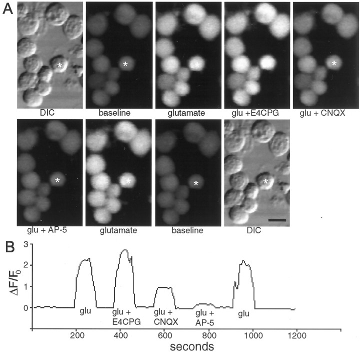
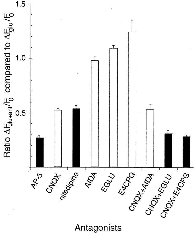
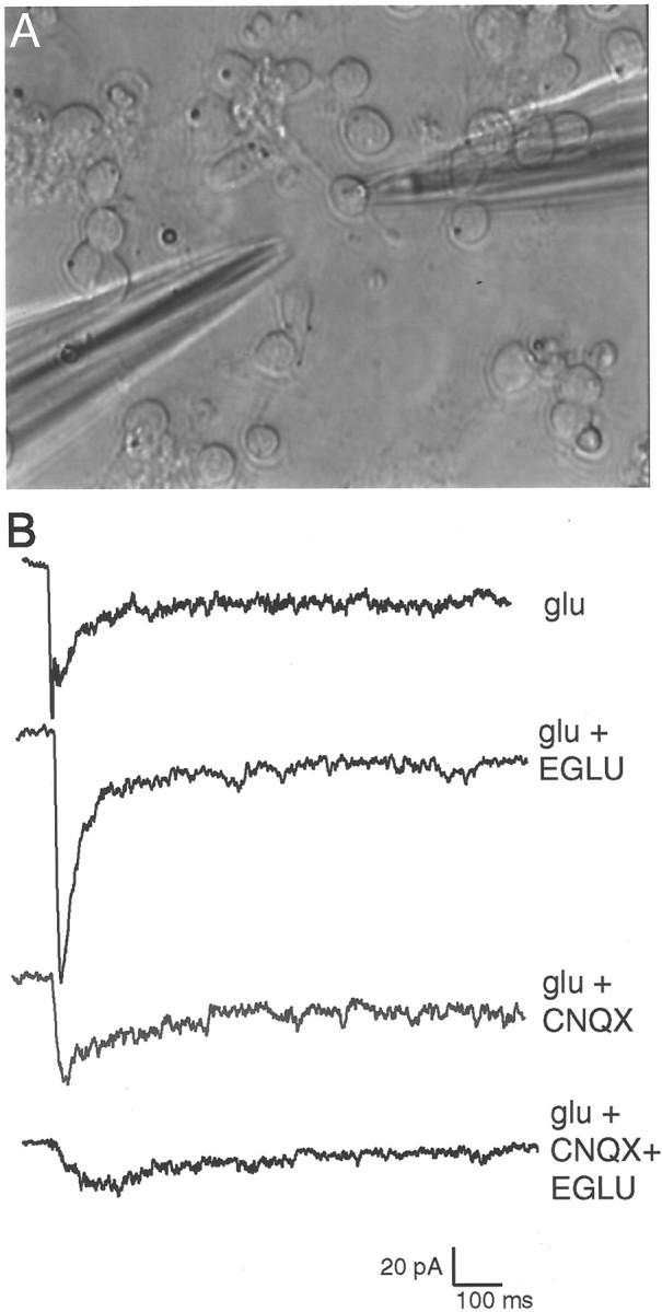
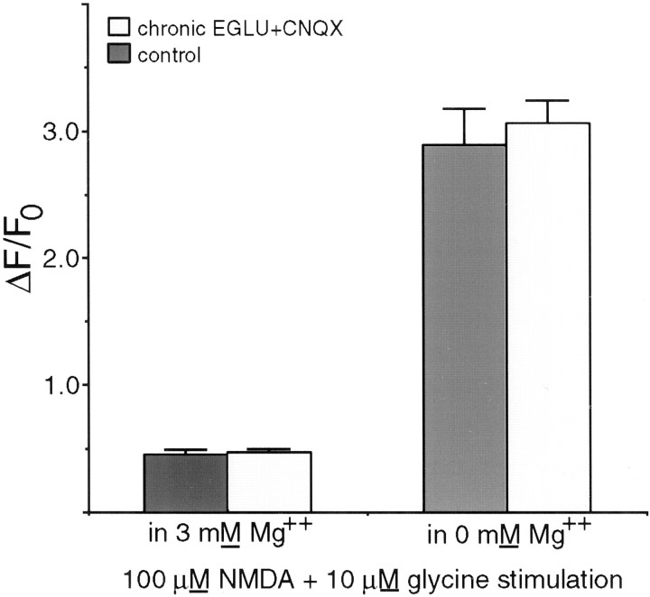
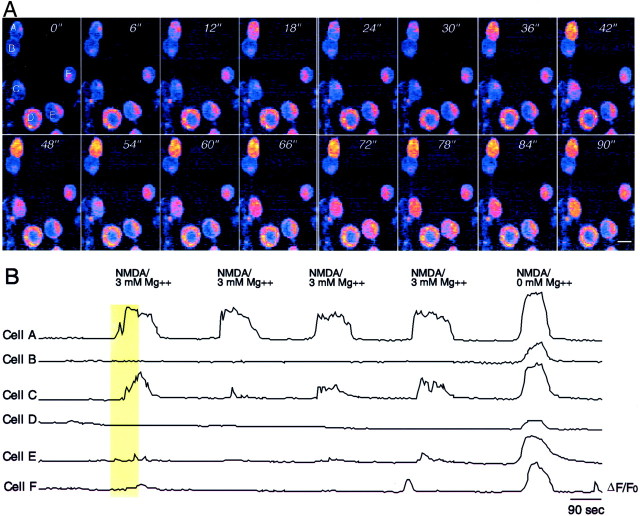
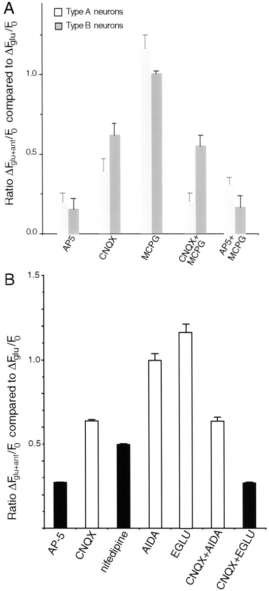
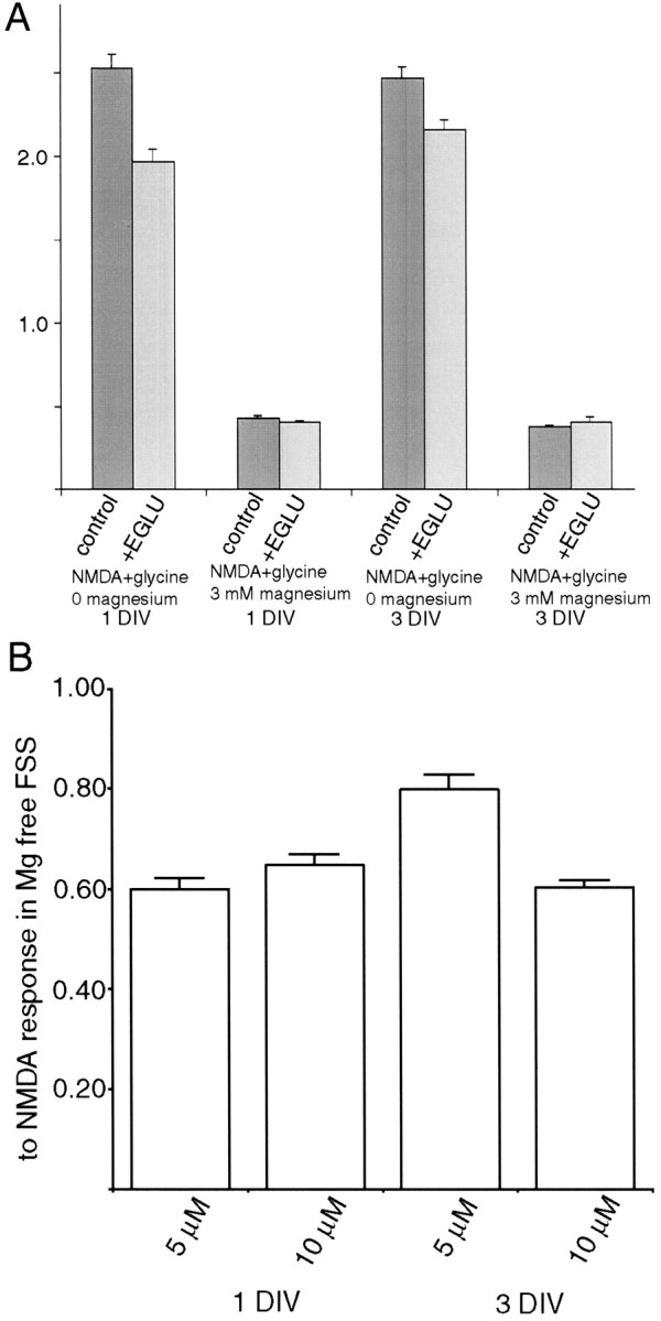
Recently, NMDA receptors (NMDARs) have been implicated in a cell contact-dependent suppression of sprouting in cultured Xenopus tectal neurons during an early period when neither AMPA/kainate (KA) receptors nor action potentials play a prominent role in cell-cell communication. We asked how the NMDA receptors function in the absence of the depolarizing effect of AMPA/KA receptor activity. We show that type II metabotropic glutamate receptors (mGluRs) can operate synergistically with NMDA receptors in the absence of AMPA/KA receptor function to suppress an early neurite sprouting response of the tectal neurons. Calcium imaging with fluo-3 AM and morphological analyses after exposure to glutamate receptor antagonists show that a combination of AMPA/KA receptor and type II mGluR blockers mimics the decrease in intracellular free calcium in response to glutamate and the structural effects produced by NMDA receptor antagonists in these cultures. Patch-clamp analyses confirmed a decrease in NMDA receptor-mediated currents with type II mGluR blockade, and 8-bromo cAMP application produced a decrease in NMDA receptor-mediated calcium influx. These data suggest that type II mGluRs potentiate NMDA receptor function by decreasing cAMP levels in tectal neurons. We also show that NMDARs exhibit low magnesium sensitivity in tectal neurons during the first few days in culture. Thus both metabotropic and ionotropic glutamate receptors can play a role in the contact-mediated suppression of ongoing sprouting at early neuron-neuron contacts before action potential activity.


Figures

Fig. 1.
Fig. 1.
Chronically applied AMPA/KAR and type II mGluR antagonists increase sprouting in contacted tectal neurons by 3 DIV. A, Quantification of the average number of free neurite ends on contacted (black bars) and isolated (white bars) cells in the same fields, along with their SEs in the presence of various glutamate receptor antagonists. Each of the four experiments used cells from a single dissociation, and cells from two coverslips in the dissociation were analyzed for each antagonist treatment. A two-tailed Student'st test was performed between contacted and isolated cells on the same coverslip to compare the number of neurites per cell, and mANOVA analysis using a Tukey post hoc test compared differences between the number of neurites per contacted cell under different treatments within a given experiment. Statistics were not applied across experiments because of the dissociation-associated differences in overall sprouting. Multiple ANOVA analysis of free neurite ends among different treatments within a given experiment consistently revealed no changes in sprouting of isolated cells across the control and treatment groups. B, Differential interference contrast (DIC) images of dissociated tectal neurons in cultures treated with 20 μm CNQX and 0.2 mmEGLU show significant sprouting from both isolated and contacted cells. Isolated cells are labeled with i, contacted cells are labeled with c, and free neurite ends are marked with awhite dot. C, Control image showing more free neurite ends on isolated compared with contacted cells. Scale bar, 10 μm.
Fig. 2.
Fig. 2.
Fluo-3 AM imaging of the calcium response to glutamate and its reduction by glutamate receptor antagonists. A, Representative DIC and fluorescent images of Xenopus tectal neurons under various stimulation conditions. CNQX and AP-5 reduce the fluorescence increase in response to glutamate, whereas E4CPG increases the calcium response without permanently altering the baseline fluorescence or impairing the ability of the cell to respond to subsequent applications of glutamate after removal of the antagonists. Scale bar, 10 μm.B, Graph of ΔF/F0 versus time for the single cell marked by the asterisks in Ashowing the quantitation for the glutamate-induced calcium-dependent fluorescence increases in the presence of various glutamate receptor antagonists.
Fig. 3.
Fig. 3.
Quantitation of the calcium response to glutamate in the presence of glutamate receptor antagonists or calcium channel blockers. The figure shows the calcium response of cells treated with glutamate plus various antagonists (ΔFglu+ant) as a proportion of the cellular response to glutamate alone (ΔFglu) at 3 DIV. Type I (AIDA) mGluR blocker alone does not alter calcium flux (n = 34 cells), although drugs that block the type II mGluR response (EGLU and E4CPG) significantly increase calcium influx (n = 43 and 34 cells, respectively) when applied individually. CNQX and nifedipine decrease calcium flux by 48 and 46% (n = 140 cells each), whereas AP-5 treatment decreases calcium flux by 73% (n = 140 cells). The reduction caused by the type I mGluR blocker (AIDA) plus CNQX is not different from the reduction with CNQX alone (47% decrease; n = 34 cells), whereas CNQX plus either the type I/II (E4CPG) or type II (EGLU) mGluR antagonists show a calcium flux that is decreased by ∼70% compared with glutamate alone (n = 140 and 43 cells, respectively). ANOVA analysis of the various treatment groups shows that AP-5, CNQX + E4CPG, and CNQX + EGLU produce significantly lower calcium fluxes than other treatments (p < 0.02; Tukey post hoc tests). Black bars denote those antagonist treatments that increase the sprouting of contacted tectal neurons at 3 DIV.
Fig. 4.
Fig. 4.
A, A 3 DIV Xenopus culture with a perforated-patch recording electrode (right side) and a pipette containing glutamate to be puffed onto the cell (left side). Spontaneous synaptic currents are not detectable at this stage in culture. B, Electrophysiological recordings from a single cell in response to identically puffed glutamate in the presence of various glutamate receptor blockers. Blockade of type II mGluRs alone using EGLU causes a substantial increase in glutamate response in this neuron, whereas CNQX decreases the glutamate response. Combined type II mGluR and AMPA/KAR blockade (EGLU+CNQX) decreases glutamate response by a larger amount than CNQX alone. These results are consistent with the calcium imaging results reported in Figure 3, and the results of recording from several cells are summarized in Table 1.
Fig. 5.
Fig. 5.
Chronic treatment of dissociatedXenopus tectal neurons with the type II mGluR antagonist EGLU and AMPA/KAR blocker (CNQX) for the first 3 DIV does not change the NMDAR-mediated calcium response of cultured tectal neurons to NMDA and glycine. The two sets of cultures had indistinguishable responses regardless of whether magnesium ions were present in the testing medium.
Fig. 6.
Fig. 6.
NMDARs on some early Xenopus tectal neurons have low magnesium sensitivity. A, Frames showing a field of six neurons at 1 DIV after loading with fluo-3 AM. Images collected at 6 sec intervals illustrate the NMDA-induced calcium fluorescence in a recording solution containing 3 mmmagnesium. Two cells (A, C) have significant calcium influx in response to 100 μm NMDA + glycine in 3 mm magnesium, whereas four other cells show little or no response. B, Plot of ΔF/F0 for the same cells over a longer time interval as well as their response to NMDA in magnesium-free solution. The vertical yellow bar shows the frames of the record illustrated in A. Scale bar, 10 μm.
Fig. 7.
Fig. 7.
Alterations of the calcium response to glutamate in the presence of glutamate receptor antagonists at 1 DIV.A, Comparison of neurons showing normal (Type A neurons) or reduced (Type B neurons) magnesium sensitivity in response to NMDA + glycine stimulation. As expected, CNQX is less effective at decreasing the calcium influx in cells with low magnesium sensitivity compared with its effect on cells with normal magnesium sensitivity. The effect of type I/II mGluR and AMPA/KAR blockade (CNQX+MCPG) on the calcium response to glutamate is also more pronounced in type A cells showing a normal magnesium sensitivity. MCPG alone actually increases the calcium response to glutamate in these type A neurons. MCPG + AP-5 treatment yields a higher calcium influx through type A neurons compared with AP-5 alone, consistent with a normal weak suppression of calcium entry through AMPA/KAR receptors by type II mGluRs. B, Quantitation of the reduction of the calcium response to glutamate by glutamate receptor and calcium channel antagonists at 1 DIV. CNQX is relatively less effective at 1 DIV (n = 57 cells) than at 3 DIV (Fig. 3) in blocking the glutamate-induced calcium response of tectal neurons. Nifedipine, however, depresses the calcium response to the same extent at both 1 (n = 57 cells) and 3 DIV. The type II mGluR antagonist EGLU (but not the type I antagonist AIDA) in combination with CNQX further decreases the calcium entry in response to glutamate (n = 48 cells). Neither mGluR antagonist alone reduces the calcium response to glutamate, and in fact the calcium response to glutamate when both ionotropic receptor types are functioning is actually increased in the presence of EGLU (ANOVA Tukey post hoc test; p = 0.015).Black columns denote antagonists that resulted in increased free neurite ends among contacted cells after 3 d of chronic application in vitro.
Fig. 8.
Fig. 8.
Regulation of NMDAR-mediated calcium influx by acute blockade of type II mGluRs. A, EGLU presented with NMDA + glycine in magnesium-free solution, but not in 3 mmmagnesium, reduces the neuronal calcium response to NMDA. This response is most pronounced at 1 DIV (n = 78 cells;p < 0.0001; Student's t test) but is still significant at 3 DIV (n = 94 cells;p = 0.0039; Student's t test), suggesting that type II mGluR activity normally facilitates calcium influx through the NMDAR using a mechanism that does not alter magnesium sensitivity. B, The cell-permeable cAMP analog 8-Br-cAMP decreases calcium influx through the NMDAR. This decrease is seen at both 1 and 3 DIV, but cells appear to be more sensitive to 8-Br-cAMP at 1 DIV on the basis of the relative responses at each age to 5 μm 8-Br-cAMP (p < 0.0001; ANOVA Tukey post hoc test).n = 104 cells at 1 DIV, 5 μm;n = 107 cells at 1 DIV, 10 μm;n = 88 cells at 3 DIV, 5 μm; andn = 136 cells at 3 DIV, 10 μm.

Similar articles

Cited by

References

    1. Aniksztejn L, Bregestovski P, Ben-Ari Y. Selective activation of quisqualate metabotropic receptor potentiates NMDA but not AMPA responses. Eur J Pharmacol. 1991;205:327–328. - PubMed
    1. Banke TG, Bowie D, Lee H-K, Huganir RL, Schousboe A, Traynelis SF. Control of gluR1 AMPA receptor function by cAMP-dependent protein kinase. J Neurosci. 2000;20:89–102. - PMC - PubMed
    1. Beaver CJ, Ji Q, Daw NW. Effect of the group II metabotropic glutamate agonist, 2R,4R-APDC, varies with age, layer, and visual experience in the visual cortex. J Neurophysiol. 1999;82:86–93. - PubMed
    1. Ben-Ari Y. Developing networks play a similar melody. Trends Neurosci. 2001;24:353–360. - PubMed
    1. Burnashev N. Calcium permeability of ligand-gated channels. Cell Calcium. 1998;24:325–332. - PubMed

Publication types

MeSH terms

LinkOut - more resources