. 2002 Jan 1;22(1):350-6.
doi: 10.1523/JNEUROSCI.22-01-00350.2002.

Circadian rhythms in isolated brain regions

Affiliations

Circadian rhythms in isolated brain regions

Michikazu Abe et al. J Neurosci. .

Abstract

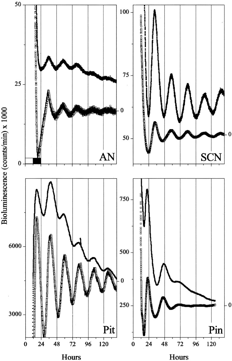
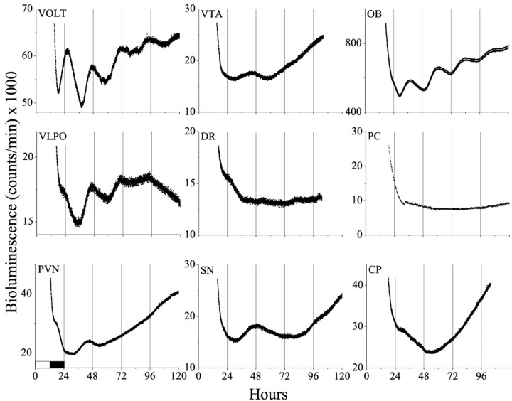
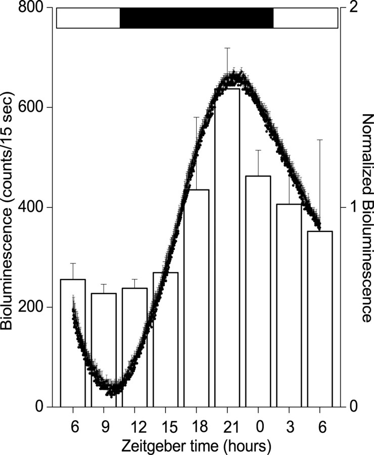
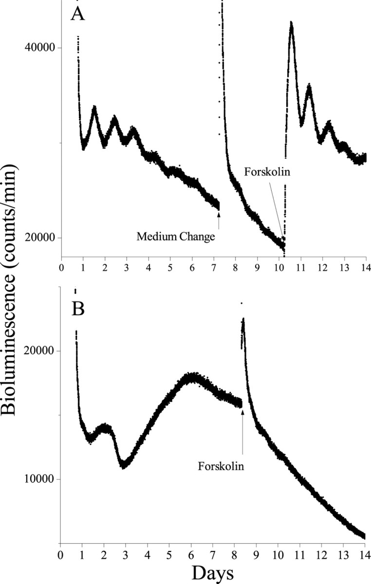
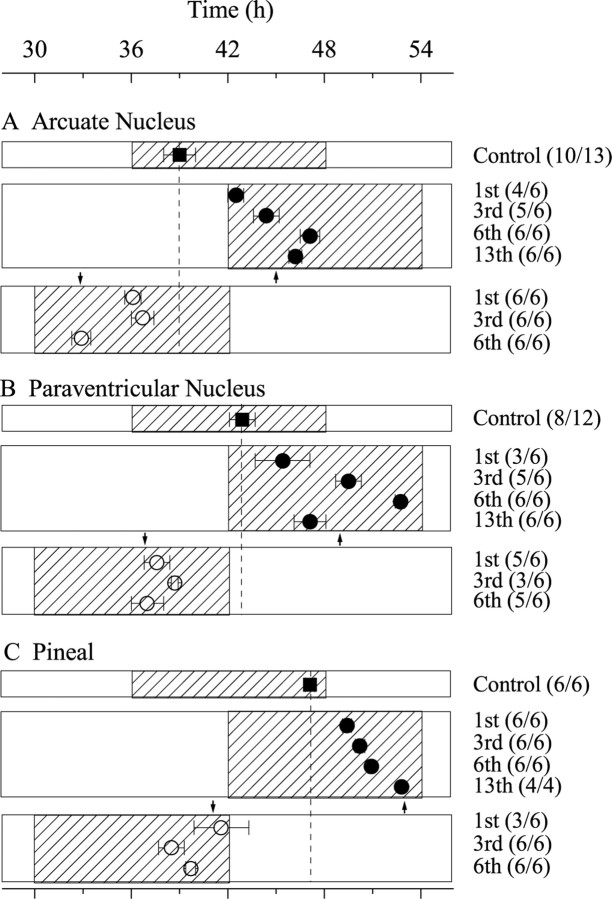
The suprachiasmatic nucleus (SCN) of the mammalian hypothalamus has been referred to as the master circadian pacemaker that drives daily rhythms in behavior and physiology. There is, however, evidence for extra-SCN circadian oscillators. Neural tissues cultured from rats carrying the Per-luciferase transgene were used to monitor the intrinsic Per1 expression patterns in different brain areas and their response to changes in the light cycle. Although many Per-expressing brain areas were arrhythmic in culture, 14 of the 27 areas examined were rhythmic. The pineal and pituitary glands both expressed rhythms that persisted for >3 d in vitro, with peak expression during the subjective night. Nuclei in the olfactory bulb and the ventral hypothalamus expressed rhythmicity with peak expression at night, whereas other brain areas were either weakly rhythmic and peaked at night, or arrhythmic. After a 6 hr advance or delay in the light cycle, the pineal, paraventricular nucleus of the hypothalamus, and arcuate nucleus each adjusted the phase of their rhythmicity with different kinetics. Together, these results indicate that the brain contains multiple, damped circadian oscillators outside the SCN. The phasing of these oscillators to one another may play a critical role in coordinating brain activity and its adjustment to changes in the light cycle.


Figures

Fig. 1.
Fig. 1.
The SCN differs from other brain areas in the phase and amplitude of its circadian rhythmicity.Per1-luc rhythms from isolated tissues show that, whereas the SCN peaks during the subjective day, the Pin gland, Pit, and AN peak in the subjective night. In addition, circadian expression damps out in the extra-SCN regions, whereas rhythmicity persists within the cultured SCN. Tissues were explanted from Per1-luctransgenic rats 1 hr before light offset (whiteand black bars in the top left plot indicate light and dark periods in the animal colony). Shown are the raw (filled circles) and detrended bioluminescence (open squares; see Materials and Methods) for representative cultures.
Fig. 2.
Fig. 2.
Circadian rhythmicity is not ubiquitous in the brain. Brain areas of diencephalic origin, such as the VOLT, VLPO, and PVN, showed damped circadian rhythmicity for one to four cyclesin vitro, whereas most others did not (e.g., VTA, DR, SN, PC, and CP). The OB was the only telencephalic structure tested that showed circadian rhythmicity.
Fig. 3.
Fig. 3.
Pineal rhythmicity is similar in vivo and in vitro. Reporter construct activity was assayed from explanted pineal glands, either acutely at different times in the light cycle (open bars; mean ± SEM;n = 4 at each time point) or from cultured glands in continuous darkness (filled circles;n = 6). Bioluminescence profiles from cultured pineals were normalized to their peak emission. Acute and continuous measurements peaked ∼3 hr before light onset.
Fig. 4.
Fig. 4.
Damped circadian rhythmicity can be reinstated in tissues that were previously rhythmic in vitro. Although changing the medium had little effect on bioluminescence in transgenic AN cultures, exposure to forskolin (5 min, 10 μm) evoked circadian oscillations that damped over the following 4 d (A). In contrast, rhythmicity was not seen in cultured NA, even after forskolin treatment (B). These results indicate that these tissues are healthy in vitro and retain different abilities to express autonomous circadian rhythmicity.
Fig. 5.
Fig. 5.
Brain areas reentrain at different rates after a shift in the light schedule. Relative to tissues taken from unshifted animals (filled squares), tissues taken from animals that experienced a 6 hr delay (filled circles) or advance (open circles) took many days to resume their normal phase relationship to the light cycle. Plotted against time since the last light onset of the control group, rhythmicity in the arcuate nucleus (A) peaked 3 hr later than usual after 1 d in the delayed light cycle and appeared close to entrained after 3 d in the new schedule (numbers in parentheses indicate rhythmic cultures per number of cultures tested, and arrowsindicate the point of complete phase shift). After a 6 hr advance, the AN assumed its normal phase after 6 d in the novel light cycle. In contrast, the PVN (B) and pineal (C) appeared to reentrain more rapidly to the advance but took much longer to synchronize to the delayed schedule.

References

    1. Albrecht U, Sun ZS, Eichele G, Lee CC. A differential response of two putative mammalian circadian regulators, mper1 and mper2, to light. Cell. 1997;91:1055–1064. - PubMed
    1. Aschoff J, Wever RA. Human circadian rhythms: a multioscillator system. Fed Proc. 1976;35:2326–2332. - PubMed
    1. Aschoff J, Wever RA. Handbook of behavioral neurology. Plenum; New York: 1981. The circadian system of man. pp. 311–331.
    1. Bae K, Jin X, Maywood ES, Hastings MH, Reppert SM, Weaver DR. Differential functions of mPer1, mPer2, and mPer3 in the SCN circadian clock. Neuron. 2001;30:525–536. - PubMed
    1. Balsalobre A, Damiola F, Schibler U. A serum shock induces circadian gene expression in mammalian tissue culture cells. Cell. 1998;93:929–937. - PubMed

Publication types

MeSH terms

LinkOut - more resources