. 2002 Jan;22(2):555-66.
doi: 10.1128/MCB.22.2.555-566.2002.

A DNA damage-regulated BRCT-containing protein, TopBP1, is required for cell survival

Affiliations

A DNA damage-regulated BRCT-containing protein, TopBP1, is required for cell survival

Kazuhiko Yamane et al. Mol Cell Biol. 2002 Jan.

Abstract

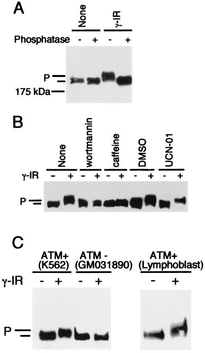
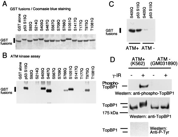
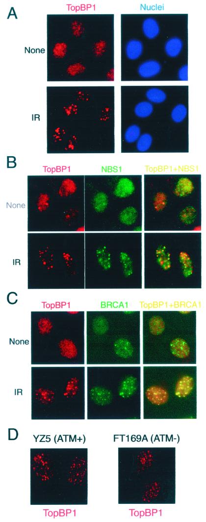
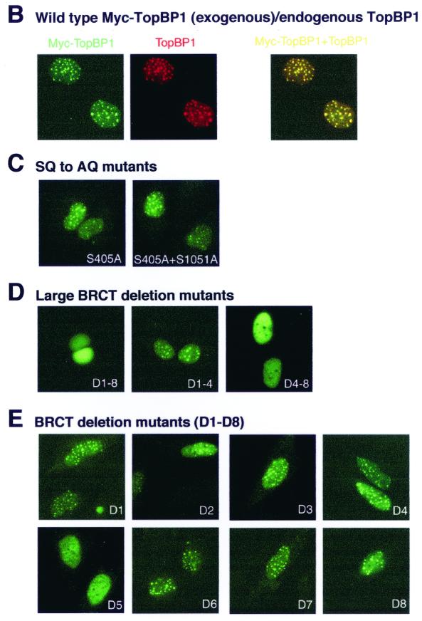
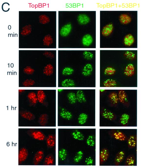
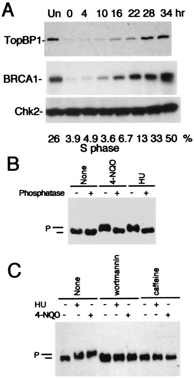
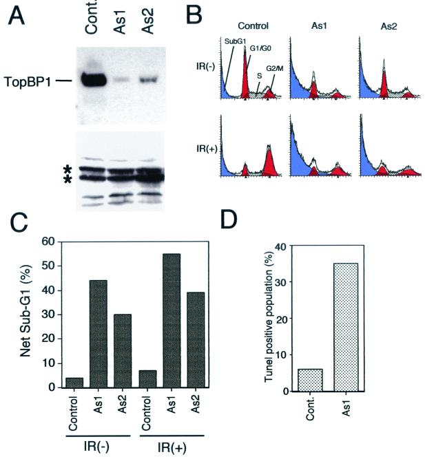
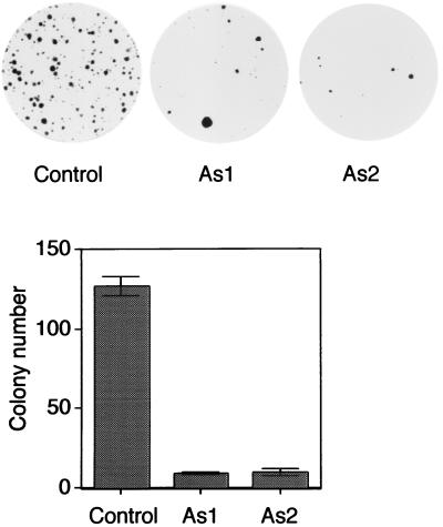
BRCA1 carboxyl-terminal (BRCT) motifs are present in a number of proteins involved in DNA repair and/or DNA damage-signaling pathways. Human DNA topoisomerase II binding protein 1 (TopBP1) contains eight BRCT motifs and shares sequence similarity with the fission yeast Rad4/Cut5 protein and the budding yeast DPB11 protein, both of which are required for DNA damage and/or replication checkpoint controls. We report here that TopBP1 is phosphorylated in response to DNA double-strand breaks and replication blocks. TopBP1 forms nuclear foci and localizes to the sites of DNA damage or the arrested replication forks. In response to DNA strand breaks, TopBP1 phosphorylation depends on the ataxia telangiectasia mutated protein (ATM) in vivo. However, ATM-dependent phosphorylation of TopBP1 does not appear to be required for focus formation following DNA damage. Instead, focus formation relies on one of the BRCT motifs, BRCT5, in TopBP1. Antisense Morpholino oligomers against TopBP1 greatly reduced TopBP1 expression in vivo. Similar to that of ataxia telangiectasia-related protein (ATR), Chk1, or Hus1, downregulation of TopBP1 leads to reduced cell survival, probably due to increased apoptosis. Taken together, the data presented here suggest that, like its putative counterparts in yeast species, TopBP1 may be involved in DNA damage and replication checkpoint controls.


Figures

FIG. 1.
FIG. 1.
TopBP1 is phosphorylated in response to DNA damage. (A) K562 cells were treated with γ irradiation (γ-IR) (10 Gy) or left untreated (None). After 1 h, whole-cell lysates were immunoprecipitated with anti-TopBP1 antibody. Duplicate samples were treated with lambda phosphatase (+) or left untreated (−). Western blotting was performed with anti-TopBP1 antibody. (B) Treatments of cells with wortmannin (100 μM), caffeine (10 mM), or UCN-01 (1 μM) were performed 30 min before γ irradiation. Dimethyl sulfoxide (DMSO) was the solvent for wortmannin and was used here as a negative control. Duplicate samples were treated with γ irradiation (+) (10 Gy) or left untreated (−) and analyzed by Western blotting. (C) Cells expressing wild-type ATM (K562 and normal lymphoblast) and ATM-deficient cells (GM031890) were treated with γ irradiation (+) (10 Gy) or left untreated (−) and collected 1 h later. Western blotting was performed using anti-TopBP1 antibody. Phosphorylated (P) and unphosphorylated (unlabeled bar) TopBP1 bands are indicated for all panels.
FIG. 2.
FIG. 2.
ATM is required for phosphorylation of TopBP1. (A) GST fusion proteins with corresponding SQ or TQ mutation sites of TopBP1 were expressed in Escherichia coli and purified on glutathione-Sepharose beads. Proteins were eluted with SDS loading buffer, separated by gel electrophoresis, and visualized with Coomassie blue staining. (B) ATM kinase assays were performed with the purified GST fusion proteins as substrates, which were prepared as described for panel A, and ATM kinase immunoprecipitated from K562 lysates. (C) ATM kinase assays were performed with anti-ATM immunoprecipitates prepared from K562 cells (containing wild-type ATM) (ATM+) or from ATM-deficient GM031890 cells (ATM−). (D) Anti-TopBP1 immunoprecipitates were prepared as described in the legend to Fig. 1A. A phosphospecific antibody raised against the phospho-Ser405 of TopBP1 (anti-phospho-TopBP1 antibody) was used for Western blotting to detect phosphorylated TopBP1 following ionizing radiation. Anti-TopBP1 and anti-phosphotyrosine antibodies were used here as the positive and negative controls, respectively. +, γ irradiated; −, untreated; unlabeled bar, unphosphorylated TopBP1.
FIG. 3.
FIG. 3.
TopBP1 forms foci and colocalizes with NBS1 and BRCA1 after ionizing irradiation. U2OS cells were treated with γ irradiation (IR) (10 Gy) or left untreated (None) and incubated for 6 (A and B) or 20 (C) h. Cells were fixed and stained with anti-TopBP1 (red, TopBP1) antibody and Hoechst dye (blue, Nuclei) (A), anti-TopBP1 (red, TopBP1) and anti-NBS1 (green, NBS1) antibodies (B), or anti-TopBP1 (red, TopBP1) and anti-BRCA1 (green, BRCA1) antibodies (C). (B and C) Two images were merged on the unlabeled right panels, and yellow dots indicate colocalization of TopBP1 with NBS1 or BRCA1. (D) YZ5 cells containing wild-type ATM and ATM-deficient FT169 cells were treated with γ irradiation (10 Gy) and incubated for 6 h before being fixed and stained with anti-TopBP1 antibody (red).
FIG. 4.
FIG. 4.
BRCT5 is required for IRIF localization of TopBP1. (A) Schematic diagram of wild-type TopBP1 and its derivatives. All mutations were generated with the QuickChange mutagenesis protocol (Stratagene). Thick lines indicate remaining regions in TopBP1. Results of focus formation as described below are summarized. (B to E) Plasmids carrying Myc-epitope-tagged wild-type or mutant TopBP1 were transfected into SaOS2 cells. The cells were treated with γ irradiation (10 Gy), incubated for 6 h, and fixed and stained with anti-Myc (green, Myc-TopBP1) and anti-TopBP1 (red, TopBP1) antibodies. Only transfected cells showed moderate or strong positive signals for anti-Myc antibody, whereas untransfected cells showed weak, uniform nuclear staining. (B) Two images were merged on the unlabeled right panel, and yellow dots indicate colocalization of TopBP1 with Myc-TopBP1.
FIG. 5.
FIG. 5.
TopBP1 associates with 53BP1 and colocalizes with 53BP1 following DNA damage. (A) Cell lysates prepared from HBL100 cells were immunoprecipitated with the indicated antibodies, and Western blotting was performed with anti-53BP1 or anti-TopBP1 antibodies. In each panel, 10% of the immunoprecipitates was applied in the two left lanes. (B) TopBP1 was translated and 35S labeled in vitro by using the T7 quick-coupled transcription-translation system (Promega). The products were incubated with GST-53BP1 fusion proteins immobilized on beads. The beads were washed, and the proteins bound to the beads were eluted in SDS sample buffer, separated by SDS-PAGE, and visualized by autoradiography. Fragments 1, 2, 3, 4, 5, and 6 correspond to 53BP1 residues 1 to 346, 339 to 671, 628 to 962, 956 to 1354, 1331 to 1664, and 1657 to 1972, respectively. (C) U2OS cells were left untreated (0 min) or treated with γ irradiation at 10 Gy, incubated for the indicated times, and fixed and immunostained with anti-TopBP1 (red, TopBP1) and anti-53BP1 (green, 53BP1) antibodies. Two images were merged on the unlabeled right panels, and yellow dots indicate colocalization of TopBP1 with 53BP1.
FIG. 6.
FIG. 6.
Regulation of TopBP1 in S-phase cells. (A) MCF7 cells were serum starved for 2 days. The cells were then sequentially released from serum starvation for the indicated times. Western blotting was performed with anti-TopBP1, anti-BRCA1 (MS110), and anti-Chk2 (monoclonal antibody no. 7) antibodies, respectively. The percentages of the cell populations in the S phase were analyzed by FACS and are indicated at the bottom of the panel. Un, unsynchronized cells. (B) K562 cells were left untreated (None) or treated with HU (1 mM) or 4-NQO (2 μg/ml). After 1 h, whole-cell lysates were prepared and immunoprecipitated with anti-TopBP1 antibody. Portions of samples were treated with lambda phosphatase. Western blotting was performed with anti-TopBP1 antibody. Phosphorylated (P) and unphosphorylated (unlabeled bar) TopBP1 bands are indicated. (C) K562 cells were treated with wortmannin (100 μM) or caffeine (10 mM) for 30 min before the procedures described above were performed. (D) U2OS cells were treated with 4-NQO or HU, fixed 1 h later, and stained with anti-TopBP1 (red, TopBP1) and anti-PCNA (green, PCNA) antibodies. Two images were merged on the unlabeled right panels, and yellow dots indicate colocalization of TopBP1 with PCNA.
FIG. 7.
FIG. 7.
Antisense Morpholino oligomers for TopBP1 mRNA strongly inhibit TopBP1 expression and induce apoptosis. Two different antisense Morpholino oligomers, As1 and As2, for the TopBP1 mRNA 5′ region were transfected into HeLa cells by electroporation. More than 95% of the cells were transfected as suggested, with fluorescein-conjugated Morpholino oligomers being used as a control (data not shown). After 3 days, cells were collected and equal amounts of whole-cell lysates were analyzed by Western blotting using anti-TopBP1 antibody. (A) Although TopBP1 levels were reduced in the cells transfected with As1 or As2, the intensities of several cross-reacting bands (indicated by stars) in the lower portion of the panel were not altered. (B) Cells transfected with control or As1 or As2 Morpholino oligomers were fixed with ethanol. Apoptosis and cell cycle distribution were analyzed by FACS analysis. (C) The percentages of the net sub-G1 populations are shown; from each of these values, the value representing the sub-G1 population derived from electroporation (∼20%) has been subtracted. IR(−), unirradiated; IR(+), irradiated. (D) Cells transfected with control or As1 Morpholino oligomers were fixed with formaldehyde, double-stained with propidium iodide, and analyzed by the TUNEL method using fluorescein-labeled dUTP. Percentages of TUNEL-positive cell populations are shown. Cont, control.
FIG. 8.
FIG. 8.
Reduction of TopBP1 expression leads to reduced cell survival. Colony formation assays were performed with HeLa cells cotransfected with a vector carrying a neomycin-resistant gene and the control, As1, or As2 Morpholino oligomers. Transfected cells were selected with neomycin-containing medium. Colonies were stained and counted. Representative plates are shown in the top panel. Averages of results from three independent experiments (plus or minus standard errors of the mean) are summarized in the bottom panel.

Similar articles

Cited by

References

    1. Anderson, L., C. Henderson, and Y. Adachi. 2001. Phosphorylation and rapid relocalization of 53BP1 to nuclear foci upon DNA damage. Mol. Cell. Biol. 21:1719–1729. - PMC - PubMed
    1. Araki, H., S. H. Leem, A. Phongdara, and A. Sugino. 1995. Dpb11, which interacts with DNA polymerase II(epsilon) in Saccharomyces cerevisiae, has a dual role in S-phase progression and at a cell cycle checkpoint. Proc. Natl. Acad. Sci. USA 92:11791–11795. - PMC - PubMed
    1. Banin, S., L. Moyal, S. Shieh, Y. Taya, C. W. Anderson, L. Chessa, N. I. Smorodinsky, C. Prives, Y. Reiss, Y. Shiloh, and Y. Ziv. 1998. Enhanced phosphorylation of p53 by ATM in response to DNA damage. Science 281:1674–1677. - PubMed
    1. Blasina, A., I. V. de Weyer, M. C. Laus, W. H. Luyten, A. E. Parker, and C. H. McGowan. 1999. A human homologue of the checkpoint kinase Cds1 directly inhibits Cdc25 phosphatase. Curr. Biol. 9:1–10. - PubMed
    1. Blasina, A., B. D. Price, G. A. Turenne, and C. H. McGowan. 1999. Caffeine inhibits the checkpoint kinase ATM. Curr. Biol. 9:1135–1138. - PubMed

Publication types

MeSH terms