. 2002 Jan;22(2):567-77.
doi: 10.1128/MCB.22.2.567-577.2002.

PAK5, a new brain-specific kinase, promotes neurite outgrowth in N1E-115 cells

Affiliations

PAK5, a new brain-specific kinase, promotes neurite outgrowth in N1E-115 cells

Chuntao Dan et al. Mol Cell Biol. 2002 Jan.

Abstract

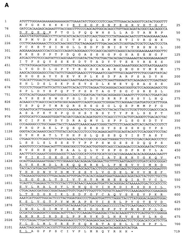
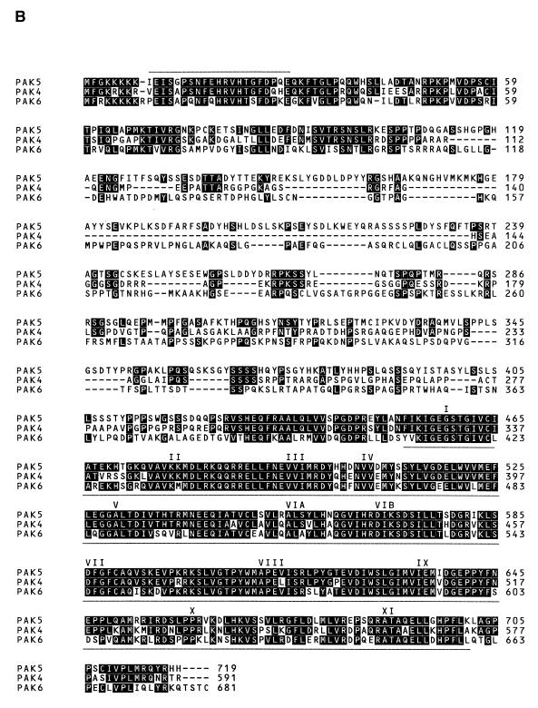
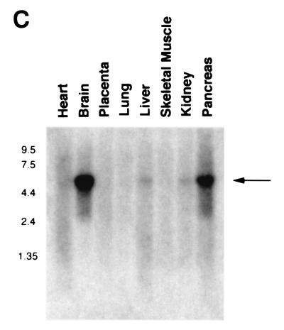
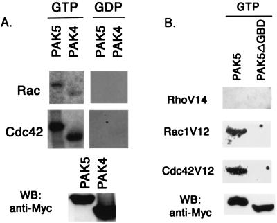
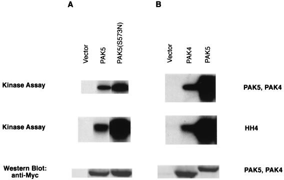
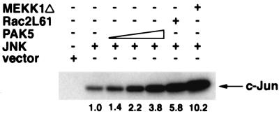
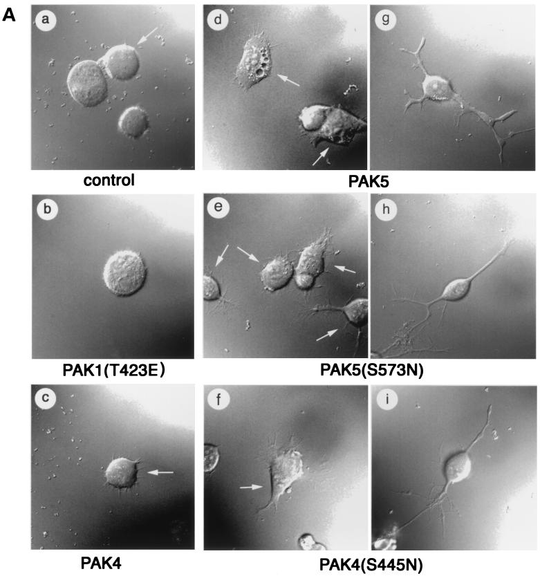
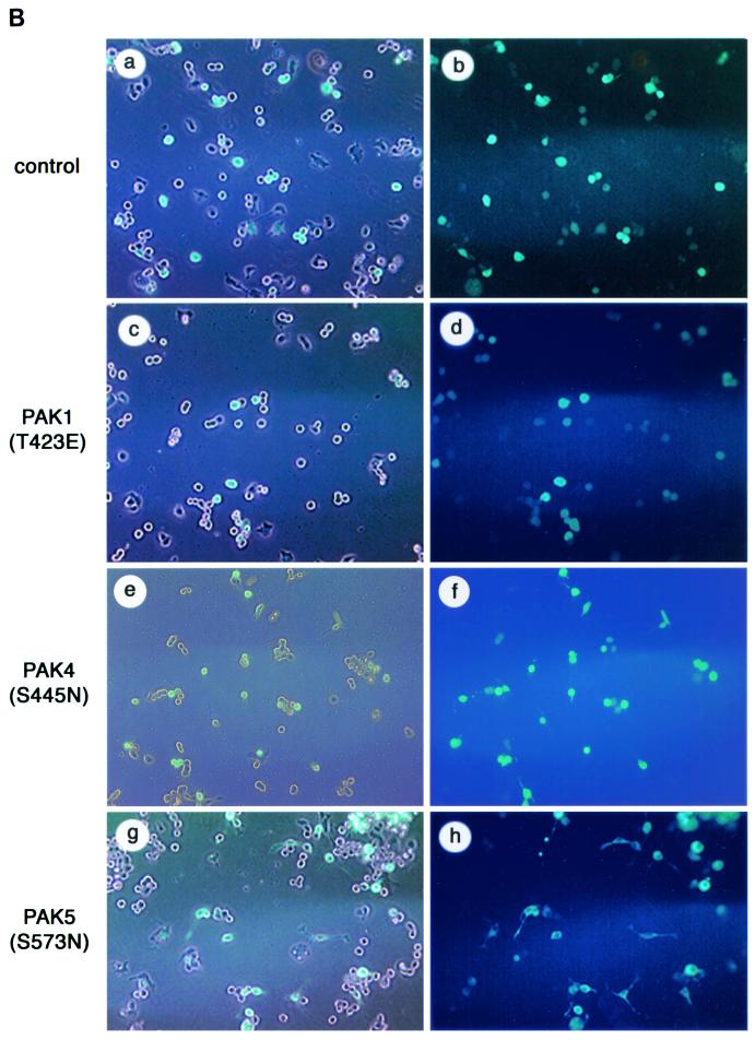
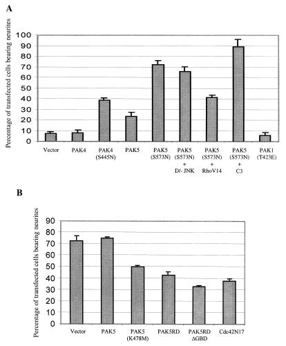
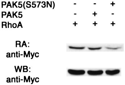
We have characterized a new member of the mammalian PAK family of serine/threonine kinases, PAK5, which is a novel target of the Rho GTPases Cdc42 and Rac. The kinase domain and GTPase-binding domain (GBD) of PAK5 are most closely related in sequence to those of mammalian PAK4. Outside of these domains, however, PAK5 is completely different in sequence from any known mammalian proteins. PAK5 does share considerable sequence homology with the Drosophila MBT protein (for "mushroom body tiny"), however, which is thought to play a role in development of cells in Drosophila brain. Interestingly, PAK5 is highly expressed in mammalian brain and is not expressed in most other tissues. We have found that PAK5, like Cdc42, promotes the induction of filopodia. In N1E-115 neuroblastoma cells, expression of PAK5 also triggered the induction of neurite-like processes, and a dominant-negative PAK5 mutant inhibited neurite outgrowth. Expression of activated PAK1 caused no noticeable changes in these cells. An activated mutant of PAK5 had an even more dramatic effect than wild-type PAK5, indicating that the morphologic changes induced by PAK5 are directly related to its kinase activity. Although PAK5 activates the JNK pathway, dominant-negative JNK did not inhibit neurite outgrowth. In contrast, the induction of neurites by PAK5 was abolished by expression of activated RhoA. Previous work has shown that Cdc42 and Rac promote neurite outgrowth by a pathway that is antagonistic to Rho. Our results suggest, therefore, that PAK5 operates downstream to Cdc42 and Rac and antagonizes Rho in the pathway, leading to neurite development.


Figures

FIG. 1.
FIG. 1.
Sequence and expression pattern of PAK5. (A) Sequence of human PAK5. (B) Alignment of PAK5 with PAK4 and PAK6. The overscored region is the GBD. The underlined region is the kinase domain. The 11 subdomains conserved throughout serine/threonine kinases are indicated. (C) A human multiple tissue mRNA Northern blot was probed with a cDNA containing part of the kinase domain of PAK5. A band of ∼5.5 kb is indicated.
FIG. 1.
FIG. 1.
Sequence and expression pattern of PAK5. (A) Sequence of human PAK5. (B) Alignment of PAK5 with PAK4 and PAK6. The overscored region is the GBD. The underlined region is the kinase domain. The 11 subdomains conserved throughout serine/threonine kinases are indicated. (C) A human multiple tissue mRNA Northern blot was probed with a cDNA containing part of the kinase domain of PAK5. A band of ∼5.5 kb is indicated.
FIG. 2.
FIG. 2.
PAK4 and PAK5 interact with activated Rac and Cdc42. (A) 293 cells were transfected with equal amounts of expression vectors containing wild-type Myc-tagged PAK4 or PAK5. After transient expression, Myc-PAK4 and Myc-PAK5 were immunoprecipitated from whole-cell lysates by using a mouse anti-Myc antibody and protein A-Sepharose. The immunocomplexes were separated by SDS-PAGE and transferred to a PVDF membrane. The membranes were then probed with purified wild-type Rac or Cdc42 that was preloaded with either [γ-32P]GTP or [β-32P]GDP as described in Materials and Methods (see top two panels). A portion of the lysate was used for Western blot analysis (WB) with anti-Myc antibody to detect the PAK4 and PAK5 expression levels (bottom panel). (B) 293 cells were transfected with equal amounts of expression vectors containing Myc-tagged wild-type PAK5 or Myc-tagged PAK5ΔGBD. After transient transfection, PAK5 and PAK5ΔGBD were immunoprecipitated from whole-cell lysates and transferred to a PVDF membrane as described above, and the membranes were probed with purified RhoV14, Rac1V12, or Cdc42V12 that was preloaded with [γ-32P]GTP as described above. PAK5 and PAK5ΔGBD expression in whole-cell lysates was detected by Western blots (WB) by using anti-Myc antibody, as shown in the bottom panel.
FIG. 3.
FIG. 3.
PAK5 autophosphorylates and phosphorylates HH4. 293 cells were transfected with equal amounts of either GFP vector or expression vectors containing Myc-tagged PAK4, Myc-tagged PAK5, or Myc-tagged PAK5(S573N). After transient expression, the amounts of Myc-PAK4 and Myc-PAK5 were normalized by Western blots probed with a mouse anti-Myc antibody. Approximately equal amounts of Myc-PAK4, Myc-PAK5, and Myc-PAK5(S573N) were immunoprecipitated from whole-cell lysates by using a mouse anti-Myc antibody and protein A-Sepharose. The immunocomplexes were then incubated with HH4 and [γ-32P]ATP in in vitro kinase buffer. Substrate phosphorylation and autophosphorylation were analyzed after SDS-PAGE (15% gel) and autoradiography. Both the autophosphorylation of PAK4, PAK5, and PAK5(S573N) and the phosphorylation of HH4 are indicated. The gel was exposed for 15 min (A) or 2 h (B). Western blots showing expression of Myc-PAK5 and Myc-PAK4 are shown in the bottom panel (10% gel).
FIG. 4.
FIG. 4.
PAK5 activates the JNK pathway. 293 cells were transfected with either empty vector or 5 μg of expression vector containing HA-tagged JNK (HA-JNK) either alone or with increasing doses of expression vectors containing Myc-tagged PAK5 (1, 3, and 5 μg) or expression vectors containing Rac2L61 (2.5 μg) or MEKK1Δ (2.5 μg). After transient expression, cells were lysed, and the amount of HA-JNK was normalized by Western blots probed with a mouse anti-HA antibody. Equal amounts of HA-JNK were then immunoprecipitated from whole-cell lysates by using a mouse anti-HA antibody and protein A-Sepharose. The immunoprecipitates were then incubated with recombinant GST–c-Jun in the presence of [γ-32P]ATP in in vitro kinase buffer. Substrate phosphorylation was analyzed after SDS-PAGE and autoradiography. The phosphorylation of GST–c-Jun is indicated. The numbers indicate the extent (fold) of activation of JNK by PAK5, Rac2L61, and MEKK1Δ as quantitated by phosphorimager analysis.
FIG. 5.
FIG. 5.
PAK5 induces neurite outgrowth and filopodia. (A) Expression vectors containing EGFP (control) or equal amounts of PAK5, PAK5(S573N), PAK4, PAK4(S445N), or PAK1(T423E) were transfected into N1E-115 cells. Vectors containing PAK4 and PAK5 were either cotransfected with an EGFP vector at a 1:3 EGFP/PAK5 ratio or expressed as EGFP fusions, with similar results. PAK1(T423E) was cotransfected with EGFP. Cells were visualized by fluorescence microscopy at 20 h after transfection. Cells were then photographed with a ×100 objective lens. Where more than one cell is shown, the transfected cells, as observed by fluorescence microscopy, are indicated by an arrow. Cells transfected with empty vector EGFP, PAK1(T423E), or PAK4 are shown in panels a, b, and c. Representative examples of the PAK5-, PAK5(S573N)-, and PAK4(S445N)-expressing cells that had filopodia but not neurites are shown in panels d, e, and f. Representative examples of PAK5-, PAK5(S573N)-, and PAK4(S445N)-expressing cells that had differentiated and extended neurites are shown in panels g, h, and i. (B) Representative fields of cells transfected with empty EGFP vector, PAK1(T423E), PAK4(S445N), or PAK5(S573N) were taken by using a ×10 objective. The left side shows cells that were viewed under visible light and UV light together. The right side shows the same field that was viewed under only UV light.
FIG. 5.
FIG. 5.
PAK5 induces neurite outgrowth and filopodia. (A) Expression vectors containing EGFP (control) or equal amounts of PAK5, PAK5(S573N), PAK4, PAK4(S445N), or PAK1(T423E) were transfected into N1E-115 cells. Vectors containing PAK4 and PAK5 were either cotransfected with an EGFP vector at a 1:3 EGFP/PAK5 ratio or expressed as EGFP fusions, with similar results. PAK1(T423E) was cotransfected with EGFP. Cells were visualized by fluorescence microscopy at 20 h after transfection. Cells were then photographed with a ×100 objective lens. Where more than one cell is shown, the transfected cells, as observed by fluorescence microscopy, are indicated by an arrow. Cells transfected with empty vector EGFP, PAK1(T423E), or PAK4 are shown in panels a, b, and c. Representative examples of the PAK5-, PAK5(S573N)-, and PAK4(S445N)-expressing cells that had filopodia but not neurites are shown in panels d, e, and f. Representative examples of PAK5-, PAK5(S573N)-, and PAK4(S445N)-expressing cells that had differentiated and extended neurites are shown in panels g, h, and i. (B) Representative fields of cells transfected with empty EGFP vector, PAK1(T423E), PAK4(S445N), or PAK5(S573N) were taken by using a ×10 objective. The left side shows cells that were viewed under visible light and UV light together. The right side shows the same field that was viewed under only UV light.
FIG. 6.
FIG. 6.
Quantification of the effects of PAK5 on neurite formation. (A) Cells were transfected with expression vectors containing EGFP (control), EGFP-PAK5, EGFP-PAK5(S573N), PAK4, PAK4(S445N), or PAK1(T423E) and EGFP. EGFP-PAK5(S573N) was also transfected with either empty vector or with a threefold excess of dominant-negative JNK, RhoV14, or C3 transferase and then grown in the presence of serum. Cells bearing neurites were counted, and the percentages of transfected cells that had neurites are indicated. Cotransfection of PAK5(S573N) vector with EGFP vector gave similar results as EGFP-PAK5(S573N) (data not shown). (B) Cells were transfected with the indicated expression vectors. At 24 h after transfection the cells were incubated in serum-free medium. After 72 h, immunofluorescence assays were performed with anti-Myc antibody to identify transfected cells. Transfected cells bearing neurites were counted, and the percentages of transfected cells that had neurites are indicated. Approximately 100 transfected cells were counted in each experiment. The results are an average of at least two independent experiments for each condition.
FIG. 7.
FIG. 7.
Activated PAK5 inhibits Rho activity. 293 cells were transfected with wild-type Myc-RhoA expression vector together with equal amounts of either empty vector, wild-type EGFP-PAK5, or EGFP-PAK5(S573N) expression vectors (without Myc tags). After transient expression, cell lysates were incubated with GST-Rhotekin glutathione agarose complexes, which bind specifically to GTP-loaded RhoA (40). Complexes were then washed and separated by SDS-PAGE, and the GTP RhoA content was analyzed by Western blot analysis by using anti-Myc antibody (top panel). The total RhoA content in an aliquot of whole-cell lysates is shown in the bottom panel.

Similar articles

Cited by

References

    1. Abo, A., J. Qu, M. S. Cammarano, C. Dan, A. Fritsch, V. Baud, B. Belisle, and A. Minden. 1998. PAK4, a novel effector for Cdc42Hs, is implicated in the reorganization of the actin cytoskeleton and in the formation of filopodia. EMBO J. 17:6527–6540. - PMC - PubMed
    1. Bagrodia, S., B. Derijard, R. J. Davis, and R. A. Cerione. 1995. Cdc42 and PAK-mediated signaling leads to Jun kinase and p38 mitogen-activated protein kinase activation. J. Biol. Chem. 270:27995–27998. - PubMed
    1. Berninger, B., and M. Poo. 1996. Fast actions of neurotrophic factors. Curr. Opin. Neurobiol. 6:324–330. - PubMed
    1. Brown, J. L., L. Stowers, M. Baer, J. Trejo, S. Coughlin, and J. Chant. 1996. Human Ste20 homologue hPAK1 links GTPases to the JNK MAP kinase pathway. Curr. Biol. 6:598–605. - PubMed
    1. Brown, M. D., B. J. Cornejo, and J. R. Bamburg. 2000. Cdc42 stimulates neurite outgrowth and formation of growth cone filopodia and lamellipodia. J. Neurobiol 43:352. - PubMed

Publication types

MeSH terms

Substances