. 2002 Jan;22(2):587-98.
doi: 10.1128/MCB.22.2.587-598.2002.

Transactivation of Fra-1 and consequent activation of AP-1 occur extracellular signal-regulated kinase dependently

Affiliations

Transactivation of Fra-1 and consequent activation of AP-1 occur extracellular signal-regulated kinase dependently

Matthew R Young et al. Mol Cell Biol. 2002 Jan.

Abstract

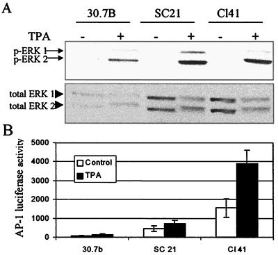
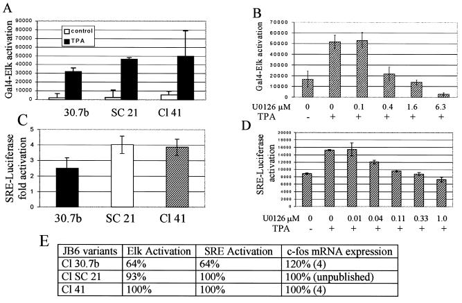
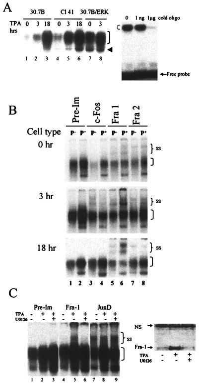
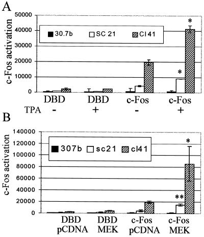
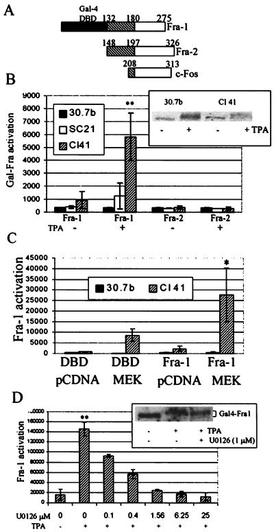
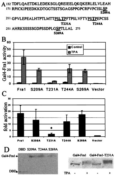
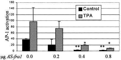
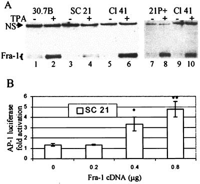
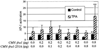
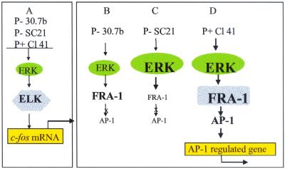
Mitogen-activated protein (MAP) kinase, extracellular-signal-regulated kinases (ERKs) play an important role in activating AP-1-dependent transcription. Studies using the JB6 mouse epidermal model and a transgenic mouse model have established a requirement for AP-1-dependent transcription in tumor promotion. Tumor promoters such as 12-O-tetradecanoylphorbol-13-acetate (TPA) and epidermal growth factor induce activator protein 1 (AP-1) activity and neoplastic transformation in JB6 transformation-sensitive (P(+)) cells, but not in transformation-resistant (P(-)) variants. The resistance in one of the P(-) variants can be attributed to the low levels of the MAP kinases, ERKs 1 and 2, and consequent nonresponsiveness to AP-1 activation. The resistant variant is not deficient in c-fos transcription. The purpose of these studies was to define the targets of activated ERK that lead to AP-1 transactivation. The results establish that the transactivation domain of Fra-1 can be activated, that activation of Fra-1 is ERK dependent, and that a putative ERK phosphorylation site must be intact for activation to occur. Fra-1 was activated by TPA in ERK-sufficient P(+) cells but not in ERK-deficient P(-) cells. A similar activation pattern was seen for c-Fos but not for Fra-2. Gel shift analysis identified Fra-1 as distinguishing mitogen-activated (P(+)) from nonactivated (P(-)) AP-1 complexes. A second AP-1-nonresponsive P(-) variant that underexpresses Fra-1 gained AP-1 response upon introduction of a Fra-1 expression construct. These observations suggest that ERK-dependent activation of Fra-1 is required for AP-1 transactivation in JB6 cells.


Figures

FIG. 1.
FIG. 1.
AP-1-nonresponsive JB6 P variants include both ERK-deficient and ERK-sufficient cells. (A) Whole-cell extracts from JB6 cells serum starved for 24 h and treated with TPA (10 ng/ml) or DMSO for 30 min were analyzed by Western blot analysis with antibody specific to phosphorylated (p) ERK 1 and 2 or to total ERK. An equal amount of total protein was added to each lane. Independent experiments show similar results. (B) JB6 cells were transiently transfected with a 4× AP-1 luciferase reporter. Cells were serum starved for 24 h and then treated with TPA for an additional 16 h. Cells were lysed, and luciferase activity (relative luminescence units) was determined with a Dynex 96-well luminometer.
FIG. 2.
FIG. 2.
MAPK-regulated serum response pathway is functional in ERK-deficient JB6 cells. (A) Activation of Elk-1 is not lacking in ERK-deficient cells. JB6 cells transiently transfected with a Gal4-Elk-1 fusion and a Gal4-luciferase reporter were serum starved for 24 h and then treated with TPA for 6 h. Cell extracts were analyzed for luciferase activity. (B) Inhibition of ERK activity blocks activation of Elk. Cl 41 cells transiently transfected with Gal4-Elk-1 and Gal4-luciferase were treated with the MEK inhibitor U0126 (Promega) or DMSO 1 h before TPA exposure. Luciferase activity was determined 6 h after TPA exposure. (C) ERK levels have little effect on activation of SRE. JB6 cells were transiently transfected with an SRE-luciferase reporter, serum starved for 24 h, and then treated with TPA for 3 h. (D) SRE activation is ERK activation dependent. Cl 41 cells transiently transfected with SRE-luciferase were treated with the MEK inhibitor U0126 or DMSO for 1 h before TPA treatment. Luciferase activity was measured 3 h later. Fold activation is defined as TPA-induced luciferase activity relative to the uninduced level. The average of three transfections is shown. Similar results were seen in multiple experiments. (E) c-fos mRNA expression is not limiting in ERK-deficient P cells. The activation of Elk-1, SRE, and c-fos expression relative to that in the ERK-sufficient Cl 41 cells is shown. The values for Elk-1 and SRE activation are from panels A and C above. The values for c-fos expression are from Ben-Ari et al. (4) and data not shown.
FIG. 3.
FIG. 3.
(A) AP-1 DNA binding is enhanced by ERK activation. The left panel shows 30.7B, Cl 41, and 30.7B/ERK cells that were serum starved for 24 h and then treated with TPA for 0, 3, and 18 h. Nuclear extracts were isolated and analyzed for DNA-binding activity by EMSA. The right panel shows the specificity of DNA binding. Unlabeled (cold) AP-1-binding oligonucleotide (1 ng and 1 μg) was used to compete with the labeled oligonucleotide. The bracket marks the broad (multiband) AP-1 complex. The arrowhead shows a faster-migrating band seen predominantly in ERK-sufficient cells. Electrophoresis conditions in which the free probe was visible on the gel (right) were not sufficient to separate the faster-migrating band from the major band, as seen on the left. To separate the faster-migrating band, the free probe was run off the gel. Representative gels from multiple experiments are shown. (B) TPA-activated AP-1 complex from P+ cells contains more Fra-1 than the complex from ERK-deficient P cells. Fra-2 is also present in the AP-1 complex. Nuclear extracts were harvested from 30.7B (P) and Cl 41 (P+) cells that had been serum starved for 24 h and then treated with TPA for 0 (top), 3 (middle), and 18 (bottom) h. Extracts were analyzed by supershift EMSA with Fos-specific antibodies as indicated. Higher antibody concentrations produced no greater shifts. It should be noted that neither these antibody concentrations nor higher concentrations (8, 42, 48) of specific AP-1 antibodies produced complete shifts in previous studies of JB6 cells in our laboratory or others. Pre-Im, preimmune; SS, supershifted bands. Unlabeled brackets mark nonsupershifted AP-1 complexes. Labeled oligonucleotide was added to the nuclear extract, and the extracts were then aliquoted to tubes containing the indicated antibodies. EMSA samples were run on two gels per sample time point. All six gels were run simultaneously. The 0-h and 3-h autoradiographs were analyzed by phosphorimaging. The 18-h autoradiograph was exposed to X-ray film and digitally rearranged for clarity. A representative autoradiogram of multiple EMSAs is shown. (C) Fra-1 recruitment to AP-1 complex is ERK dependent. JB6 cells treated for 1 h with 5 μM MEK inhibitor U0126 or DMSO were exposed to TPA for 3 h. Nuclear extracts were harvested and analyzed for AP-1 binding by supershift EMSA or for Fra-1 expression by Western blot. The left panel shows the antibody-induced supershift EMSA with preimmune (lanes 1 to 3), Fra-1 (lanes 4 to 6), and Jun-D (lanes 7 to 9) antibodies. SS, supershifted band; the bracket indicates unshifted complexes. The right panel shows the Western blot analysis of nuclear extracts detected with anti-Fra-1. NS, nonspecific cross-reactive protein to the Fra-1 antibody. Equal protein levels, as determined by the BSA assay, were loaded.
FIG. 4.
FIG. 4.
c-Fos protein is activated by TPA or MEK-1 in ERK-sufficient but not ERK-deficient cells. (A) JB6 cells transiently transfected with a Gal4-luciferase and either a Gal4-c-Fos fusion or the empty Gal4 vector pFCMV (DBD) were serum starved for 24 h and then treated with TPA or DMSO. Luciferase activity was measured 6 h later. (B) JB6 cells transiently transfected with a Gal4-luciferase, Gal4-c-Fos, or empty pFCMV (DBD) plus activated MEK or pcDNA3 were serum starved for 24 h, and luciferase activity was measured. The average for three transfections + standard deviation is shown. Similar results were seen in multiple experiments. Activation of the Gal4-Elk-1 fusion (see Fig. 2) was used as a control for mitogen activation. * and **, statistically significant difference between TPA- or MEK-induced c-Fos-transfected cells and uninduced cells as determined by Student’s t test (*, P < 0.05; **, P < 0.01).
FIG. 5.
FIG. 5.
Fra-1 is activated by TPA or MEK-1 in ERK-sufficient cells but not ERK-deficient cells. (A) Gal4 fusions. The Fra-1 and Fra-2 Gal4 fusions contain the yeast Gal4 DNA-binding domain (DBD) (solid box) fused to C-terminal residues of the rat Fra-1 (aa 132 to 275; NCBI accession no. NP037085) or Fra-2 protein (aa 148 to 326; accession no. P51145). Gal4-c-Fos is encoded by pFA-Fos and contains the C-terminal transactivation domain from c-Fos (aa 208 to 313) fused to the Gal4 DNA-binding domain (Stratagene). The hatched regions show the homologous domain in Fra-1, Fra-2, and c-Fos. (B) Fra-1 activation. JB6 cells transiently transfected with a Gal4-Fra-1 or Gal4-Fra-2 fusion and a Gal4-luciferase reporter were serum starved for 24 h and then treated with TPA for 6 h. Cell extracts were analyzed for luciferase activity. The inset shows a Western blot of 30.7b and Cl 41 cells transiently transfected with Gal4-Fra-1 and detected with anti-Gal4 antibody. (C) JB6 cells transiently transfected with Gal4-Fra-1 or empty pFCMV (DBD), activated MEK-1, or pcDNA3 and Gal4-luciferase were serum starved for 24 h, and luciferase activity was measured. (D) Fra-1 activation is ERK dependent. Cl 41 cells were transfected with Gal4-Fra-1 fusion and the Gal4-luciferase vector, serum starved for 24 h, and treated with MEK inhibitor U0126 or DMSO for 1 h before being exposed to TPA for 6 h. Cell extracts were analyzed for luciferase activity. Assays were done in triplicate, and a representative of multiple assays is shown. Activation of the Gal4-Elk-1 fusion (see Fig. 2) was used as a control for mitogen activation. The inset shows a Western blot of Cl 41 cells transfected with Gal4-Fra-1 and detected with anti-Gal4 antibody. * and **, statistically significant difference between TPA- or MEK-induced Gal4-Fra-1-transfected cells and uninduced cells as determined by Student’s t test (*, P < 0.05; **, P < 0.01). The P values for the inhibition of Fra-1 activation with U0126 were all <0.001.
FIG. 6.
FIG. 6.
Thr-231 is required for activation of Fra-1. (A) Sequence of the C terminus of Fra-1 that comprises the Gal4-Fra-1 fusion. Ser/Thr-Pro sites are underlined. Mutated residues are in boldface and indicated under the sequence. (B) Fra-1 activation. Cl 41 cells cotransfected with Gal4 fusions containing wild-type or mutant Fra-1 and the Gal4-luciferase reporter were serum starved for 24 h and then untreated or treated with TPA (10 ng/ml) for 6 h. Cells were harvested, and luciferase activity was determined. A representative assay, the average of three tranfections, is shown. (C) Fold activation of Fra-1 activity was determined from the average of three individual assays. The fold activation for three separate experiments of three transfections each is shown. *, statistically significant difference between wild-type Ga14-Fra-1 and mutant Ga14-Fra-1-T231A as determined by Student’s t test (P < 0.011). (D) Expression of Gal4-Fra-1 and Gal4-Fra-1 mutants in Cl 41 cells. Cl 41 cells were transfected with Gal4-Fra-1 or Gal4-Fra-1 mutants, and nuclear extracts were harvested 48 h later (DBD) (pFCMV) (left panel). Cl 41 cells transfected with Gal4-Fra-1 or Gal4-Fra-1-T231A and serum starved overnight were treated with TPA for 6 h. Nuclear extracts were harvested and analyzed by Western blot (right panel). Gal4 fusions were detected with anti-Gal4 antibody.
FIG. 7.
FIG. 7.
Antisense (AS) fra-1 blocks AP-1 activation in P+ cells. Cl 41 cells were cotransfected with the 4× AP-1 reporter and antisense fra-1 at the indicated concentrations. pcDNA3 was used to equilibrate the DNA concentration used for transfection. At 24 h after transfection, cells were serum starved overnight and untreated or treated with TPA for 6 h. * and **, statistically significant difference between antisense fra-1- and pcDNA3-transfected cells as determined by Student’s t test (*, P < 0.05; **, P < 0.01).
FIG. 8.
FIG. 8.
P SC21 cells are deficient in Fra-1. (A) Cl 30.7b, Cl SC21, and Cl 41 cells were serum starved for 24 h and then treated with TPA for 0 or 3 h. Nuclear extracts were harvested and analyzed for Fra-1 protein by Western blot. Lanes 1 to 6 show extracts from P 30.7b, P SC21, and P+ Cl 41 cells. Lanes 7 and 8 show extracts from SC21 cells that have spontaneously converted to P+ cells as determined by TPA-induced AP-1 activation and transformation (data not shown). NS, nonspecific cross-reactive protein to the Fra-1 antibody. Equal protein levels, as determined by BSA assay, were loaded. (B) Expression of fra-1 cDNA in SC21 cells restores AP-1 activation. SC21 cells were transiently transfected with an AP-1 luciferase reporter and increasing amounts of fra-1 cDNA. Cells were starved for 24 h and then treated with TPA for 6 h. Fold activation is defined as TPA-induced luciferase activity relative to uninduced levels. * and **, statistically significant difference between fra-1- and pcDNA3-transfected cells as determined by Student’s t test (*, P < 0.05; **, P < 0.01).
FIG. 9.
FIG. 9.
Fra-1-T231A does not activate AP-1 in JB6 cells. Cl 41 cells were cotransfected with the 4× AP-1 reporter, CMV-fra-1, and/or CMV-fra-1-T231A at the indicated concentrations. pcDNA3 was used to equalize the DNA concentration. At 24 h after transfection, cells were serum starved overnight and untreated or treated with TPA for 6 h. * and **, statistically significant difference between fra-1- and fra-1-T231A-transfected cells as determined by Student’s t test (*, P < 0.05; **, P < 0.01).
FIG. 10.
FIG. 10.
nMAPK ERK to AP-1 pathway. (A) All three JB6 variants have sufficient ERK to drive activation of Elk-1 and transactivation of the SRE promoter, leading to c-fos expression. (B) JB6 P 30.7b cells do not have sufficient ERK for activation of Fra-1 and/or transactivation of the AP-1 promoter. (C) P SC21 cells have sufficient ERK for activation of exogenously added Gal4-Fra-1 but lack sufficient endogenous Fra-1, rendering them resistant to mitogen-induced transactivation of AP-1. (D) P+ Cl 41 cells have sufficient ERK and Fra-1 protein to complete the signal cascade from ERK to AP-1. Shading indicates activated proteins.

Similar articles

Cited by

References

    1. Abate, C., D. R. Marshak, and T. Curran. 1991. Fos is phosphorylated by p34cdc2, cAMP-dependent protein kinase and protein kinase C at multiple sites clustered within regulatory regions. Oncogene 6:2179–2185. - PubMed
    1. Angel, P., and M. Karin. 1991. The role of Jun, Fos and the AP-1 complex in cell-proliferation and transformation. Biochim. Biophys. Acta 1072:129–157. - PubMed
    1. Balmanno, K., and S. J. Cook. 1999. Sustained MAP kinase activation is required for the expression of cyclin D1, p21Cip1 and a subset of AP-1 proteins in CCL39 cells. Oncogene 18:3085–3097. - PubMed
    1. Ben-Ari, E. T., L. R. Bernstein, and N. H. Colburn. 1992. Differential c-jun expression in response to tumor promoters in JB6 cells sensitive or resistant to neoplastic transformation. Mol. Carcinog. 5:62–74. - PubMed
    1. Bergers, G., P. Graninger, S. Braselmann, C. Wrighton, and M. Busslinger. 1995. Transcriptional activation of the fra-1 gene by AP-1 is mediated by regulatory sequences in the first intron. Mol. Cell. Biol. 15:3748–3758. - PMC - PubMed

MeSH terms

LinkOut - more resources