. 2002 Jan;22(2):599-613.
doi: 10.1128/MCB.22.2.599-613.2002.

Novel mechanism for gonadotropin-releasing hormone neuronal migration involving Gas6/Ark signaling to p38 mitogen-activated protein kinase

Affiliations

Novel mechanism for gonadotropin-releasing hormone neuronal migration involving Gas6/Ark signaling to p38 mitogen-activated protein kinase

Melissa P Allen et al. Mol Cell Biol. 2002 Jan.

Abstract

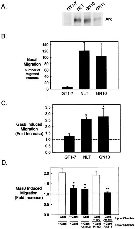
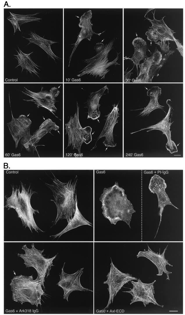
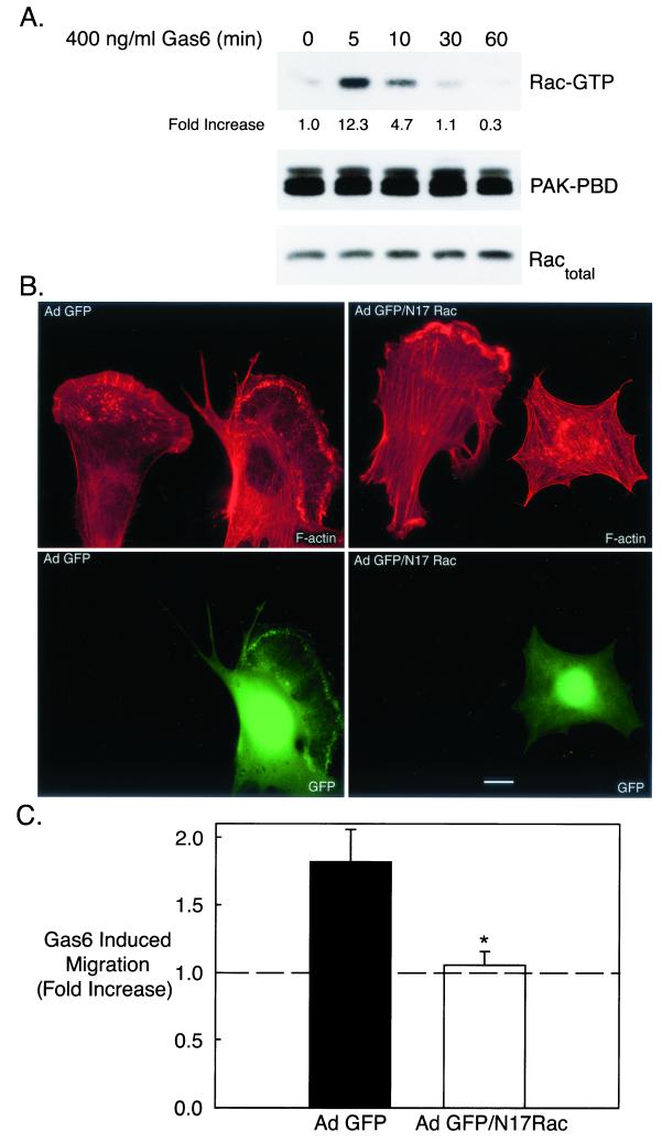
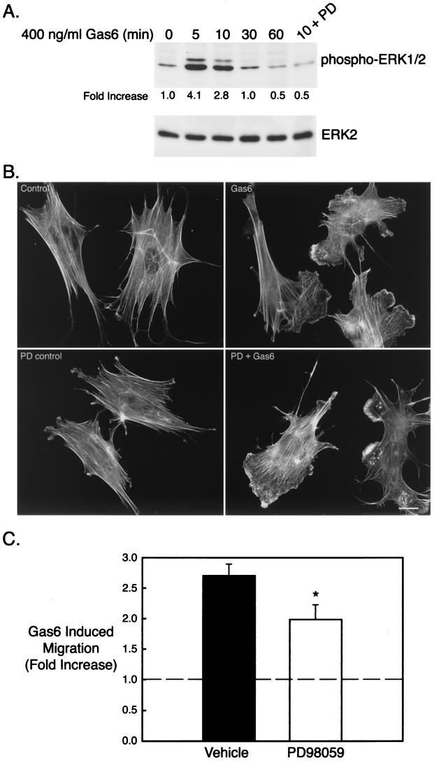
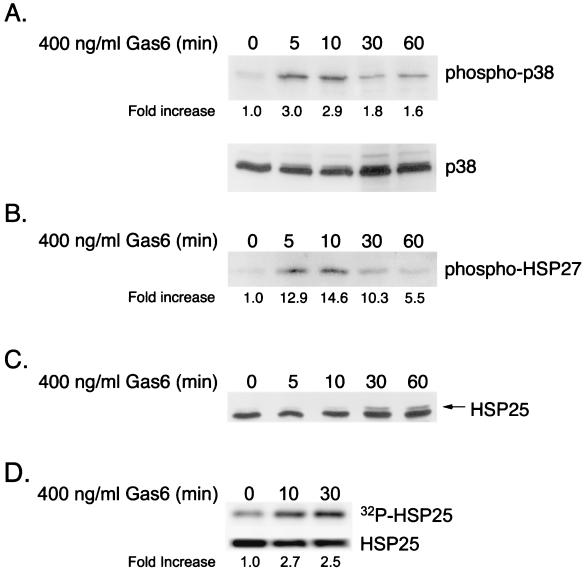
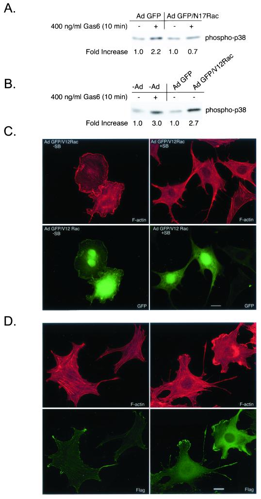
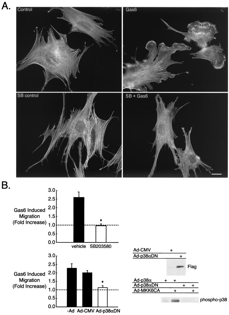
Gonadotropin-releasing hormone (GnRH) is the central regulator of the reproductive axis. Normal sexual maturation depends on the migration of GnRH neurons from the olfactory placode to the hypothalamus during development. Previously, we showed restricted expression of the membrane receptor adhesion-related kinase (Ark) in immortalized cell lines derived from migratory but not postmigratory GnRH neurons. In addition, Ark and GnRH transcripts were detected along the GnRH neuron migratory route in the E13 mouse cribriform plate. In the present study, we examined the role of Ark and its ligand, Gas6 (encoded by growth arrest-specific gene 6), in GnRH neuron migration. Gas6 stimulated lamellipodial extension, membrane ruffling, and chemotaxis of immortalized NLT GnRH neuronal cells via the Ark receptor. Gas6/Ark signaling promoted activation of the Rho family GTPase Rac, and adenoviral-mediated expression of dominant negative N17Rac abolished Gas6/Ark-induced actin cytoskeletal reorganization and migration of GnRH neuronal cells. In addition, p38 MAPK was activated downstream of Ark and Rac, and inhibition of p38 MAPK with either SB203580 or adenoviral dominant negative p38alpha also blocked Gas6/Ark-mediated migration. Finally, downstream of Rac and p38 mitogen-activated protein kinase (MAPK), Gas6/Ark signaling promoted activation of MAPK-activated protein kinase 2 and induced phosphorylation of HSP25, a known regulator of cortical actin remodeling. The data are the first to demonstrate a migratory signaling pathway downstream of Ark/Axl family receptors and suggest a previously unidentified role for p38 MAPK in neuronal migration. Furthermore, these studies support a potential role for Ark in the regulation of GnRH neuronal migration.


Figures

FIG. 1.
FIG. 1.
Gas6 stimulates migration of Ark-expressing GnRH neuronal cell lines. (A) GT1-7, NLT, GN10, and GN11 cell lysates (20 μg) were analyzed by immunoblotting with Ark318 IgG. (B to D) The migratory behavior of the cells was examined with a Boyden chamber system as described in Materials and Methods. (B) Twenty-four-hour migration in the presence of 0.5% FBS in the lower chamber. Values are as follows: GT1-7, 7 ± 2 (n = 4); NLT, 121 ± 26 (n = 4); GN10, 103 ± 42 (n = 6). (C) Twenty-four-hour migration in the presence of Gas6 (400 ng/ml) in the lower chamber (n = 4 for GT1-7 and n = 62 for NLT and GN10). *, statistically significant by the t test: P < 0.01 for NLT and P < 0.05 for GN10. (D) Effects of Gas6, recombinant Axl ECD (800 ng/ml), Ark318 IgG (10 μg/ml), and PI IgG (10 μg/ml) on migration (24 h). *, statistically significantly different from the control (Gas6 in the lower chamber only) by the Tukey-Kramer multiple comparisons test, P < 0.05 (n = 3). **, statistically different from the control (PI IgG in the upper and lower chambers) by the t test, P < 0.05 (n = 3).
FIG. 2.
FIG. 2.
Gas6 stimulates Ark-dependent formation of lamellipodia and membrane ruffles in NLT GnRH neuronal cells. (A) NLT cells were incubated with Gas6 (400 ng/ml) for the indicated times. Following fixation, F-actin was visualized with rhodamine phalloidin. Arrows indicate lamellipodia and membrane ruffles. The data are representative of three independent experiments (see Materials and Methods for quantitation of actin cytoskeletal remodeling). Bar = 20 μm. (B) NLT cells were pretreated (4 h) with the Axl ECD (800 ng/ml), Ark318 IgG (10 μg/ml), or PI IgG (10 μg/ml) followed by a 10-min stimulation with Gas6 (400 ng/ml). F-actin was visualized as in panel A. The partial effectiveness of the Ark318 IgG at blocking the Gas6 effect is indicated by the arrow. The data are representative of two independent experiments. Bar = 20 μm.
FIG. 3.
FIG. 3.
Gas6/Ark-stimulated NLT migration and cytoskeletal reorganization requires the Rho family GTPase Rac. (A) NLT cells were treated with Gas6 (400 ng/ml) for the indicated times followed by a Rac activation assay. Briefly, GTP-bound Rac (Rac-GTP) was precipitated with PAK-PBD agarose, and GTP-bound Rac was visualized by Rac immunoblotting. Equal amounts of PAK-PBD were used to precipitate Rac-GTP (PAK-PBD blot), and each sample contained a similar amount of Rac protein (for the Ractotal blot, 1/20 of each sample was loaded). The data are representative of three independent experiments. The increase in Rac-GTP was calculated by dividing the Rac-GTP density by the Ractotal density at each time point and setting the value at the zero time point to 1. (B) NLT cells were infected with Ad GFP or Ad GFP/N17Rac (see Materials and Methods), stimulated for 10 min with Gas6 (400 ng/ml), and stained with rhodamine phalloidin. Actin is shown in red and GFP is in green. The data are representative of two independent experiments. Bar = 20 μm. (C) NLT cells infected with Ad GFP or GFP/N17Rac were tested in the Boyden chamber migration assay with or without Gas6. *, statistically different from the Ad GFP value by the t test, P < 0.05 (n = 5).
FIG. 4.
FIG. 4.
The ERK pathway is not essential for development of the motile phenotype induced by Gas6/Ark signaling. (A) NLT cells were treated for the indicated times with Gas6 (400 ng/ml) and immunoblotted for phospho-ERK1/2 and total ERK2. In the last lane, cells were pretreated for 4 h with PD98059 (PD) (30 μM) and then stimulated with Gas6 for 10 min. The data are representative of two independent experiments. The increase in phospho-ERK1/2 was calculated by dividing the phospho-ERK1/2 density by the total ERK2 density at each time point and then setting the zero time point value to 1. (B) NLT cells were pretreated for 4 h with PD (30 μM), stimulated with Gas6 (10 min), and then stained with rhodamine phalloidin. The data are representative of four independent experiments. Bar = 20 μm. (C) NLT cells were subjected to the migration assay in the presence of vehicle (DMSO) or PD (30 μM) as described in Materials and Methods. The data are the fold increase in migration in the presence of Gas6 (400 ng/ml). *, P = 0.05 (n = 5, t test).
FIG. 5.
FIG. 5.
Gas6/Ark signaling activates a p38 MAPK → MAPKAP kinase 2 → HSP25 signaling cascade in NLT GnRH neuronal cells. (A) NLT cells were treated for the indicated times with Gas6 (400 ng/ml) and immunoblotted for phospho-p38 and total p38. The data are representative of three independent experiments. The increase in phospho-p38 was calculated by dividing the phospho-p38 density by the total p38 density at each time point and setting the zero time point value to 1. (B) MAPKAP kinase 2 was immunoprecipitated from Gas6-treated neuronal cells and exposed to recombinant human HSP27 in a kinase assay, and phosphorylated HSP27 was detected by phospho-HSP27 immunoblotting. The data are representative of two independent experiments. The increase in phospho-HSP27 was calculated by setting the zero time point value at 1. (C) NLT cells were treated for the indicated times with Gas6 (400 ng/ml) and immunoblotted for HSP25. The arrow indicates a slower-migrating form of HSP25 consistent with phosphorylation. The data are representative of two independent experiments. (D) NLT cells were labeled with 32Pi and stimulated with Gas6. HSP25 was immunoprecipitated from the cell lysates, transferred to PVDF, and analyzed by autoradiography (top) or HSP25 immunoblotting (bottom). The increase in 32P-labeled HSP25 was calculated by dividing the 32P-labeled-HSP25 density by the total HSP25 density at each time point and setting the zero time point value to 1. The data are representative of two experiments.
FIG. 6.
FIG. 6.
p38 activation promotes actin reorganization downstream of Rac in NLT GnRH neuronal cells. (A) NLT cells expressing Ad GFP or GFP/N17Rac were treated for 10 min with Gas6 (400 ng/ml) and then immunoblotted for phospho-p38. The data are representative of two independent experiments. The increase in phospho-p38 was calculated by setting the density of the no-Gas6 samples to 1. (B) uninfected NLT cells or those expressing Ad GFP or GFP/V12Rac were treated for 10 min with Gas6 (400 ng/ml) or vehicle and then immunoblotted for phospho-p38. The data are representative of two independent experiments. (C) NLT cells were infected with adenoviruses expressing GFP/V12Rac (see Materials and Methods), treated for 4 h with vehicle (DMSO) or SB203580 (30 μM) and then stained with rhodamine phalloidin. The data are representative of two independent experiments. Actin is shown in red and GFP is in green. Bar = 20 μm. (D) NLT cells infected with adenoviral MKK6CA and Flag-p38α were visualized with rhodamine phalloidin and anti-Flag immunocytochemistry (left panels, uninfected cells; right panels, cells infected with MKK6CA and Flag-p38α). Bar = 20 μm.
FIG. 7.
FIG. 7.
Gas6/Ark-mediated GnRH neuronal cytoskeletal remodeling and migration requires p38 MAPK. (A) NLT cells were pretreated for 4 h with SB203580 (SB) (30 μM), stimulated with Gas6 (10 min), and then stained with rhodamine phalloidin. Bar = 20 μm. The data are representative of four independent experiments. (B) (Left) The effect of Gas6 on NLT migration was examined in the presence of vehicle (DMSO) or SB (30 μM) and in neurons infected with either Ad-CMV or Ad-p38αDN as described in Materials and Methods. *, P < 0.05 for SB versus vehicle (n = 3, t test); P < 0.01 for p38αDN versus −Ad and P < 0.05 for p38αDN versus Ad-CMV (n = 4, Tukey-Kramer multiple comparisons test). (Right) Anti-Flag immunoblotting demonstrated that adenoviral Flag-p38αDN was efficiently expressed in the NLT neurons. The phospho-p38 blot demonstrated that wild-type p38α (Ad-p38α) was activated in the presence of Ad-MKK6CA, but Ad-p38αDN was not.

References

    1. Allen, M. P., M. Xu, C. Zeng, S. A. Tobet, and M. E. Wierman. 2000. Myocyte enhancer factors-2B and -2C are required for adhesion related kinase repression of neuronal gonadotropin releasing hormone gene expression. J. Biol. Chem. 275:39662–39670. - PubMed
    1. Allen, M. P., C. Zeng, K. Schneider, X. Xiong, M. K. Meintzer, P. Bellosta, C. Basilico, B. Varnum, K. A. Heidenreich, and M. E. Wierman. 1999. Growth arrest-specific gene 6 (Gas6)/adhesion related kinase (Ark) signaling promotes gonadotropin-releasing hormone neuronal survival via extracellular signal-regulated kinase (ERK) and Akt. Mol. Endocrinol. 13:191–201. - PubMed
    1. Bateman, J., H. Shu, and D. Van Vactor. 2000. The guanine nucleotide exchange factor trio mediates axonal development in the Drosophila embryo. Neuron 26:93–106. - PubMed
    1. Bellosta, P., M. Costa, D. A. Lin, and C. Basilico. 1995. The receptor tyrosine kinase ARK mediates cell aggregation by homophilic binding. Mol. Cell. Biol. 15:614–625. - PMC - PubMed
    1. Bellosta, P., Q. Zhang, S. P. Goff, and C. Basilico. 1997. Signaling through the ARK tyrosine kinase receptor protects from apoptosis in the absence of growth stimulation. Oncogene 15:2387–2397. - PubMed

Publication types

MeSH terms