. 2002 Jan;22(2):635-43.
doi: 10.1128/MCB.22.2.635-643.2002.

Obligate roles for p16(Ink4a) and p19(Arf)-p53 in the suppression of murine pancreatic neoplasia

Affiliations

Obligate roles for p16(Ink4a) and p19(Arf)-p53 in the suppression of murine pancreatic neoplasia

Nabeel Bardeesy et al. Mol Cell Biol. 2002 Jan.

Abstract

Epithelial tumors of the pancreas exhibit a wide spectrum of histologies with varying propensities for metastasis and tissue invasion. The histogenic relationship among these tumor types is not well established; moreover, the specific role of genetic lesions in the progression of these malignancies is largely undefined. Transgenic mice with ectopic expression of transforming growth factor alpha (TGF-alpha) in the pancreatic acinar cells develop tubular metaplasia, a potential premalignant lesion of the pancreatic ductal epithelium. To evaluate the cooperative interactions between TGF-alpha and signature mutations in pancreatic tumor genesis and progression, TGFalpha transgenic mice were crossed onto Ink4a/Arf and/or p53 mutant backgrounds. These compound mutant mice developed a novel pancreatic neoplasm, serous cystadenoma (SCA), presenting as large epithelial tumors bearing conspicuous gross and histological resemblances to their human counterpart. TGFalpha animals heterozygous for both the Ink4a/Arf and the p53 mutation showed a dramatically increased incidence of SCA, indicating synergistic interaction of these alleles. Inactivation of p16(Ink4a) by loss of heterozygosity, intragenic mutation, or promoter hypermethylation was a common feature in these SCAs, and correspondingly, none of the tumors expressed wild-type p16(Ink4a). All tumors sustained loss of p53 or Arf, generally in a mutually exclusive fashion. The tumor incidence data and molecular profiles establish a pathogenic role for the dual inactivation of p16(Ink4a) and p19(Arf)-p53 in the development of SCA in mice, demonstrating that p16(Ink4a) is a murine tumor suppressor. This genetically defined model provides insights into the molecular pathogenesis of SCA and serves as a platform for dissection of cell-specific programs of epithelial tumor suppression.


Figures

FIG. 1.
FIG. 1.
Tumor-free survival in the metallothionein-TGFα cohort (MT42 transgenic strain).
FIG. 2.
FIG. 2.
(A) Resemblance of human and murine SCAs. (Upper panels) Gross anatomy of human (left) and mouse (right) SCAs, showing intercystic vascular arcades. Bars, 4 and 1 cm in the human and mouse images, respectively. (Lower panels) Light micrographs showing hematoxylin and eosin staining of human (left) and mouse (right) SCAs, demonstrating characteristic low cuboidal epithelium with thin fibrous septa. (B) Structural features of murine SCA. (Upper left) SCA presenting as an encapsulated mass. (Upper right) Cross-sectional anatomy shows multilocular cystic architecture and pseudocapsule. (Lower left) Arrow depicts vascular pedicle. (Lower right) Immunohistochemical detection of limited focal mucicarmine staining in the tumors identifies these lesions as SCAs rather than mucinous cystadenomas. (C) Histological progression in the development of SCA. (i) Low-power photomicrograph depicting transition from tubular metaplasia (upper right) into SCA (left and bottom regions). (ii through iv) Immunohistochemistry shows a decreasing gradient of Pdx-1 expression in SCA progression, going from regions of tubular metaplasia (shown in a low-power view [ii] and a high-power view [ii, inset]) to the SCA tumor base (iii) and SCA growing edge (iv).
FIG. 3.
FIG. 3.
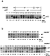
(a) LOH analysis of p53 in murine SCAs. Numbers below the gel indicate the normalized ratio of wild-type(WT) to mutant (knockout [KO]) alleles in SCAs relative to heterozygous DNA specimens isolated from normal tissue. Tumor names and mouse genotypes are shown above the gel. (b) LOH analysis of Ink4a/Arf in murine SCAs.
FIG. 4.
FIG. 4.
(a) Immunoblot for p53 expression in murine SCAs. There was no aberrant overexpression of p53 in the SCAs. Results for positive-control extracts from irradiated NIH 3T3 cells (+) and negative-control p53-null fibroblasts (−) are shown. (b) Immunoblots for p19Arf (upper panel) and p16Ink4a (lower panel) expression. The positive control (+) was a mouse melanoma cell lysate, and the negative control (−) was a lysate from Ink4a/Arf−/− fibroblasts. (c) Immunoblot shows aberrant migration of p16Ink4a expressed in tumor C7. Lysates were resolved on an 8-to-16% gradient gel. (d) RT-PCR analysis of the Ink4a mRNA. PCR of cDNAs from tumors C7 and E3 yielded products with primers amplifying Ink4a exon 1α (e1α) (upper panel) but not with primers spanning exons 1 and 2 (lower panel). RNA from a wild-type mouse embryonic fibroblast was used as a positive control. As a negative control for residual DNA contamination, RNA specimens were subjected to the same analysis without the addition of reverse transcriptase (−RT). (e) Methylation-specific PCR. DNA specimens were treated with bisulfite and analyzed by PCR using primers that recognize methylated (upper panel) or unmethylated (lower panel) sequences in the p16Ink4a regulatory region. The positive control (+) was the Sp6c murine lung cancer cell line (34), and the negative control (−) was normal pancreas DNA. The specificity of the primers was confirmed by the lack of amplification of DNA that was not subjected to bisulfite treatment (unmodified). (f) VEGF ELISA showing elevated VEGF levels in cyst fluid from SCAs.

Similar articles

Cited by

References

    1. Ahlgren, U., J. Jonsson, L. Jonsson, K. Simu, and H. Edlund. 1998. Beta-cell-specific inactivation of the mouse Ipf1/Pdx1 gene results in loss of the beta-cell phenotype and maturity onset diabetes. Genes Dev. 12:1763–1768. - PMC - PubMed
    1. Alevizopoulos, K., J. Vlach, S. Hennecke, and B. Amati. 1997. Cyclin E and c-Myc promote cell proliferation in the presence of p16INK4a and of hypophosphorylated retinoblastoma family proteins. EMBO J. 16:5322–5333. - PMC - PubMed
    1. Baba, M., S. Hirai, S. Kawakami, T. Kishida, N. Sakai, S. Kaneko, M. Yao, T. Shuin, Y. Kubota, M. Hosaka, and S. Ohno. 2001. Tumor suppressor protein VHL is induced at high cell density and mediates contact inhibition of cell growth. Oncogene 20:2727–2736. - PubMed
    1. Bardeesy, N., B. C. Bastian, A. Hezel, D. Pinkel, R. A. DePinho, and L. Chin. 2001. Dual inactivation of RB and p53 pathways in RAS-induced melanomas. Mol. Cell. Biol. 21:2144–2153. - PMC - PubMed
    1. Bockman, D. E., and G. Merlino. 1992. Cytological changes in the pancreas of transgenic mice overexpressing transforming growth factor alpha. Gastroenterology 103:1883–1892. - PubMed

Publication types

MeSH terms

Substances