. 2002 Jan 8;99(1):523-8.
doi: 10.1073/pnas.012470999. Epub 2001 Dec 26.

Dorsal anterior cingulate cortex: a role in reward-based decision making

Affiliations

Dorsal anterior cingulate cortex: a role in reward-based decision making

George Bush et al. Proc Natl Acad Sci U S A. .

Abstract

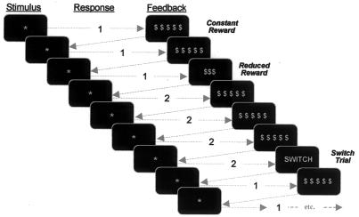
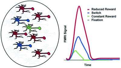
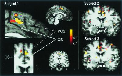
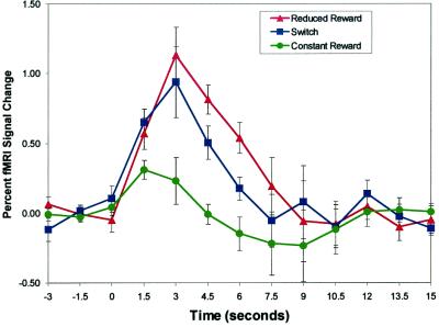
Dorsal anterior cingulate cortex (dACC) is a brain region that subserves cognition and motor control, but the mechanisms of these functions remain unknown. Human neuroimaging and monkey electrophysiology studies have provided valuable insights, but it has been difficult to link the two literatures. Based on monkey single-unit recordings, we hypothesized that human dACC is comprised of a mixture of functionally distinct cells that variously anticipate and detect targets, indicate novelty, influence motor responses, encode reward values, and signal errors. As an initial test of this conceptualization, the current event-related functional MRI study used a reward-based decision-making task to isolate responses from a subpopulation of dACC cells sensitive to reward reduction. As predicted, seven of eight subjects showed significant (P < 10(-4)) dACC activation when contrasting reduced reward (REDrew) trials to fixation (FIX). Confirmatory group analyses then corroborated the predicted ordinal relationships of functional MRI activation expected during each trial type (REDrew > SWITCH > CONrew > or = FIX). The data support a role for dACC in reward-based decision making, and by linking the human and monkey literatures, provide initial support for the existence of heterogeneity within dACC. These findings should be of interest to those studying reward, cognition, emotion, motivation, and motor control.


Figures

Figure 1
Figure 1
Reward-based decision-making task. Subjects were instructed to respond to each stimulus (*) by pressing one of two buttons based on the feedback from the previous trial (i.e., to repeat the same button press after a CONrew, and to change buttons after either a REDrew or SWITCH signal).
Figure 2
Figure 2
Schematized predicted fMRI response. Schematized representation of components most relevant to the fMRI results. A CONrew cell is depicted in green, cells responsible for the additional demands of performing SWITCH trials are depicted in blue, and cells specific to REDrew trials are depicted in red. Following Shima and Tanji (27), REDrew and CONrew cells are depicted at an approximate 5:1 ratio. Gray cells represent cells that support all trial types (e.g., anticipation, target detection) but are not the subject of immediate focus because they do not serve to differentiate fMRI responses. Qualitative predictions for fMRI responses appear on the right. FIX was predicted to produce no activation, CONrew was predicted to produce only minimal activation, and SWITCH trials (recruiting novelty detection cells and placing greater demands on response selection) were predicted to produce significantly greater activation. REDrew trials, recruiting cells involved in all previous trial types plus the very numerous REDrew sensitive cells, were predicted to produce the greatest activation.
Figure 3
Figure 3
dACC fMRI Response and ROI. Activation of dACC in response to REDrew trials (vs. FIX) is shown in three subjects. Pseudocolor-scaled statistical maps are displayed superimposed on the medial surface of the left hemisphere (sagittal view) and a coronal slice (y = 12 mm) in radiological convention (r = L) for subject 1. These areas are enlarged on the left. The dACC ROI included ACC between y = 0 and y = +30 within the cingulate sulcus (CS) for cerebral hemispheres with a single CS (Subject 1, Right), and ACC between the paracingulate sulcus (PCS) and CS, inclusive, for cerebral hemispheres with a double parallel cingulate sulcal pattern (Subject 1, Upper Left). It refers to the same cortical region that has been called the anterior cingulate cognitive division (ACcd) (1), rostral cingulate zone (9), or midcingulate cortex (3). The dACC ROI is indicated in aqua on the coronal slice enlargement (Subject 1, Lower Left). Coronal slices are shown for subjects 2 and 3.
Figure 4
Figure 4
dACC fMRI response. Group-averaged (n = 7), time-locked dACC activity for REDrew, SWITCH, and CONrew trials plotted as percent change from mean MRI signal during the first three images for each condition. Error bars indicate the SEM. As predicted, at t = 4.5 and 6 s (accounting for hemodynamic delay), REDrew > SWITCH trials > CONrew.

References

    1. Bush G, Luu P, Posner M I. Trends Cogn Sci. 2000;4:215–222. - PubMed
    1. Vogt B A, Nimchinsky E A, Vogt L J, Hof P R. J Comp Neurol. 1995;359:490–506. - PubMed
    1. Vogt B A, Vogt L J, Nimchinsky E A, Hof P R. In: Handbook of Chemical Neuroanatomy, The Primate Nervous System. Bloom F E, Björklund A, Hökfelt T, editors. Vol. 13. Amsterdam: Elsevier; 1997. , Part I, pp. 455–528.
    1. Dum R P, Strick P L. In: Neurobiology of Cingulate Cortex and Limbic Thalamus: A Comprehensive Handbook. Vogt B A, Gabriel M, editors. Boston: Birkhäuser; 1993. pp. 415–441.
    1. Van Hoesen G W, Morecraft R J, Vogt B A. In: Neurobiology of Cingulate Cortex and Limbic Thalamus: A Comprehensive Handbook. Vogt B A, Gabriel M, editors. Boston: Birkhäuser; 1993. pp. 249–284.

Publication types