. 2002 Jan 8;99(1):345-50.
doi: 10.1073/pnas.012511599. Epub 2001 Dec 26.

Macrophage migration inhibitory factor (MIF) sustains macrophage proinflammatory function by inhibiting p53: regulatory role in the innate immune response

Affiliations

Macrophage migration inhibitory factor (MIF) sustains macrophage proinflammatory function by inhibiting p53: regulatory role in the innate immune response

Robert A Mitchell et al. Proc Natl Acad Sci U S A. .

Abstract

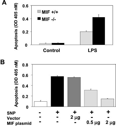
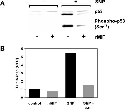
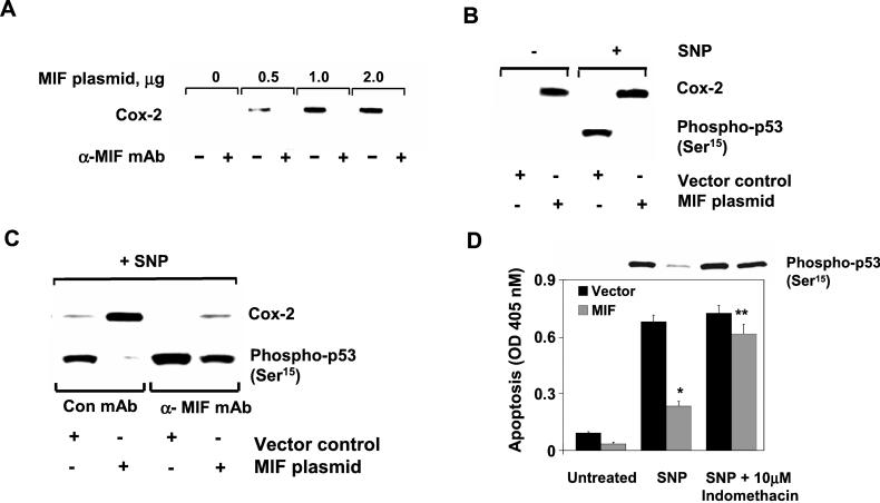
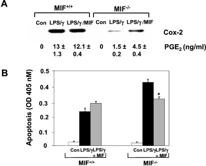
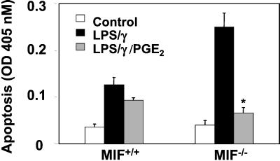
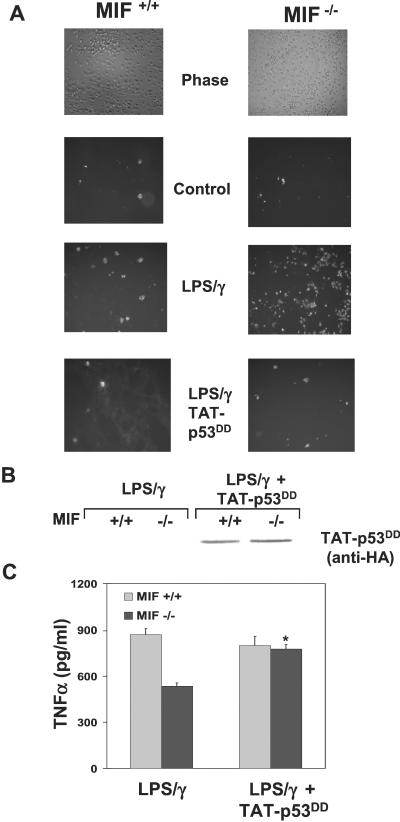
The importance of the macrophage in innate immunity is underscored by its secretion of an array of powerful immunoregulatory and effector molecules. We report herein that macrophage migration inhibitory factor (MIF), a product of activated macrophages, sustains macrophage survival and function by suppressing activation-induced, p53-dependent apoptosis. Endotoxin administration to MIF(-/-) mice results in decreased macrophage viability, decreased proinflammatory function, and increased apoptosis when compared with wild-type controls. Moreover, inhibition of p53 in endotoxin-treated, MIF-deficient macrophages suppresses enhanced apoptosis and restores proinflammatory function. MIF inhibits p53 activity in macrophages via an autocrine regulatory pathway, resulting in a decrease in cellular p53 accumulation and subsequent function. Inhibition of p53 by MIF coincides with the induction of arachidonic acid metabolism and cyclooxygenase-2 (Cox-2) expression, which is required for MIF regulation of p53. MIF's effect on macrophage viability and survival provides a previously unrecognized mechanism to explain its critical proinflammatory action in conditions such as sepsis, and suggests new approaches for the modulation of innate immune responses.


Figures

Figure 1
Figure 1
MIF modulates activation-induced macrophage apoptosis. (A) MIF-deficient mice are sensitized to LPS-induced peritoneal macrophage apoptosis. MIF+/+ and MIF−/− mice were injected i.p. with either vehicle or LPS (E. coli 0111: B4, 15 mg/kg) in 0.4 ml of sterile PBS. After 18 h, peritoneal macrophages were collected by lavage and immediately assessed for apoptosis by an ELISA that detects cytoplasmic oligonucleosomes. (B) MIF rescues RAW 264.7 macrophages from nitric oxide-induced apoptosis. RAW 264.7 macrophages were transfected with an MIF-expressing plasmid, pcDNA GS/MIF, or an empty pcDNA GS control plasmid for 20 h. The NO donor, SNP, was added at 1 mM for 16 h, and DNA fragmentation was assessed by ELISA. Data are mean ± SD of three experiments.
Figure 2
Figure 2
MIF inhibits p53 accumulation and function. (A) RAW 264.7 macrophages were pretreated overnight with 50 ng/ml rMIF. The NO donor, SNP, then was added at 1 mM for 4 h. Lysates were assessed for total p53 and p53 Ser15 phosphorylation by Western blotting. (B) RAW 264.7 macrophages were transiently cotransfected with the multimeric p53 responsive luciferase plasmid, p53-Luc, and the Renilla pRL-TK vector for 16 h. rMIF or vehicle then were added to cells for 8 h, followed by the addition of 1 mM SNP, as indicated, for 16 h. Results are expressed as fold increase over control after normalizing ratios of luciferase/Renilla luciferase from quadruplicate samples.
Figure 3
Figure 3
MIF induction of Cox-2 inhibits p53. (A) MIF induces Cox-2 expression in RAW 264.7 macrophages. Increasing concentrations of pcDNA GS/MIF plasmid or pcDNA GS vector control (lanes 1 and 2) were transfected into macrophages, and Cox-2 expression was examined. (B) Inverse relationship between MIF-induced Cox-2 and p53 expression. RAW 264.7 macrophages were transfected with either pcDNA GS vector control or pcDNA GS/MIF plasmid. SNP (1 mM) was added to cells as indicated for an additional 4 h. (C) Suppression of MIF-induced Cox-2 and p53 restoration by anti-MIF. Same as B but in the presence of anti-MIF or isotype control. (D) Inhibition of MIF-induced arachidonate metabolism restores p53 accumulation and function. Indomethacin was added at the time of transfection to inhibit the activity of cyclooxygenases. Data are the mean ± SD of three determinations and are representative of three independent experiments (*, P < 0.01, and **, P < 0.05 for MIF vs. vector).
Figure 4
Figure 4
MIF is required for LPS-induced Cox-2 expression, activity and suppression of apoptosis in murine peritoneal macrophages. (A) Resting peritoneal macrophages were stimulated for 18 h with 10 μg/ml LPS and 100 units/ml IFN-γ together with or without mouse rMIF. Results shown are the mean ± SD of duplicate samples (PGE2) and are representative of two independent experiments. (B) Restoration of Cox-2 expression and activity by rMIF rescues MIF−/− macrophages from augmented activation-induced apoptosis. Cells were treated as in A and assessed for apoptosis by ELISA of cytosolic oligonucleosomes as shown. Results shown are the mean ± SD of duplicate samples and are representative of three independent experiments. *, P < 0.04 for LPS/IFN-γ/MIF vs. LPS/IFN-γ treatment alone.
Figure 5
Figure 5
The Cox-2 product, PGE2, rescues activation-induced apoptosis in MIF−/− macrophages. Resting peritoneal macrophages from MIF+/+ and MIF−/− mice were stimulated with 10 μg/ml LPS and 100 units/ml IFN-γ in the absence or presence of exogenously added PGE2. PGE2 was added to cultures 4 h after the addition of LPS/IFN-γ to mimic relative time of PGE2 accumulation in MIF+/+ macrophages. Data shown are the mean ± SD of duplicate samples and are representative of two independent experiments. *, P < 0.03 for LPS/IFN-γ/PGE2 vs. LPS/IFN-γ alone.
Figure 6
Figure 6
Dominant-negative p53 inhibits enhanced activation-induced apoptosis in MIF−/− macrophages and restores function. Peritoneal macrophages from MIF+/+ and MIF−/− mice were treated with or without 250 nM TAT-p53DD for 15 min before stimulation with 10 μg/ml LPS and 100 units/ml IFN-γ for 24 h. (A) Activation induced apoptosis was assessed by fluorescent-annexin staining and fluorescence microscopy. (B) Anti-hemagglutinin (HA) immunoblot analysis of primary macrophages from MIF+/+ and MIF−/− mice transduced with TAT-p53DD, demonstrating equivalent transduction efficiencies. (C) Supernatants from indicated samples were assessed for TNF-α production by ELISA. Data shown are the mean ± SD of duplicate samples and are representative of two independent experiments. *, P < 0.05 for LPS/IFN-γ/TAT-p53DD vs. LPS/IFN-γ alone.

References

    1. Hoffmann J A, Kafatos F C, Janeway C A, Ezekowitz R A. Science. 1999;284:1313–1318. - PubMed
    1. Dinarello C A. Chest. 2000;118:503–508. - PubMed
    1. Ohmori Y, Hamilton T A. Pharmacol Ther. 1994;63:235–264. - PubMed
    1. Albina J E, Cui S, Mateo R B, Reichner J S. J Immunol. 1993;150:5080–5085. - PubMed
    1. Sarih M, Souvannavong V, Adam A. Biochem Biophys Res Commun. 1993;191:503–508. - PubMed

Publication types

MeSH terms