. 2002 Jan 8;99(1):467-72.
doi: 10.1073/pnas.012605299. Epub 2001 Dec 26.

A rapamycin-sensitive signaling pathway contributes to long-term synaptic plasticity in the hippocampus

Affiliations

A rapamycin-sensitive signaling pathway contributes to long-term synaptic plasticity in the hippocampus

Shao Jun Tang et al. Proc Natl Acad Sci U S A. .

Abstract

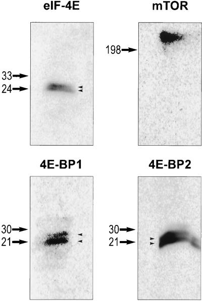
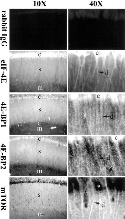
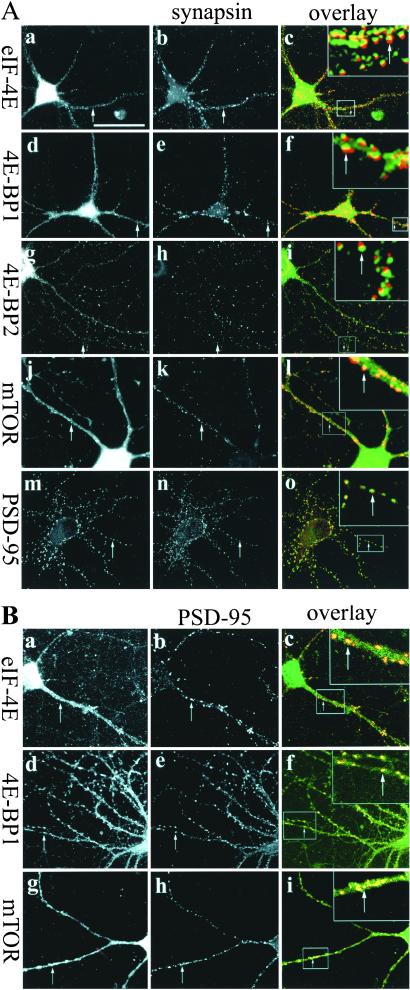
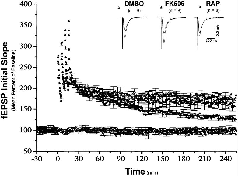
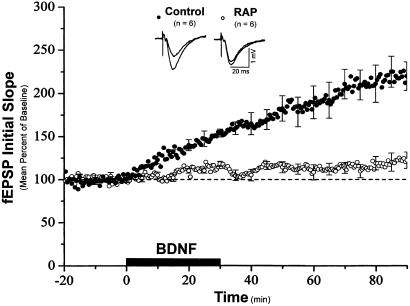
Many forms of long-lasting behavioral and synaptic plasticity require the synthesis of new proteins. For example, long-term potentiation (LTP) that endures for more than an hour requires both transcription and translation. The signal-transduction mechanisms that couple synaptic events to protein translational machinery during long-lasting synaptic plasticity, however, are not well understood. One signaling pathway that is stimulated by growth factors and results in the translation of specific mRNAs includes the rapamycin-sensitive kinase mammalian target of rapamycin (mTOR, also known as FRAP and RAFT-1). Several components of this translational signaling pathway, including mTOR, eukaryotic initiation factor-4E-binding proteins 1 and 2, and eukaryotic initiation factor-4E, are present in the rat hippocampus as shown by Western blot analysis, and these proteins are detected in the cell bodies and dendrites in the hippocampal slices by immunostaining studies. In cultured hippocampal neurons, these proteins are present in dendrites and are often found near the presynaptic protein, synapsin I. At synaptic sites, their distribution completely overlaps with a postsynaptic protein, PSD-95. These observations suggest the postsynaptic localization of these proteins. Disruption of mTOR signaling by rapamycin results in a reduction of late-phase LTP expression induced by high-frequency stimulation; the early phase of LTP is unaffected. Rapamycin also blocks the synaptic potentiation induced by brain-derived neurotrophic factor in hippocampal slices. These results demonstrate an essential role for rapamycin-sensitive signaling in the expression of two forms of synaptic plasticity that require new protein synthesis. The localization of this translational signaling pathway at postsynaptic sites may provide a mechanism that controls local protein synthesis at potentiated synapses.


Figures

Figure 1
Figure 1
Western blot analysis of eIF-4E, 4E-BP1, 4E-BP2, and mTOR proteins in hippocampal lysates. Total lysates of the rat adult hippocampus were used for Western blot analysis, with antibodies specific for eIF-4E, 4E-BP1, 4E-BP2, and mTOR (see Material and Methods for details). The size of protein markers is labeled on the left of each blot. Note the doublet bands detected by anti-eIF-4E, 4E-BP1, and 4E-BP2 (arrowheads).
Figure 2
Figure 2
Spatial distribution of eIF-4E, 4E-BP1, 4E-BP2, and mTOR in hippocampal slices. Shown are confocal images of hippocampal slices stained with primary antibodies against purified rabbit IgG (control staining), eIF4-E, 4E-BP1, 4E-BP2, or mTOR proteins, and FITC-conjugated secondary antibodies. Note the staining signals for eIF-4E, 4E-BP1, 4E-BP2, and mTOR in cell bodies and dendrites. Because mTOR is mainly a membrane protein, little signal for this protein is detected in the cytoplasm of the cell body (asterisk). c, cell body; d, dendrites; s, stratum radiatum; m, molecular layer.
Figure 3
Figure 3
Synaptic localization of eIF-4E, 4E-BP1, 4E-BP2, and mTOR. (A) Dissociated hippocampal neurons (P2; 14 DIV) were double-stained with components of the mTOR pathway (first column) including anti-eIF-4E (a), 4E-BP1 (d), 4E-BP2 (g), mTOR (j), or PSD-95 (m) antibodies and anti-synapsin I antibodies (second column) (b, e, h, and k). Signals from eIF-4E, 4E-BP1, 4E-BP2, mTOR, and PSD-95, and signals from the corresponding anti-synapsin staining on the same cells were overlaid in c, f, i, l, and o, respectively. In the overlaid images, green signals were generated by anti-eIF-4E, 4E-BP1, 4E-BP2, mTOR, or PSD-95 staining, red signals were generated by anti-synapsin staining, and yellow signals resulted from the overlapping of green and red signals. Insets in c, f, i, l, and o are the higher magnification images for the dendritic regions marked by boxes. (B) Double-labeling of eIF-4E (a), 4E-BP1 (d), mTOR (g) with PSD-95 (b, e, h). Signals from eIF-4E, 4E-BP1, and mTOR and signals from the corresponding PSD-95 staining on the same cells were overlaid in c, f, l, and i, respectively. Insets in c, f, and i are the higher magnification images for the dendritic regions marked by boxes (green signals resulted from eIF-4E, 4E-BP1, or mTOR staining; red signals from PSD-95 staining; yellow signals from the overlapping of green and red signals). Arrows indicate the synaptic regions that are double-stained. (Bar = 50 μm.)
Figure 4
Figure 4
Rapamycin decreases the magnitude of late-phase LTP in hippocampal slices. Shown are ensemble averages for all DMSO, FK506, and rapamycin experiments (n = 6, 9, and 8, respectively). All drugs were applied in the ACSF at least 30 min before the first tetanus. Mean DMSO control fEPSP slope values were 0.20 ± 0.01 mV/msec before LTP induction, and 0.35 ± 0.03 mV/msec 220–240 min after LTP induction. Mean FK506-treated fEPSP slope values were 0.21 ± 0.02 mV/msec before LTP induction, and 0.30 ± 0.09 mV/msec 220–240 min after LTP induction. Mean rapamycin-treated fEPSP slope values were 0.24 ± 0.05 mV/msec before LTP induction, and 0.31 ± 0.07 mV/msec 220–240 min after LTP induction. The control pathways for FK506 and rapamycin experiments are symbolized by the filled inverted triangle and the open circles, respectively.
Figure 5
Figure 5
Rapamycin prevents BDNF-induced synaptic potentiation in hippocampal slices. Shown are ensemble averages for all control and rapamycin experiments. BDNF (50 ng/ml) was applied in the ACSF at the time indicated by the bar. Mean control fEPSP slope values were 0.13 ± 0.03 mV/msec before BDNF application and 0.23 ± 0.04 mV/msec after BDNF application. Mean rapamycin-treated fEPSP slope values were 0.16 ± 0.01 mV/msec before BDNF application and 0.18 ± 0.01 mV/msec after BDNF application.

References

    1. Nguyen P V, Abel T, Kandel E R. Science. 1994;265:1104–1107. - PubMed
    1. Frey U, Morris R G. Trends Neurosci. 1998;21:181–188. - PubMed
    1. Kang H, Schuman E M. Science. 1996;273:1402–1406. - PubMed
    1. Montarolo P G, Goelet P, Castellucci V, Morgan J, Kandel E R, Schacher S. Science. 1986;234:1249–1254. - PubMed
    1. Davis H P, Squire L R. Psych Bull. 1984;96:518–559. - PubMed

MeSH terms

Substances