. 2002 Jan 8;99(1):303-8.
doi: 10.1073/pnas.012610299. Epub 2001 Dec 26.

Effects of peroxisome proliferator-activated receptor delta on placentation, adiposity, and colorectal cancer

Affiliations

Effects of peroxisome proliferator-activated receptor delta on placentation, adiposity, and colorectal cancer

Yaacov Barak et al. Proc Natl Acad Sci U S A. .

Abstract

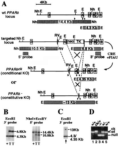
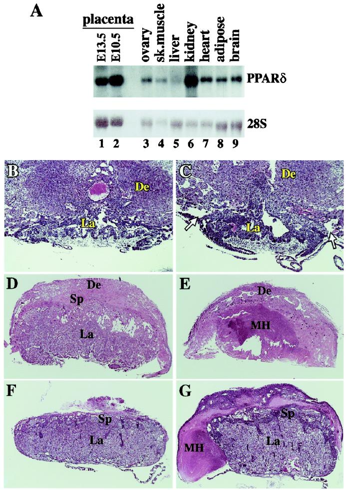
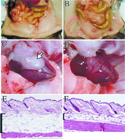
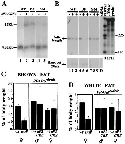
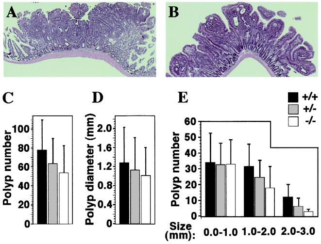
Targeting of the nuclear prostaglandin receptor peroxisome proliferator-activated receptor delta (PPARdelta) by homologous recombination results in placental defects and frequent (>90%) midgestation lethality. Surviving PPARdelta(-/-) mice exhibit a striking reduction in adiposity relative to wild-type levels. This effect is not reproduced in mice harboring an adipose tissue-specific deletion of PPARdelta, and thus likely reflects peripheral PPARdelta functions in systemic lipid metabolism. Finally, we observe that PPARdelta is dispensable for polyp formation in the intestine and colon of APC(min) mice, inconsistent with its recently proposed role in the establishment of colorectal tumors. Together, these observations reveal specific roles for PPARdelta in embryo development and adipocyte physiology, but not cancer.


Figures

Figure 1
Figure 1
PPARδ targeting strategy. (A) (Top to Bottom) The wt, primary targeted allele and the two secondary, CRE-induced recombinant alleles of PPARδ [the conditional knockout allele (PPARδck) and the constitutively null one (PPARδ)] (see Materials and Methods for details). Expected DNA fragments and their sizes are drawn as patterned bars under the respective genomic structures. Arrowheads indicate approximate location of PCR oligos. Restriction sites are: E, EcoRI; Nh, NheI; RV, EcoRV. (B) Southern blot analysis of homologous integration of the primary targeting construct shows the appropriate genomic alterations both 5′ and 3′ to the homologous recombination site in two of the targeted embryonic stem clones (T), as opposed to wt cells (+). (C) Southern blot analysis of daughter clones after CRE-mediated recombination and 1-(2-deoxy-2-fluoro-β-d-arabinofuranosyl)-5-iodouracil selection of the primary targeted embryonic stem cells. Arrow indicates clone A4, which contains a mixture of cells carrying either the constitutive knockout allele (13-kb fragment) or the PPARδck allele (see D). (D) PCR analysis of mice carrying various PPARδ allele combinations. Bottom band (≈240 bp), null allele; middle band (≈360 bp), wt; top band (≈400 bp), the PPARδck allele. Deduced genotypes are indicated on top.
Figure 2
Figure 2
Pathologies of PPARδ null placentas. (A) Mouse PPARδ tissue blot. Note the high PPARδ levels in two different stages of placental development (E10.5, E13.5), superior to most tissues, except kidney. (B and C) wt and PPARδ null placentas at E9.5. Arrows in C denote the abnormal detachment of the PPARδ null placenta from the decidua. (D and E) wt and PPARδ null placentas at E12.5. The inner core of the mutant placenta contains a massive maternal hematoma (MH) and is completely devoid of trophoblast cells. (F and G) wt and PPARδ null placentas at E14.5. The mutant placenta is surrounded by a maternal hematoma (MH). De, decidua; Sp, spongiotrophoblast layer; La, placental labyrinth; Th, thrombus. (Magnifications: B and C: ×20; D and E: ×11; F and G: ×7.)
Figure 3
Figure 3
Compromised adipose stores in PPARδ mutants. (A and B) Abdominal fat depots of wt (A) and PPARδ null mice (B), exhibiting a substantial compromise in the amount of fat tissue in a null mouse. (C and D) At 4 months the pericardial white fat depots are routinely found in wt mice (arrow in C) but absent from PPARδ null ones (D). (E and F) Hematoxylin and eosin-stained paraffin sections of skin from the lower back of wt (E) and PPARδ null mice (F). The subcutaneous fat layer is dramatically shrunk in the mutant, reflecting a combined effect of reduced adipocyte number and size. (Magnifications: E and F: ×15.)
Figure 4
Figure 4
Adipocyte-specific PPARδ knockout does not affect adipose tissue mass. (A) Southern blot analysis of epidydimal white fat pad (WF), interscapular brown fat pad (BF), and thigh skeletal muscle (SM) from mice carrying the PPARδck/ck allele and an aP2-CRE transgene. A 4.35-kb EcoRI fragment represents the nonexcised PPARδck allele, whereas CRE-mediated recombination yields a 13-kb EcoRI fragment (see Fig. 1 and Materials and Methods for further detail). Notice that CRE-dependent conversion into the 13-kb band is substantial in white and brown fat (lanes 2 and 3), and residual in skeletal muscle (lane 5). (B) RNase protection analysis of PPARδ transcript structure in the same mice. CRE-mediated deletion modifies a 210-nt-long protected fragment representing the full-length transcript (see arrow) into a 70-nt-long fragment representing a functionally null transcript devoid of its fourth exon (arrow, Bottom). The assay reveals ≈50% aP2CRE-mediated deletion of the full-length PPARδ mRNA in white fat (lanes 2 and 3 vs. lane 1), >80% in brown fat (lanes 5 and 6 vs. lane 4) and only a marginal one in skeletal muscle (lanes 8 and 9 vs. lane 7). Specificity controls with yeast tRNA and no RNase are shown in lanes 10 and 12, respectively. (C and D) Relative weights of interscapular brown fat pads (C) and epidydimal white fat pads (D) in wt and PPARδ null mice, and in PPARδck/ck mice in the absence (−) or presence of the aP2-CRE transgene. The 2.5- to 3-fold differences in adipose mass between wt and null mice (Left) are not recapitulated by an adipose-specific gene knockout (aP2-CRE vs. −, Right).
Figure 5
Figure 5
Intestinal polyp analysis in PPARδ-deficient APCmin mice. Histology of representative polyps from wt (A) and PPARδ−/− (B) APCmin mutants. Both adenomas display a similar, benign, tubular phenotype, regardless of the genetic status of PPARδ. (Magnification: ×16.) (C) Average polyp numbers in APCmin females carrying PPARδ+/+ (black bars; 77.6 ± 32.3), PPARδ+/− (gray bars; 63.6 ± 26.4), and PPARδ−/− genotypes (white bars; 54.0 ± 28.7). Differences between these values are statistically insignificant. (D) Median polyp diameters in PPARδ+/+ (1.28 ± 0.75 mm), PPARδ+/− (1.13 ± 0.68 mm), and PPARδ−/− (1.01 ± 0.59 mm). Differences between these values are statistically insignificant. (E) Intestinal polyp size distribution. Polyps in each of the three PPARδ genotypes were classified into three size ranges (0–1.0 mm; 1.0–2.0 mm; 2.0–3.0 mm). Note that wt PPARδ allele dosage is directly related to the incidence of larger polyps (>1.0 mm). However, differences are still statistically insignificant according to Student's t test.

Similar articles

Cited by

References

    1. Mangelsdorf D J, Thummel C, Beato M, Herrlich P, Schutz G, Umesono K, Blumberg B, Kastner P, Mark M, Chambon P, et al. Cell. 1995;83:835–839. - PMC - PubMed
    1. Kliewer S A, Forman B M, Blumberg B, Ong E S, Borgmeyer U, Mangelsdorf D J, Umesono K, Evans R M. Proc Natl Acad Sci USA. 1994;91:7355–7359. - PMC - PubMed
    1. Kliewer S A, Xu H E, Lambert M H, Willson T M. Recent Prog Horm Res. 2001;56:239–263. - PubMed
    1. Pineda Torra I, Gervois P, Staels B. Curr Opin Lipidol. 1999;10:151–159. - PubMed
    1. Tontonoz P, Hu E, Spiegelman B M. Cell. 1994;79:1147–1156. - PubMed

Publication types

MeSH terms

Substances