. 2001;9(6):237-48.
doi: 10.3727/000000001783992542.

Adenovirus-mediated increase of HNF-3 levels stimulates expression of transthyretin and sonic hedgehog, which is associated with F9 cell differentiation toward the visceral endoderm lineage

Affiliations

Adenovirus-mediated increase of HNF-3 levels stimulates expression of transthyretin and sonic hedgehog, which is associated with F9 cell differentiation toward the visceral endoderm lineage

Y Tan et al. Gene Expr. 2001.

Abstract

Retinoic acid-induced differentiation of mouse F9 embryonal carcinoma cells toward the visceral endoderm lineage is accompanied by increased expression of the Forkhead Box (Fox) transcription factors hepatocyte nuclear factor 3a (HNF-3alpha) and HNF-3beta, suggesting that they play a crucial role in visceral endoderm development. Retinoic acid stimulation results in a cascade of HNF-3 induction in which HNF-3alpha is a primary target for retinoic acid action and its increase is required for subsequent induction of HNF-3beta expression. Increased expression of HNF-3beta precedes activation of its known target genes, including transthyretin (TTR), Sonic hedgehog (Shh), HNF-1alpha, HNF-1beta, and HNF-4alpha. In order to examine whether increased HNF-3 expression is sufficient to induce expression of its downstream target genes without retinoic acid stimulation, we have used adenovirus-based expression vectors to increase HNF-3 protein levels in F9 cells. We demonstrate that adenovirus-mediated increase of HNF-3alpha levels in F9 cells is sufficient to induce activation of endogenous HNF-3beta levels followed by increased TTR and Shh expression. Furthermore, we show that elevated HNF-3beta levels stimulate expression of endogenous TTR and Shh without retinoic acid stimulation. Moreover, ectopic HNF-3 levels in undifferentiated F9 cells are insufficient to induce HNF-3alpha, HNF-1alpha, HNF-1beta, and HNF-4alpha expression, suggesting that their transcriptional activation required other regulatory proteins induced by the retinoic acid differentiation program. Finally, our studies demonstrate the utility of cell infections with adenovirus expressing distinct transcription factors to identify endogenous target genes, which are assembled with the appropriate nucleosome structure.


Figures

Figure 1
Figure 1
Construction of adenovirus vectors expressing HNF-3α and HNF-3β. (A) Structure of adenovirus recombination pAdCL-XEB. Schematically shown is the adenovirus recombination plasmid containing the expression cassette consisting of the CMV promoter, RNA splice site, EcoRI site for insertion of the rat HNF-3 and HNF-3 cDNAs, and the SV-40 polyadenylation sequence. The expression cassette was bounded on both sides by adenovirus sequences 1–355 and 3333–5788, which are required for adenovirus recombination to reconstitute the full-length virus. (B) Schematic representation of the replication-deficient recombinant adenoviruses in which the HNF-3 cDNAs replaces the E1 region. Note that this figure is not drawn to scale.
Figure 2
Figure 2
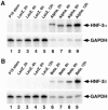
Detection of adenovirus-derived HNF-3 mRNAs. (A) Time course of adenovirus-derived HNF-3α mRNA. To determine HNF-3α mRNA levels, total RNA was prepared from P19 stem cells at various intervals following infection with adenovirus (at a multiplicity of 20 pfu/cell) and RNase protection assays were performed with antisense rat HNF-3α RNA probe as described in Materials and Methods. Shown are RNase protection assays with either P19 stem cell mRNA (lane 1) or AdLacZ-infected P19 cell mRNA isolated at 2 h (lane 2), 4 h (lane 3), 6 h (lane 4), and 12 h (lane 5) postinfection (PI). In addition, RNase protection assays with AdHNF3α-infected P19 cell mRNA isolated at 2 h (lane 6), 4 h (lane 7), 6 h (lane 8), and 12 h (lane 9) PI. For normalization purposes, signals of GAPDH-specific mRNA are shown in the bottom panel. (B) Temporal profile of adenovirus-derived HNF-3β mRNA. To determine HNF-3β mRNA levels, RNase protection assays were performed as described in (A) with antisense rat HNF-3β RNA probe. RNase protection assays with AdHNF3β-infected P19 cell mRNA isolated 2 h (lane 6), 4 h (lane 7), 6 h (lane 8), and 12 h (lane 9) PI and controls are described in (A).
Figure 3
Figure 3
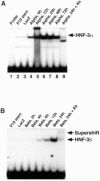
DNA binding properties of adenovirus-derived HNF-3 proteins. (A) Time course of adenovirus-derived HNF-3α protein. To determine HNF-3α mRNA levels, nuclear extracts were prepared from P19 stem cells at various intervals following infection with adenovirus (at a multiplicity of 20 pfu/cell) and used for gel shift assays with an HNF-3 binding site from the TTR promoter as described in Materials and Methods. Shown are gel shift assays using nuclear extracts isolated from either P19 stem cells (lane 2) or AdLacZ-infected P19 cells isolated at 24 h PI (lane 3) or AdH NF3α-infected P19 cells isolated at 6 h (lane 4), 12 h (lane 5), 24 h (lane 6), 48 h (lane 7), and 72 h (lane 8) PI. Lane 1 contains probe only. Lane 9 depicts a gel shift reaction carried out with extract derived from AdHNF3α-infected P19 cells (prepared 24 h PI) and HNF-3α-specific antiserum, which disrupted HNF-3α protein–DNA complex formation. (B) Time course of adenovirus-derived HNF-3β protein. To determine HNF-3β protein levels, gel shift assays were performed as described in (A). Shown are gel shift assays using nuclear extracts isolated from either P19 stem cells (lane 1), AdLacZ-infected P19 cells isolated at 24 h PI (lane 2) or AdHNF3β-infected P19 cells isolated at 2 h (lane 3), 4 h (lane 4), 6 h (lane 5), 12 h (lane 6), and 24 h (lane 7) PI. Lane 8 depicts a gel shift assay performed with nuclear extracts prepared from P19 cells infected with AdHNF3β at 24 h PI and HNF-3β-specific antiserum, which supershifted the HNF-3β protein–DNA complex.
Figure 4
Figure 4
Activation of HNF-3 binding site-containing promoter by AdHNF3α and AdHNF3β. A CAT reporter construct that contained four contiguous HNF-3 binding sites fused to the minimal TATA sequence from the CMV promoter was transfected into P19 cells. Sixteen hours later, the transfected cells were infected with AdLacZ (lane 2), AdHNF3α (lane 3), or AdHNF3β (lane 4) at a multiplicity of 20 pfu/cell. Twenty-four hours after the viral infections, cell extracts were prepared and CAT assays were performed as detailed in Materials and Methods. Lane 1 depicts a CAT assay carried out with cell extract prepared from transfected cells that had not been infected.
Figure 5
Figure 5
HNF-3α induces HNF-3β Shh and TTR levels in F9 cells. F9 stem cells were infected with AdLacZ or AdHNF3α at a multiplicity of 20 pfu/cell and total RNA was isolated at various intervals PI. RNase protection assays were performed by hybridizing 5 μg of RNA with antisense RNA probes specific for either mouse HNF-3β, mouse transthyretin (TTR), mouse sonic hedgehog (Shh,) or rat HNF-3α as described in Materials and Methods. (A) HNF-3α induces HNF-3β and TTR expression in F9 cells. RNase protection assays with indicated probes and AdHNF3α -infected F9 cell mRNA isolated at 12 h (lane 3), 1 day (lane 4), 2 days (lane 5), 3 days (lane 6), 4 days (lane 7), 5 days (lane 8), and 6 days (lane 9) PI. Control lanes include RNase protection assays from F9 stem cells (lane 1) or AdLacZ-infected F9 cells isolated 3 days PI (lane 2). For normalization purposes, GAPDH mRNA was detected by RNase protection (bottom panel). (B) HNF-3β induces Shh expression in F9 cells but not in fibroblasts. RNase protection assays with indicated probes and AdHNF3α-infected F9 cell mRNA isolated at 12 h (lane 3), 1 day (lane 4), 2 days (lane 5), 3 days (lane 6), 4 days (lane 7), 5 days (lane 8), and 6 days (lane 9) PI. RNase protection assays with indicated probes and AdHNF3α-infected fibroblast cell mRNA isolated at 12 h (lane 11), 1 day (lane 12) and 2 days (lane 13) PI. Controls lanes are identical to those described in (A).
Figure 6
Figure 6
HNF-3β stimulates TTR and Shh expression but not HNF-3α in F9 cells. F9 stem cells were infected with AdLacZ or AdHNF3β at a multiplicity of 20 pfu/cell and RNA was isolated at various intervals postinfection (PI). RNase protection assays were performed by hybridizing 5 μg of RNA with antisense probes for either mouse TTR, Shh, or rat HNF-3β as described in Materials and Methods. (A) HNF-3β stimulates TTR expression in F9 cells. RNase protection assays with indicated probes and AdHNF3β -infected F9 cell mRNA isolated at 12 h (lane 3), 1 day (lane 4), 2 days (lane 5), 3 days (lane 6), 4 days (lane 7), 5 days (lane 8), and 6 days (lane 9) PI. (B) HNF-3β stimulates Shh expression in F9 cells. RNase protection assays with indicated probes and AdHNF3β-infected F9 cell mRNA isolated at 12 h (lane 2), 1 day (lane 3), 2 days (lane 4), 3 days (lane 5), and 4 days (lane 6) PI. (C) Overexpression of HNF-3β in F9 cells does not activate HNF-3α. RNase protection assays with indicated probes and AdHNF3β-infected F9 cell mRNA isolated 12 h (lane 2), 1 day (lane 3), 2 days (lane 4), 3 days (lane 5), and 4 days (lane 6) after adenoviral infection. Controls are described in Figure 5 legend.

Similar articles

Cited by

References

    1. Andrews N. C.; Faller D. V. A rapid micropreparation technique for extraction of DNA-binding proteins from limiting numbers of mammalian cells. Nucleic Acids Res 19:2499; 1991. - PMC - PubMed
    1. Ang S. L.; Rossant J. HNF-3β is essential for node and notochord formation in mouse development. Cell 78:561–574; 1994. - PubMed
    1. Ang S. L.; Wierda A.; Wong D.; Stevens K. A.; Cascio S.; Rossant J.; Zaret K. S. The formation and maintenance of the definitive endoderm lineage in the mouse: Involvement of HNF3/forkhead proteins. Development 119:1301–1315; 1993. - PubMed
    1. Chang B. E.; Blader P.; Fischer N.; Ingham P. W.; Strahle U. Axial (HNF3beta) and retinoic acid receptors are regulators of the zebrafish sonic hedgehog promoter. EMBO J. 16:3955–3964; 1997. - PMC - PubMed
    1. Clark K. L.; Halay E. D.; Lai E.; Burley S. K. Co-crystal structure of the HNF-3/fork head DNA-recognition motif resembles histone H5. Nature 364:412–420; 1993. - PubMed

Publication types

MeSH terms