

A case of spontaneous pneumomediastinum and pneumopericardium in a young adult
- PMID: 11769580
- PMCID: PMC4531731
- DOI: 10.3904/kjim.2001.16.3.205
A case of spontaneous pneumomediastinum and pneumopericardium in a young adult
Abstract
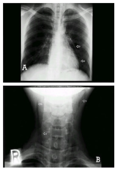
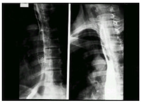
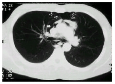
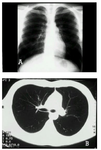
Spontaneous medialstinal emphysema (pneumomediastinum) and pneumopericardium may be defined as the presence of free air or gas in the mediastinal structures and in the pericardial sac without an apparent precipitating cause. It most frequently occurs in young healthy adults without serious underlying pulmonary disease. Although pneumomediastinum and pneumopericardium is often asymptomatic, it may cause pain in the neck and chest, dysphonia and shortness of breath. Treatment is supportive unless the patient has a history of trauma from foreign body aspiration. The course of spontaneous pneumomediastinum and pneumopericardium is usually benign and self-limited. A case of spontaneous pneumomediastinum, pneumopericardium and subcutaneous emphysema in a 20-year-old male is reported in this paper.
Figures
References
-
- Munsell WP. Pneumomediastinum: A report of 28 cases and a review of the literature. J Am Med Assoc. 1967;202:689–693. - PubMed
-
- Bejvan SM, Godwin JD. Pneumomediastinum: Old signs and new signs. Am J Roentgenol. 1996;166:1041–1048. - PubMed
-
- Macklin CC. Transport of air along sheaths of pulmonic blood vessels from alveoli to mediastinum : clinical implications. Arch Intern Med. 1939;64:913–926.
-
- Maunder RJ, Pierson DJ, Hudson LD. Subcutaneous and mediastinal emphysema: pathophysiology, diagnosis and management. Arch Intern Med. 1984;144:1447–1453. - PubMed
Publication types
MeSH terms
LinkOut - more resources
Full Text Sources