Blisters in the small intestinal mucosa of coeliac patients contain T cells positive for cyclooxygenase 2
- PMID: 11772972
- PMCID: PMC1773065
- DOI: 10.1136/gut.50.1.84
Blisters in the small intestinal mucosa of coeliac patients contain T cells positive for cyclooxygenase 2
Abstract
Background and aims: Coeliac disease is characterised by atrophy of the villi and hyperplasia of the crypts in the mucosa of the small intestine. It is caused by an environmental trigger, cereal gluten, which induces infiltration of the mucosa by inflammatory cells. We hypothesised that these inflammatory cells express cyclooxygenase 2 (COX-2), an enzyme that contributes to the synthesis of pro and anti-inflammatory prostaglandins and is known to be expressed at sites of inflammation in the stomach and colon. We have investigated expression of COX-2 in the coeliac disease affected small intestinal mucosa where it may be an indicator of either disease induction or mucosal restoration processes.
Patients and methods: Small intestinal biopsy samples from 15 coeliac patients and 15 non-coeliac individuals were stained immunohistochemically for COX-2. Samples from 10 of the patients were also stained after these patients had been on a gluten free diet for 6-24 months. Various cell type marker antigens were used for immunohistochemical identification of the type of cell that expressed COX-2. To further verify colocalisation of the cell type marker and COX-2, double immunoperoxidase and immunofluorescence methods were employed. Immunoelectron microscopy was used to investigate the subcellular location of COX-2.
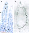
Results: In all samples taken from coeliac patients, clusters of cells with strong immunoreactivity for COX-2 were found in those areas of the lamina propria where the epithelium seemed to blister or was totally detached from the basement membrane. These clusters were reduced in number or totally absent in samples taken after a gluten free diet. No such clusters were seen in any control samples. The density of COX-2 positive cells lining the differentiated epithelium decreased significantly from 13.5 (5.1) cells/10(5) microm(2) (mean (SD)) in the untreated patient samples to 6.5 (2.0) cells/10(5) microm(2) after a gluten free diet (p<0.001), and was 3.3 (1.9) cells/10(5) microm(2) in control samples (p<0.001 compared with untreated or diet treated coeliac samples). Staining for COX-2 was localised to CD3+ T cells and CD68+ macrophages in the mucosal lesions but not all of these cells were positive for COX-2. Immunoelectron microscopy revealed that the ultrastructure of the COX-2 positive cells resembled that of lymphocytes, and the immunoreaction was localised to the rough endoplasmic reticulum and the nuclear envelope.
Conclusions: Our results show that in coeliac disease, blistering of small intestinal epithelial cells is associated with accumulation of COX-2 positive T cells, and the number of these cells decreases after a gluten free diet. These observations suggest that COX-2 mediated prostanoid synthesis contributes to healing of the coeliac mucosa and may be involved in maintenance of intestinal integrity.
Figures


References
-
- Versteeg HH, van Bergen en Henegouwen PM, van Deventer SJ, et al. Cyclooxygenase-dependent signalling: molecular events and consequences. FEBS Lett 1999;445:1–5. - PubMed
-
- Crofford LJ. COX-1 and COX-2 tissue expression: implications and predictions. J Rheumatol 1997;24:15–19. - PubMed
-
- Komhoff M, Grone HJ, Klein T, et al. Localization of cyclooxygenase-1 and -2 in adult and fetal human kidney: implication for renal function. Am J Physiol 1997;272:F460–8. - PubMed
-
- Sakuma K, Fujimori T, Hirabayashi K, et al. Cyclooxygenase (COX)-2 immunoreactivity and relationship to p53 and Ki-67 expression in colorectal cancer. J Gastroenterol 1999;34:189–94. - PubMed
-
- Ristimäki A, Honkanen N, Jankala H, et al. Expression of cyclooxygenase-2 in human gastric carcinoma. Cancer Res 1997;57:1276–80. - PubMed
Publication types
MeSH terms
Substances
LinkOut - more resources
Full Text Sources
Medical
Research Materials
