. 2002 Mar 22;277(12):10114-20.
doi: 10.1074/jbc.M110527200. Epub 2001 Dec 28.

Molecular basis of evolutionary loss of the alpha 1,3-galactosyltransferase gene in higher primates

Affiliations

Molecular basis of evolutionary loss of the alpha 1,3-galactosyltransferase gene in higher primates

Chihiro Koike et al. J Biol Chem. .

Abstract

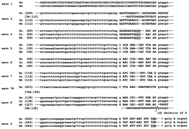
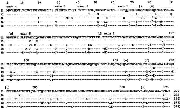
Galactose-alpha1,3-galactose (alphaGal) epitopes, the synthesis of which requires the enzyme product of alpha1,3-galactosyltransferase (alpha1,3GT), are sugar chains on the cell surface of most mammalian species. Notable exceptions are higher primates including Old World monkeys, apes, and humans. The alphaGal-negative species as well as mice with deletion of the alpha1,3GT gene produce abundant anti-alphaGal antibodies. The evolutionary loss of alphaGal epitopes has been attributed to point mutations in the coding region of the gene. Because no transcripts could be found in the higher primate species with Northern blot analysis, a potential alternative explanation has been loss of upstream regulation of the gene. Here, we have demonstrated that the rhesus promoter is functional. More importantly, a variety of full-length transcripts were detected with sensitive PCR-based methods in the tissues of rhesus monkeys, orangutans, and humans. Five crucial mutations were delineated in the coding region of the human and rhesus and three in the orangutan, any one of which could be responsible for inactivation of the alpha1,3GT gene. Two of the mutations were shared by all three higher primates. These findings, which elucidate the molecular basis for the evolutionary loss of alphaGal expression, may have implications in medical research.


Figures

Fig. 1
Fig. 1. Genomic organization on a kb scale of the α1,3GT gene of the human (above horizontal line) and rhesus (below horizontal line)
The numbered boxes indicate the approximate location of exons within the locus of the two species but do not accurately reflect the length of each exon. The translation start and stop codons are located in exons 4 and 9. The horizontal thick bar above exon 9 denotes the GenBank data for human (J05421, 794 bp) contributed by Larsen et al. (5), and the short thick bar below exon 9 denotes the data for rhesus (M73306, 371 bp) contributed by Galili and Swanson (6). Shown below the gene locus representation is a contig of overlapping subgenomic clones derived from GenomeWalker libraries (horizontal arrows) or from long PCR experiments (horizontal lines).
Fig. 2
Fig. 2
A, electrophoresis of PCR (ac) and Northern blot hybridization (d). Lane 1, electrophoresis results of RT-PCR in the human, orangutan, and rhesus cDNA libraries, respectively, using a pair of primers spanning the coding region. Lane M, DNA size marker ladder (Invitrogen, 1-kb DNA size ladder). d, Northern blot of total RNA from bovine (lane 1) or rhesus spleen tissue (lane 2) hybridized with a probe derived from rhesus exon 9 fragment. As expected, with in-species versus cross-species hybridization, gene expression was lower in the bovine than in the monkey spleen cells. B, schematic splicing variations and cryptic exons of human (a), orangutan (b), and rhesus (c) α1,3GT gene. The closed boxes denote exons in the respective species. The open boxes with arrows denote cryptic exons that were not observed in the αGal-positive species. Exon 6Rc in rhesus has exon 6, intron 6, and exon 7 (a retained intron).
Fig. 3
Fig. 3. Comparison of exon-intron boundaries of the human (H), orangutan (Ou), and rhesus (R) α1,3GT gene
Capital or lowercase letters indicate the nucleotides of the exon or intron in each species, respectively. The numbers in parentheses indicate exon size in bp. The start codon in exon 4 and the authentic stop codon in exon 9 are underlined.
Fig. 4
Fig. 4. Exon 1 vicinity of α1,3GT gene of rhesus (R) compared with pig (P)
The rhesus exon 1 and its potential regulatory region (capital letters) are aligned with the corresponding porcine exon 1 sequence (12). The lowercase letters denote intron 1. An asterisk indicates that the nucleotide is identical between both species.
Fig. 5
Fig. 5
Luciferase assay for promoter activity of the rhesus α1,3GT gene
Fig. 6
Fig. 6. Comparisons of deduced amino acid sequences of the α1,3GT gene in marmoset (M), cebus (C), rhesus (R), orangutan (O), and human (H)
Nucleotide sequences are shown from the start codon to the stop codon of the marmoset monkey cDNA in one-letter abbreviations. Dashed horizontal lines indicate that each nucleotide is identical to the corresponding nucleotide of the marmoset base-line sequence. The single asterisk in cebus denotes a triplet nucleotide deletion not present in the marmoset. A single nucleotide deletion, depicted by a slash (/), was observed at positions [a], [b], [e], and [g]. Premature termination, depicted by an X, was observed at positions [c], [f], and [h].
Fig. 7
Fig. 7. Comparison of crucial point mutations of the α1,3GT gene in primates
The numbered open boxes (top) are exons in which crucial mutations in the primates are shown with the corresponding nucleotide sequence. No mutations were observed in αGal-positive primates. Crucial mutations were observed in exons 7 and 9 of the three higher primate species examined (lower boxes). Note that the mutations at position [b] and [f] (lower closed boxes with thick lines) are shared by rhesus, orangutan, and human. (F/S), frameshift; (Stop), stop codon.

Similar articles

Cited by

References

    1. Galili U, Shohet SB, Kobrin E, Stults CLM, Macher BA. J Biol Chem. 1988;263:17755–17762. - PubMed
    1. Galili U, Clark MR, Shohet SB, Buehler J, Macher BA. Proc Natl Acad Sci U S A. 1987;84:1369–1373. - PMC - PubMed
    1. Cooper DKC, Koren E, Oliol R. Lancet. 1993;342:682–683. - PubMed
    1. Joziasse DH, Shaper JH, Van den Eijnden DH, Van Tunen AJ, Shaper NL. J Biol Chem. 1989;264:14290–14297. - PubMed
    1. Larsen RD, Rivera-Marrero CA, Ernst LK, Cummings RD, Lowe JB. J Biol Chem. 1990;265:7055–7061. - PubMed

Publication types

MeSH terms

Substances

LinkOut - more resources