. 2002 Feb;76(3):1043-50.
doi: 10.1128/jvi.76.3.1043-1050.2002.

Human cytomegalovirus UL47 tegument protein functions after entry and before immediate-early gene expression

Affiliations

Human cytomegalovirus UL47 tegument protein functions after entry and before immediate-early gene expression

Jill T Bechtel et al. J Virol. 2002 Feb.

Abstract

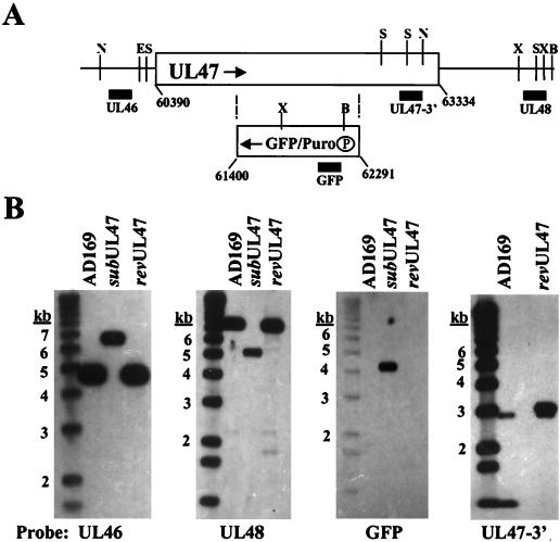
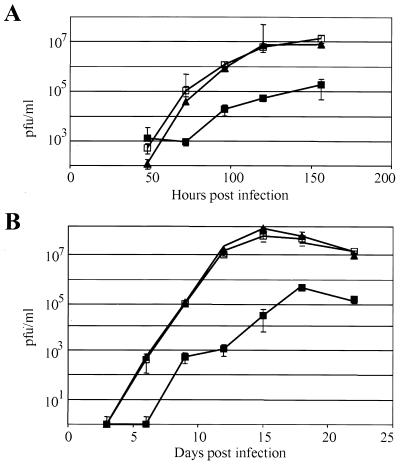
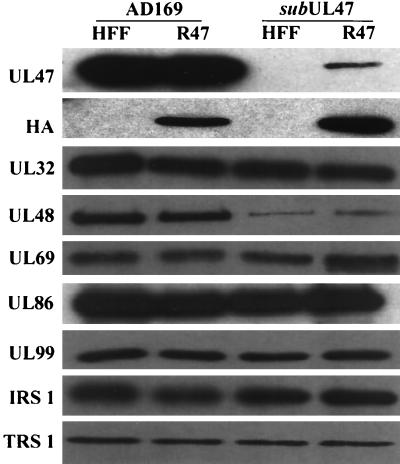
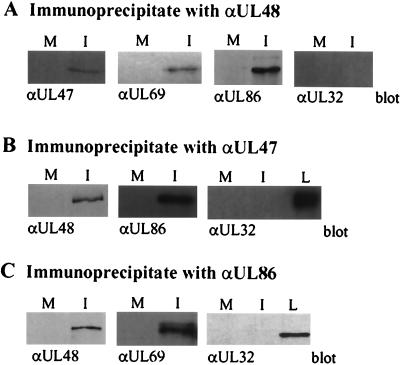
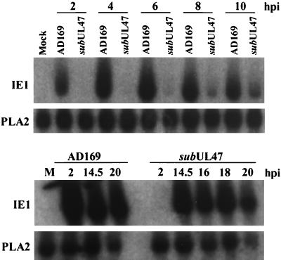
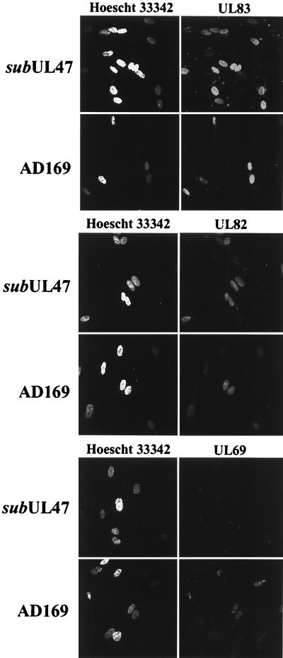
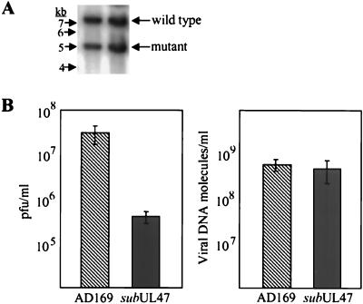
The human cytomegalovirus UL47 open reading frame encodes a 110-kDa protein that is a component of the virion tegument. We have constructed a cytomegalovirus mutant, ADsubUL47, in which the central portion of the UL47 open reading frame has been replaced by two marker genes. The mutant replicated to titers 100-fold lower than those for wild-type virus after infection at either a high or a low input multiplicity in primary human fibroblasts but was substantially complemented on cells expressing UL47 protein. A revertant virus in which the mutation was repaired, ADrevUL47, replicated with wild-type kinetics. Mutant virions lacked UL47 protein and contained reduced amounts of UL48 protein. The mutant was found to be less infectious than wild-type virus, and a defect very early in the replication cycle was observed. Transcription of the viral immediate-early 1 gene was delayed by 8 to 10 h. However, this delay was not the result of a defect in virus entry or of the inability of virion proteins to transactivate the major immediate-early promoter. We also show that the UL47 protein coprecipitated with the UL48 and UL69 tegument proteins and the UL86-encoded major capsid protein. We propose that a UL47-containing complex is involved in the release of viral DNA from the disassembling virus particle and that the loss of UL47 protein causes this process to be delayed.


Figures

FIG. 1.
FIG. 1.
Construction of ADsubUL47. (A) Schematic representation of the UL47 locus and the ADsubUL47 substitution mutation. The rectangular box represents the UL47 coding sequence with the relevant nucleotide numbers (6) shown. The GFP/Puro box represents the DNA recombined into the UL47 ORF, with the boundaries of the UL47 nucleotides replaced given below the box. The circled P represents the promoter for the GFP/Puro cassette. Arrows indicate the opposite directions of transcription for UL47 and the GFP/Puro cassette. Vertical lines represent restriction enzyme recognition sites (B, BamHI; E, EcoRI; N, EcoNI; S, StyI; X, XmnI). Solid boxes indicate locations of probes used in Southern blotting. (B) Southern blots of AD169, ADsubUL47, and RUL47 viral DNA digested with EcoNI (leftmost panel), BamHI and EcoRI (center panels), and StyI (rightmost panel). Probes are shown below the blots: UL46 probe (nucleotides 60388 to 60562), UL48 probe (nucleotides 65548 to 66150), GFP probe, and UL47 3′ probe (nucleotides 62352 to 62853). Sizes of DNA markers are shown to the left of each blot.
FIG. 2.
FIG. 2.
Growth of wild-type AD169, ADsubUL47, and ADrevUL47. HFFs were infected at a multiplicity of 2 (A) or 0.001 (B) PFU/cell with AD169 (▴), ADsubUL47 (▪), or ADrevUL47 (squlo). Samples were removed at the indicated times after infection and titered by plaque assay on R47 HFFs.
FIG. 3.
FIG. 3.
Composition of ADsubUL47 virions. HFFs or R47 HFFs were infected with wild-type AD169 or ADsubUL47 at a multiplicity of 2 PFU/cell; virions were purified from the culture medium and lysed; and equal amounts of proteins were subjected to Western blot analysis using antibodies to the indicated viral proteins or to the influenza virus HA epitope tag.
FIG. 4.
FIG. 4.
Synthesis of UL48 mRNA and protein. (A) HFFs and R47 HFFs were either mock infected or infected with either wild-type AD169 or ADsubUL47 at a multiplicity of 2 PFU/cell, and protein extracts were prepared at the indicated times. Forty micrograms of protein was analyzed by Western blotting using pUL48- and pUL69-specific monoclonal antibodies. (B) RNA was isolated at various times postinfection from mock-infected cells or from cells infected at a multiplicity of 2 PFU/cell with AD169 or ADsubUL47 and was analyzed by Northern blotting using a probe to the UL48 gene. The cellular PLA2 mRNA was used as a loading control. Arrows indicate the migration of the UL47-, UL48-, and ADsubUL47-specific (*) transcripts.
FIG. 5.
FIG. 5.
Interaction of pUL47 with pUL48, pUL69, and pUL86. R47 HFFs were mock infected or infected with wild-type AD169 at a multiplicity of 2 PFU/cell; after 72 h, protein lysates were prepared and then immunoprecipitated with antibodies against pUL48 (A), pUL47 (HA epitope) (B), or pUL86 (C), separated by SDS-polyacrylamide gel electrophoresis, and blotted to nitrocellulose membranes. Blots were probed with antibodies recognizing pUL47 (HA epitope), pUL48, pUL69, pUL82, and pUL32. M, mock-infected cells; I, HCMV-infected cells; L, lysate.
FIG. 6.
FIG. 6.
IE1 mRNA expression in ADsubUL47-infected cells. Cells were either mock infected or infected at a multiplicity of 2 PFU/cell with either wild-type AD169 or ADsubUL47; and RNA was harvested at various times and analyzed by Northern blotting. Blots were probed with 32P-labeled IE1 or PLA2 (loading control) cDNAs. M, mock-infected cells.
FIG. 7.
FIG. 7.
Tegument protein release in ADsubUL47-infected cells. HFFs were plated onto coverslips and infected with wild-type AD169 at a multiplicity of 20 PFU/cell or with ADsubUL47 at a multiplicity of 5 PFU/cell. Coverslips were fixed and stained for pUL69, pUL82, or pUL83 at 4 hpi. Nuclei were visualized by staining with Hoescht 33342.
FIG. 8.
FIG. 8.
Relative particle-to-PFU ratios. Wild-type AD169 and ADsubUL47 virus stocks were prepared, and small aliquots were titered to determine the concentration of infectious virus. Then equal volumes of the two stocks were mixed, virions were partially purified, and their DNA was extracted. (A) Viral DNA was digested with BamHI and EcoRI and subjected to a Southern blot assay using a 32P-labeled UL48-specific probe. (B) Comparison of the virus titers for AD169 and ADsubUL47 to the relative amounts of the two DNAs, determined by phosphorimager quantification of the bands visualized in panel A.

References

    1. Baldick, C. J., Jr., A. Marchini, C. E. Patterson, and T. Shenk. 1997. Human cytomegalovirus tegument protein pp71 (UL82) enhances the infectivity of viral DNA and accelerates the infectious cycle. J. Virol. 71:4400–4408. - PMC - PubMed
    1. Baldick, C. J., Jr., and T. Shenk. 1996. Proteins associated with purified human cytomegalovirus particles. J. Virol. 70:6097–6105. - PMC - PubMed
    1. Batterson, W., D. Furlong, and B. Roizman. 1983. Molecular genetics of herpes simplex virus. VIII. Further characterization of a temperature-sensitive mutant defective in the release of viral DNA and in other stages of the viral reproductive cycle. J. Virol. 45:397–407. - PMC - PubMed
    1. Bresnahan, W. A., G. E. Hultman, and T. Shenk. 2000. Replication of wild-type and mutant cytomegalovirus in life-extended diploid fibroblasts. J. Virol. 74:10816–10818. - PMC - PubMed
    1. Bresnahan, W. A., and T. E. Shenk. 2000. UL82 virion protein activates expression of immediate early viral genes in human cytomegalovirus-infected cells. Proc. Natl. Acad. Sci. USA 97:14506–14511. - PMC - PubMed

Publication types

Substances

LinkOut - more resources