. 2002 Feb;76(3):1265-72.
doi: 10.1128/jvi.76.3.1265-1272.2002.

Detection of a novel unglycosylated form of hepatitis C virus E2 envelope protein that is located in the cytosol and interacts with PKR

Affiliations

Detection of a novel unglycosylated form of hepatitis C virus E2 envelope protein that is located in the cytosol and interacts with PKR

Nicole Pavio et al. J Virol. 2002 Feb.

Abstract

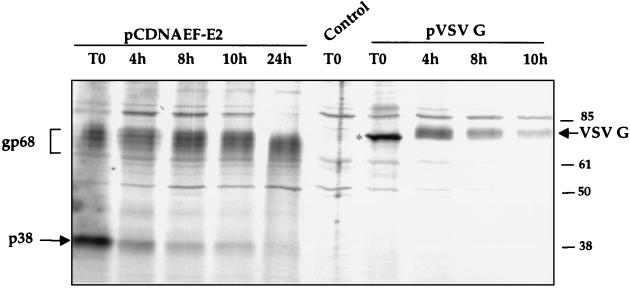
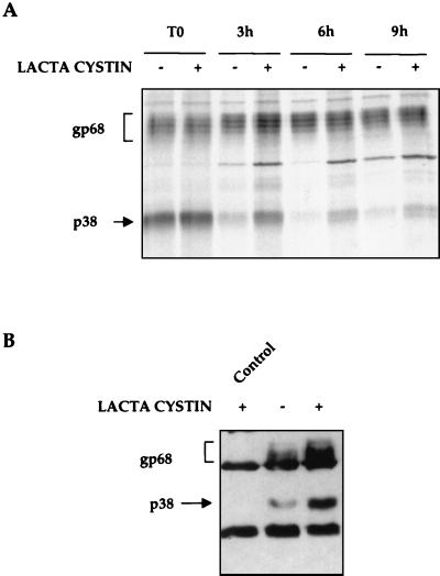
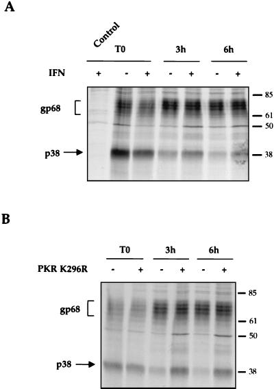
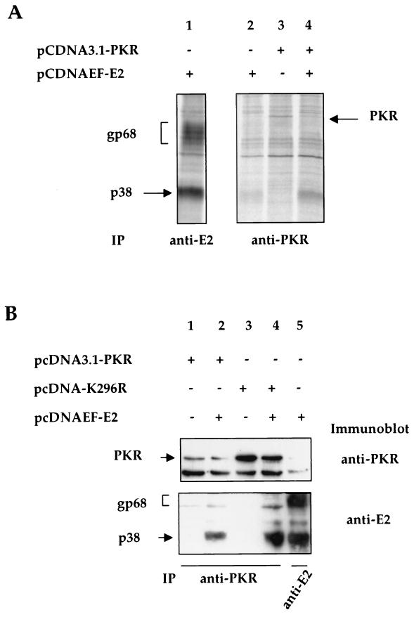
The hepatitis C virus (HCV) envelope protein E2 has been shown to accumulate in the lumen of the endoplasmic reticulum (ER) as a properly folded glycoprotein as well as large aggregates of misfolded proteins. In the present study, we have identified an additional unglycosylated species, with an apparent molecular mass of 38 kDa (E2-p38). In contrast to the glycosylated E2, E2-p38 is significantly less stable and is degraded through the proteasome pathway. Correspondingly, E2-p38 is found to be ubiquitinated. E2-p38 is localized mostly in the cytosol, in contrast to the glycosylated form, which is exclusively membrane associated. Alpha interferon (IFN-alpha) treatment or overexpression of the double-stranded RNA-activated protein kinase (PKR) significantly increased the stability of E2-p38, consistent with a previous report (D. R. Taylor, S. T. Shi, P. R. Romano, G. N. Barber, and M. M. Lai, Science 285:107-110, 1999) that E2 interacts with PKR and inhibits its kinase activity. Direct interaction between PKR and E2-p38, but not the glycosylated form of E2, was also observed. These results show that E2-p38 is the form of E2 that interacts with PKR in the cytosol and may contribute to the resistance of HCV to IFN-alpha. Thus, an ER protein can exist in the cytosol as an unglycosylated species and impair cellular functions.


Figures

FIG. 1.
FIG. 1.
Detection of an unglycosylated form of E2 in mammalian cells. (A) Detection of E2 in 293T cells transfected with pcDNAEF-E2. Cells were radiolabeled with [35S]methionine and -cysteine and immunoprecipitated with different anti-E2 monoclonal antibodies. The control was cells transfected with the empty vector pcDNAEF and immunoprecipitated with H62. (B) Same as for panel A but in HeLa cells. (C) Immunoprecipitation of radiolabeled E2 after tunicamycin treatment or digestion with PNGase F or Endo H glycosidases. (D) Detection by immunoblotting, using H62 monoclonal antibody, of immunoprecipitated E2 after tunicamycin treatment or glycosidase digestions. Control lane was cells transfected with an empty vector; the two bands correspond to the light (≈25 kDa) and heavy (≈55 kDa) chains of the antibody used for immunoprecipitation. Exposure time for the control lane was longer to identify their positions. Markers are indicated in kilodaltons. The glycosylated form of E2 is termed gp68, the unglycosylated forms of E2 are termed p38 and p42, and the partially deglycosylated E2 is termed p44.
FIG. 2.
FIG. 2.
Kinetics of degradation of E2 and VSV G proteins. Cells were pulse-labeled with [35S]methionine and -cysteine for 5 min and chased for various lengths of time as indicated in hours. Immunoprecipitations of E2 and VSV G were performed with their respective monoclonal antibodies H62 and P5D4. Control cells were transfected with pcDNAEF and immunoprecipitated with P5D4. Marker is indicated in kilodaltons. VSV G is indicated by an arrow and its unglycosylated form is marked by an asterisk.
FIG. 3.
FIG. 3.
Degradation of E2. (A) Stability of E2 in the presence of a proteasome inhibitor. Cells were pulse-labeled for 15 min (T0), chased with (+) or without (−) lactacystin for various lengths of time in hours, and immunoprecipitated with anti-E2 antibody. (B) Transfected cells were treated with (+) or without (−) 20 μM lactacystin for 1 h. Cell lysates were immunoprecipitated with a mixture of anti-ubiquitin mouse monoclonal (1:500) and anti-ubiquitin rabbit polyclonal FL-76 (1:500) antibodies. Proteins were immunoblotted for E2. The control was cells transfected with the empty vector and treated with lactacystin.
FIG. 4.
FIG. 4.
Analysis by membrane flotation gradients of the subcellular distribution of E2. Transfected 293T cells were radiolabeled for 15 min, and cellular extracts were subjected to centrifugation on sucrose gradients (10 to 75% [wt/wt] sucrose) at 38,000 rpm for 14 h. Fractions were collected from the top and each fraction was immunoprecipitated using anti-E2 antibody H62.
FIG. 5.
FIG. 5.
Stability of E2 after IFN-α treatment or cotransfection with PKR K296R. (A) IFN treatment. Transfected cells were treated with or without 1,000 IU of IFN-α per ml for 18 h, pulse-labeled for 15 min (T0), and then chased for various lengths of times as indicated. Control with empty vector pcDNAEF is included. (B) PKR K296R. Cells were transfected with E2 alone (−) or cotransfected with PKR K296R (+) (ratio, 1:1). A pulse-chase experiment was done as for panel A.
FIG. 6.
FIG. 6.
Coimmunoprecipitation of E2-p38 with PKR. (A) Cells transfected with different plasmids were radiolabeled and precipitated with the anti-E2 (lane 1) or anti-PKR (lanes 2 to 4) antibodies. (B) Protein extracts of cells transfected with different plasmids were first immunoprecipitated with an anti-PKR antibody (lanes 1 to 4) or with an anti-E2 antibody (lane 5). After SDS-PAGE analysis, proteins were immunoblotted with an anti-PKR antibody (upper panel) or an anti-E2 antibody (lower panel). In the top panel, the lower band corresponds to the heavy chain (≈55 kDa) of the antibody used and the upper one corresponds to PKR.

Similar articles

Cited by

References

    1. Choukhi, A., S. Ung, C. Wychowski, and J. Dubuisson. 1998. Involvement of endoplasmic reticulum chaperones in the folding of hepatitis C virus glycoproteins. J. Virol. 72:3851–3858. - PMC - PubMed
    1. Cocquerel, L., J. C. Meunier, A. Pillez, C. Wychowski, and J. Dubuisson. 1998. A retention signal necessary and sufficient for endoplasmic reticulum localization maps to the transmembrane domain of hepatitis C virus glycoprotein E2. J. Virol. 72:2183–2191. - PMC - PubMed
    1. Cocquerel, L., C. Wychowski, F. Minner, F. Penin, and J. Dubuisson. 2000. Charged residues in the transmembrane domains of hepatitis C virus glycoproteins play a major role in the processing, subcellular localization, and assembly of these envelope proteins. J. Virol. 74:3623–3633. - PMC - PubMed
    1. Colombo, M. 1998. The role of hepatitis C virus in hepatocellular carcinoma. Recent Results Cancer Res. 154:337–344. - PubMed
    1. Deleersnyder, V., A. Pillez, C. Wychowski, K. Blight, J. Xu, Y. S. Hahn, C. M. Rice, and J. Dubuisson. 1997. Formation of native hepatitis C virus glycoprotein complexes. J. Virol. 71:697–704. - PMC - PubMed

Publication types

LinkOut - more resources