. 2002 Feb;76(3):1273-84.
doi: 10.1128/jvi.76.3.1273-1284.2002.

The cytoplasmic tail of infectious bronchitis virus E protein directs Golgi targeting

Affiliations

The cytoplasmic tail of infectious bronchitis virus E protein directs Golgi targeting

Emily Corse et al. J Virol. 2002 Feb.

Abstract

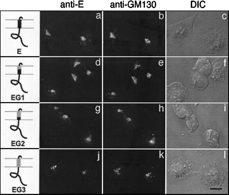
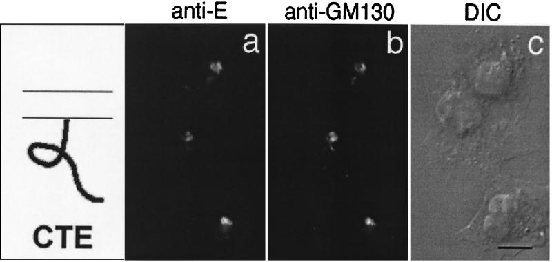
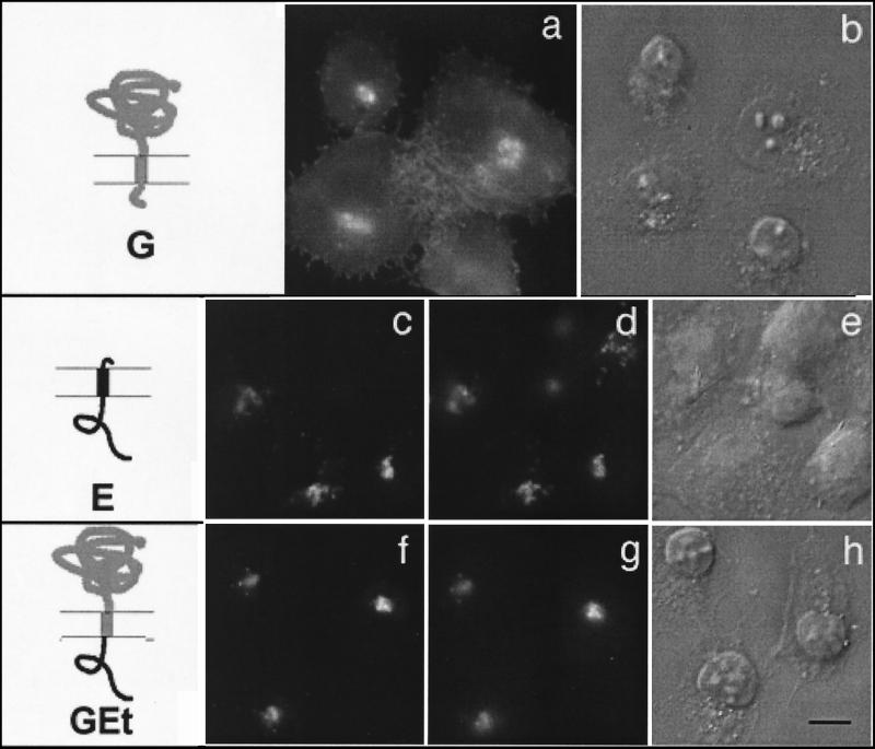
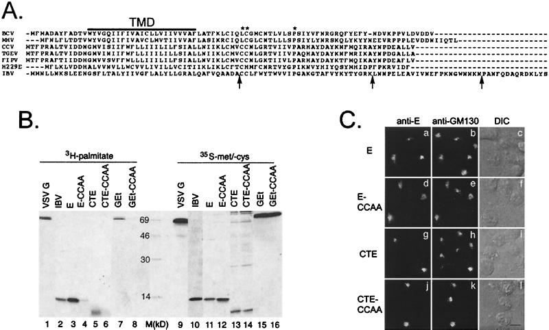
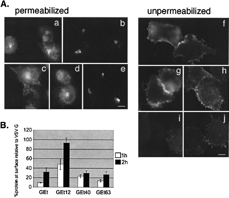
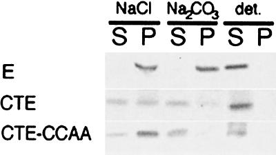
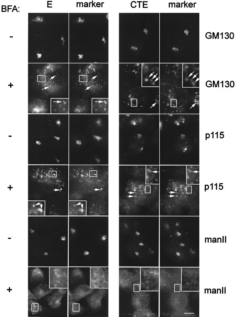
We have previously shown that the E protein of the coronavirus infectious bronchitis virus (IBV) is localized to the Golgi complex when expressed exogenously from cDNA. Here, we report that neither the transmembrane domain nor the short lumenal domain of IBV E is required for Golgi targeting. However, an N-terminal truncation containing only the cytoplasmic domain (CTE) was efficiently localized to the Golgi complex, and this domain could retain a reporter protein in the Golgi. Thus, the cytoplasmic tail of the E protein is necessary and sufficient for Golgi targeting. The IBV E protein is palmitoylated on one or two cysteine residues adjacent to its transmembrane domain, but palmitoylation was not required for proper Golgi targeting. Using C-terminal truncations, we determined that the IBV E Golgi targeting information is present between tail amino acids 13 and 63. Upon treatment with brefeldin A, both the E and CTE proteins redistributed to punctate structures that colocalized with the Golgi matrix proteins GM130 and p115 instead of being localized to the endoplasmic reticulum like Golgi glycosylation enzymes. This suggests that IBV E is associated with the Golgi matrix through interactions of its cytoplasmic tail and may have interesting implications for coronavirus assembly in early Golgi compartments.


Figures

FIG. 1.
FIG. 1.
Transmembrane domain of IBV E is not required for Golgi targeting. BHK cells infected with vvE (a to c), vvEG1 (d to f), vvEG2 (g to i), or vvEG3 (j to l) were fixed for immunofluorescence at 4 h postinfection, permeabilized, and double labeled with rabbit anti-E antibody (a, d, g, and j) and mouse anti-GM130 antibody (b, e, h, and k). Secondary antibodies were fluorescein-conjugated donkey anti-rabbit immunoglobulin (IgG) and Texas Red-conjugated goat anti-mouse IgG. The third image in each row (c, f, i, and l) is a differential interference contrast (DIC) image of the labeled cells. The diagrams indicate IBV E sequence in black and VSV sequence in gray. Bar, 10 μm.
FIG. 2.
FIG. 2.
Cytoplasmic tail of IBV E is targeted to the Golgi complex. BHK cells infected with vvCTE were fixed for immunofluorescence at 6 h postinfection, permeabilized, and double labeled with rabbit anti-E antibody (a) and mouse anti-GM130 antibody (b). Secondary antibodies were fluorescein-conjugated donkey anti-rabbit IgG and Texas Red-conjugated goat anti-mouse IgG. A DIC image of the labeled cells is shown in panel c. Bar, 10 μm.
FIG. 3.
FIG. 3.
Cytoplasmic tail of IBV E is sufficient to retain a reporter protein in the Golgi complex. BHK cells expressing wild-type VSV G (a and b), wild-type IBV E (c to e), or a chimeric protein consisting of the lumenal head and transmembrane domain of VSV G and the cytoplasmic tail of E (GEt; f to h) were fixed for immunofluorescence, permeabilized, and stained with anti-G (a) or double labeled with anti-E (c and f) and anti-GM130 (d and g) antibodies. Secondary antibodies were fluorescein-conjugated donkey anti-rabbit IgG and Texas Red-conjugated goat anti-mouse IgG. DIC images of the labeled cells are shown in panels b, e, and h. The diagrams indicate IBV E sequence in black and VSV sequence in gray. Bar, 10 μm.
FIG. 4.
FIG. 4.
GEt chimera is partially processed by late Golgi enzymes. BHK cells expressing wild-type G or GEt were pulse-labeled with [35S]methionine-cysteine, chased for 0, 15, or 30 min, lysed, and immunoprecipitated with anti-VSV antibody. The immunoprecipitates were mock treated (lanes −) or treated with endo H (lanes +), and subjected to SDS-10% PAGE and fluorography to determine the extent of endo H resistance and sialylation.
FIG. 5.
FIG. 5.
Conserved features of coronavirus E tails are dispensable for Golgi targeting. (A) An alignment of coronavirus E proteins from different species. The line labeled TMD shows the location of the transmembrane domain. The conserved cysteine(s) and proline residues that are present in all sequenced coronavirus E proteins are marked with asterisks. Arrows indicate the locations of inserted stop codons in the IBV E tail truncations. BCV, bovine coronavirus; MHV, mouse hepatitis virus; CCV, canine coronavirus; TGEV, transmissible gastroenteritis virus; FIPV, feline infectious peritonitis virus; H229E, human coronavirus strain 229E. (B) IBV-infected Vero cells or transfected BHK cells expressing the indicated proteins were labeled with [3H]palmitic acid or [35S]methionine-cysteine, lysed, and immunoprecipitated with anti-E or anti-VSV G antibodies. The immunoprecipitates were analyzed by SDS-17.5% PAGE and fluorography. Lane M, size markers (in kilodaltons). (C) BHK cells expressing wild-type E (a to c), E-CCAA (d to f), CTE (g to i), or CTE-CCAA (j to l) were fixed for immunofluorescence, permeabilized with Triton X-100, and stained with anti-E and anti-GM130 antibodies. Secondary antibodies were fluorescein-conjugated donkey anti-rabbit IgG and Texas Red-conjugated goat anti-mouse IgG. DIC images of the labeled cells are shown in panels c, f, i, and l. Bar, 10 μm.
FIG. 6.
FIG. 6.
Golgi targeting information in the IBV E protein is between residues 12 and 63 of the cytoplasmic tail. (A) BHK cells expressing VSV G (a and f), GEt (b and i), GEt12 (c and g), GEt40 (d and h), or GEt63 (e and j) were fixed for immunofluorescence and permeabilized (a to e) or fixed and left unpermeabilized (f to j) and stained with an antibody to the ectodomain of VSV G. Secondary antibody was Texas Red-conjugated goat anti-mouse IgG. (B) BHK cells expressing GEt, GEt12, GEt40, or GEt63 were pulse-labeled with [35S]methionine-cysteine and chased for 1 h (white bars) or 2 h (black bars). Intact cells were incubated with polyclonal anti-VSV antibody for 2 h at 4°C. After cell lysis, antibody complexes were collected, and supernatants were incubated with anti-VSV to immunoprecipitate intracellular proteins. Samples were run on SDS-PAGE, and quantitation of surface and internal proteins was performed. A ratio of the amount of surface to internal protein was calculated for GEt and truncation derivatives. Shown on the graph is this ratio taken as a percentage of the surface/internal ratio of wild-type VSV G protein at the corresponding time point. Each value represents an average of three experiments ± SEM.
FIG. 7.
FIG. 7.
CTE protein is peripherally associated with Golgi membranes. BHK cells expressing E, CTE, or CTE-CCAA were labeled with [35S]methionine-cysteine, and microsomes were prepared from homogenized cells. The microsomes were extracted with 0.1 M NaCl, 0.1 M Na2CO3 (pH 11.5), or detergent as indicated, and membranes were pelleted. Pellets (P) and supernatants (S) were immunoprecipitated with anti-É antibodies in the presence of detergent, and immunoprecipitates were analyzed by SDS-17.5% PAGE and fluorography.
FIG. 8.
FIG. 8.
IBV E and CTE are localized with Golgi remnants after BFA treatment. BHK cells expressing E or CTE by infection with recombinant vaccinia viruses were treated with BFA for 2 h at 3 and 5 h postinfection, respectively, fixed for immunofluorescence, permeabilized, and double labeled with rabbit anti-E antibody and anti-GM130 antibody or mouse anti-p115 antibody, or rat anti-E antibody and rabbit anti-mannosidase II (manII) antibody as indicated. Secondary antibodies were fluorescein-conjugated donkey anti-rabbit IgG and Texas Red-conjugated goat anti-mouse IgG, or fluorescein-conjugated goat anti-rat IgG and Texas Red-conjugated goat anti-mouse IgG. Boxed regions are enlarged in the insets of the BFA-treated panels. Bar, 10 μm.
FIG. 9.
FIG. 9.
Conserved residues in the IBV E and Uukuniemi virus G1 cytoplasmic tails. Cytoplasmic tail sequences were aligned using ClustalW. Identical residues are shaded in black, and similar residues are shaded in gray. The brackets indicate the regions of each protein that contain Golgi targeting information.

Similar articles

Cited by

References

    1. Andersson, A. M., L. Melin, A. Bean, and R. F. Pettersson. 1997. A retention signal necessary and sufficient for Golgi localization maps to the cytoplasmic tail of a Bunyaviridae (Uukuniemi virus virus) membrane glycoprotein. J. Virol. 71:4717–4727. - PMC - PubMed
    1. Andersson, A. M., and R. F. Pettersson. 1998. Targeting of a short peptide derived from the cytoplasmic tail of the G1 membrane glycoprotein of Uukuniemi virus virus (Bunyaviridae) to the Golgi complex. J. Virol. 72:9585–9596. - PMC - PubMed
    1. Barr, F. A., N. Nakamura, and G. Warren. 1998. Mapping the interaction between GRASP65 and GM130, components of a protein complex involved in the stacking of Golgi cisternae. EMBO J. 17:3258–3268. - PMC - PubMed
    1. Barr, F. A., M. Puype, J. Vandekerckhove, and G. Warren. 1997. GRASP65, a protein involved in the stacking of Golgi cisternae. Cell 91:253–262. - PubMed
    1. Baudoux, P., C. Carrat, L. Besnardeau, B. Charley, and H. Laude. 1998. Coronavirus pseudoparticles formed with recombinant M and E proteins induce alpha interferon synthesis by leukocytes. J. Virol. 72:8636–8643. - PMC - PubMed

Publication types

MeSH terms

LinkOut - more resources