. 2002 Feb;76(3):1328-38.
doi: 10.1128/jvi.76.3.1328-1338.2002.

The rat cytomegalovirus R33-encoded G protein-coupled receptor signals in a constitutive fashion

Affiliations

The rat cytomegalovirus R33-encoded G protein-coupled receptor signals in a constitutive fashion

Yvonne K Gruijthuijsen et al. J Virol. 2002 Feb.

Abstract

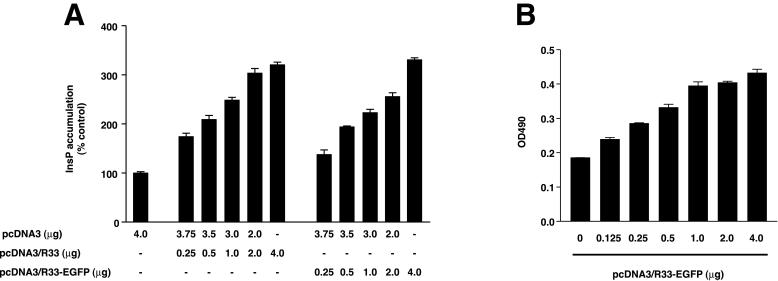
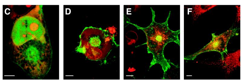
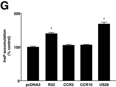
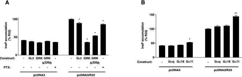
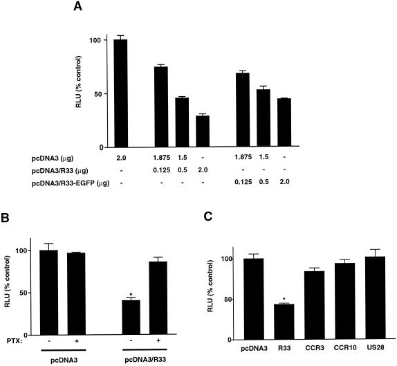
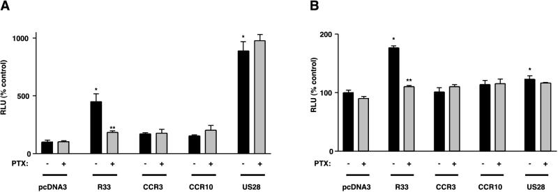
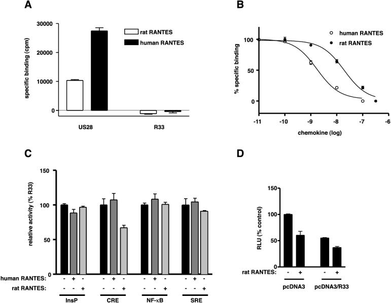
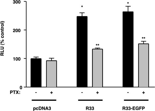
The rat cytomegalovirus (RCMV) R33 gene is conserved among all betaherpesviruses and encodes a protein (pR33) that shows sequence similarity with chemokine-binding G protein-coupled receptors (GPCRs). Previously, the physiological significance of the R33 gene was demonstrated by the finding that an RCMV strain with R33 deleted is severely attenuated in vivo and is unable to either enter or replicate in the salivary glands of infected rats. Here, we report that RCMV pR33 is expressed as a functional GPCR that signals in an agonist-independent manner in both COS-7 and Rat2 cells. Transient expression of pR33 in COS-7 cells results in constitutive activation of phospholipase C (PLC) due to coupling to G proteins of the G(q) class. Interestingly, PLC activation is partially inhibited by cotransfection with G(alpha)-transducin subunits, which indicates the involvement of G(betagamma) as well as Galpha subunits in pR33-mediated signaling. Surprisingly, PLC activation is also partially inhibited by addition of pertussis toxin (PTX), suggesting that pR33 activates not only G(q) but also G(i/0) proteins. The constitutive activation of G(i/0) proteins by pR33 is further demonstrated by the PTX-sensitive decrease of CRE-mediated transcription and the PTX-sensitive increase of both NF-kappaB- and SRE-mediated transcription. In contrast to its homolog of human herpesvirus 6B (pU12), pR33 does not bind RANTES.


Figures

FIG. 1.
FIG. 1.
pR33- and pR33-EGFP-mediated induction of inositol phosphate (InsP) accumulation. (A) COS-7 cells (106) were transiently transfected with either pcDNA3, pcDNA3/R33, or pcDNA3/R33-EGFP. At 48 h after transfection, the InsP accumulation was measured. (B) Expression of pR33-EGFP. COS-7 cells were transiently transfected with various concentrations of pcDNA3/R33-EGFP. After permeabilization, the cells were immunologically stained using an anti-EGFP polyclonal antiserum. The staining was quantified by measuring the optical density at 490 nm (OD490) by spectrophotometry. (C and D) Cellular localization of EGFP (C) and pR33-EGFP (D). COS-7 cells were transiently transfected with either pcDNA3/EGFP or pcDNA3/R33-EGFP, fixed, stained using propidium iodide (to visualize nucleic acids), and subjected to confocal microscopy at 488 and 568 nm. (E) Cellular localization of pR33-EGFP and the ER. pcDNA3/R33-EGFP-transfected cells were stained using SERCA 2-specific antibodies. (F) Cellular localization of pR33-EGFP and the Golgi apparatus. pcDNA3/R33-EGFP-transfected cells were stained using a trans-Golgi marker. Bars = 5 μm. (G) Modulation of InsP accumulation by various receptors. The InsP accumulation was determined after transient transfection of COS-7 cells with either pcDNA3, pcDNA3/R33 (R33), pcDNA3/CCR33 (CCR3), pcDNA3/CCR10 (CCR10), or pcDNA3/US28 (US28) at 0.5 μg of DNA/106 cells. A representative experiment performed in triplicate is shown. Each experiment was repeated at least twice. The asterisks indicate a statistically significant difference (P < 0.05) with data from pcDNA3-transfected cells. Error bars, standard error of the mean.
FIG. 1.
FIG. 1.
pR33- and pR33-EGFP-mediated induction of inositol phosphate (InsP) accumulation. (A) COS-7 cells (106) were transiently transfected with either pcDNA3, pcDNA3/R33, or pcDNA3/R33-EGFP. At 48 h after transfection, the InsP accumulation was measured. (B) Expression of pR33-EGFP. COS-7 cells were transiently transfected with various concentrations of pcDNA3/R33-EGFP. After permeabilization, the cells were immunologically stained using an anti-EGFP polyclonal antiserum. The staining was quantified by measuring the optical density at 490 nm (OD490) by spectrophotometry. (C and D) Cellular localization of EGFP (C) and pR33-EGFP (D). COS-7 cells were transiently transfected with either pcDNA3/EGFP or pcDNA3/R33-EGFP, fixed, stained using propidium iodide (to visualize nucleic acids), and subjected to confocal microscopy at 488 and 568 nm. (E) Cellular localization of pR33-EGFP and the ER. pcDNA3/R33-EGFP-transfected cells were stained using SERCA 2-specific antibodies. (F) Cellular localization of pR33-EGFP and the Golgi apparatus. pcDNA3/R33-EGFP-transfected cells were stained using a trans-Golgi marker. Bars = 5 μm. (G) Modulation of InsP accumulation by various receptors. The InsP accumulation was determined after transient transfection of COS-7 cells with either pcDNA3, pcDNA3/R33 (R33), pcDNA3/CCR33 (CCR3), pcDNA3/CCR10 (CCR10), or pcDNA3/US28 (US28) at 0.5 μg of DNA/106 cells. A representative experiment performed in triplicate is shown. Each experiment was repeated at least twice. The asterisks indicate a statistically significant difference (P < 0.05) with data from pcDNA3-transfected cells. Error bars, standard error of the mean.
FIG. 2.
FIG. 2.
(A) Effects of Gα transducin subunits, GRKs, and PTX on pR33-mediated InsP accumulation. COS-7 cells were transiently transfected with either pcDNA3 or pcDNA3/R33 in the presence of either pcDNA3/Gαt (Gαt), pcDNA3/GRK2 (GRK2), or pcDNA/GRK2K220R (GRK2K220R) (each construct at 2.0 μg of DNA/106 cells). The InsP accumulation was subsequently assessed either in the presence (+) or absence (−) of PTX in the culture medium. (B) Effects of Gαq, Gα16, and Gα11 on pR33-mediated InsP accumulation. COS-7 cells were transiently transfected with either pcDNA3 or pcDNA3/R33 in the presence of either pcDNA3/Gαq, pC1H1/Gα16, or pCMV5/Gα11, and the InsP accumulation was assessed after 48 h. Representative experiments performed in triplicate are shown. Each experiment was repeated at least three times. The asterisks indicate statistically significant differences (P < 0.05) between specific results and the result from pcDNA3/R33-transfected cells. Statistically significant differences (P < 0.05) were not found between results from the pcDNA3-transfected cells. Error bars, standard error of the mean.
FIG. 3.
FIG. 3.
pR33-mediated inhibition of forskolin-induced, CRE-driven luciferase expression. (A) COS-7 cells (106) were transiently transfected with reporter plasmid pTLNC-21CRE (5.0 μg of DNA/106 cells) and with various amounts of either pcDNA3, pcDNA3/R33, or pcDNA3/R33-EGFP (in total, 2.0 μg of DNA/106 cells). When amounts of pcDNA3/R33 or pcDNA3/R33-EGFP lower than 2.0 μg were used, pcDNA3 was added to include a total of 2 μg of pcDNA3-derived plasmid per transfection. At 18 h after transfection, the cells were stimulated with forskolin. Six hours later, the CRE-driven luciferase expression and activity was quantified, and expressed as relative light units (RLU). (B) Influence of PTX on the pR33-mediated inhibition of CRE-driven expression. PTX (100 ng/ml) was added to the culture medium at 2 h after transfection. (C) Influence of various receptors on forskolin-stimulated, CRE-driven expression. The luciferase activity was determined similarly as described above, after transient transfection of COS-7 cells with either pcDNA3, pcDNA3/R33 (R33), pcDNA3/CCR3 (CCR3), pcDNA3/CCR10 (CCR10), or pcDNA3/US28 (US28) at 0.5 μg of DNA/106 cells. Data are presented as percentages of the result for the control (pcDNA3-transfected cells in the absence of PTX). Representative experiments performed in triplicate are shown, and each experiment was repeated at least three times. An asterisk indicates data that are statistically significantly different (P < 0.05) from that for the control. Error bars, standard error of the mean.
FIG. 4.
FIG. 4.
pR33-mediated NF-κB and SRE activation. COS-7 cells (106) were transiently transfected with either reporter plasmid pNF-κB-Luc (A) or pTLNC-3SRE (B) (5.0 μg DNA/106 cells) and with either pcDNA3, pcDNA3/R33 (R33), pcDNA3/CCR3 (CCR3), pcDNA3/CCR10 (CCR10), or pcDNA3/US28 (US28) at 0.5 μg of DNA/106 cells. At 48 h after transfection, luciferase activity was measured. The experiments were carried out either in the presence (+) or absence (−) of PTX in the culture medium. Representative experiments performed in triplicate are shown, and each experiment was repeated at least three times. Single asterisks indicate differences statistically significant (P < 0.05) from data from pcDNA3-transfected cells (in the absence of PTX). Double asterisks (**) indicate statistically significant differences (P < 0.05) between data from PTX-treated and -untreated cells that were transfected with the same expression construct. Error bars, standard error of the mean.
FIG. 5.
FIG. 5.
pR33 does not bind rat RANTES. (A) Binding of human and rat RANTES by pUS28 and pR33. COS-7 cells were transiently transfected with either pcDNA3, pcDNA3/US28 (US28), or pcDNA3/R33 (R33). Binding was performed 48 h after transfection on whole cells using 6 nM 125I-labeled human or rat RANTES. Nonspecific binding was determined in the presence of 0.1 μM unlabeled competitor (human or rat RANTES). (B) Human and rat RANTES competition curves on pUS28. COS-7 cells were transiently transfected with pcDNA3/US28 and incubated with 0.6 nM 125I-labeled human RANTES and various amounts of unlabeled human or rat RANTES. (C) Influence of human and rat RANTES on pR33-mediated constitutive signaling. COS-7 cells were transiently transfected with either pcDNA3 or pcDNA3/R33. Human or rat RANTES was added to the supernatants, both in the [3H]inositol phosphate production assay (InsP) and in the reporter gene assays (CRE, NF-κB, and SRE). Data are presented as percentages of pR33 activity (100%). (D) Influence of rat RANTES on pcDNA3- and pcDNA3/R33-transfected cells in the CRE assay. Error bars, standard error of the mean.
FIG. 6.
FIG. 6.
pR33- and pR33-EGFP-mediated SRE activation in Rat2 cells. Rat2 cells (106) were transiently transfected with reporter plasmid pTLNC-3SRE (5.0 μg of DNA/106 cells) and with either pcDNA3, pcDNA3/R33 (R33), or pcDNA3/R33-EGFP (R33-EGFP) at 0.5 μg of DNA/106 cells. At 48 h after transfection, luciferase activity was measured. The experiment was done in six replicates, either in the presence (+) or absence (−) of PTX in the culture medium. Single asterisks indicate differences statistically significant (P < 0.05) from data from pcDNA3-transfected cells (in the absence of PTX). Double asterisks indicate statistically significant differences (P < 0.05) between data from PTX-treated and -untreated cells that were transfected with the same expression construct. Error bars, standard error of the mean.

Similar articles

Cited by

References

    1. Arai, H., and I. F. Charo. 1996. Differential regulation of G-protein-mediated signaling by chemokine receptors. J. Biol. Chem. 271:21814–21819. - PubMed
    1. Arvanitakis, L., E. Geras-Raaka, A. Varma, M. C. Gershenghorn, and E. Cesarman. 1997. Human herpesvirus KSHV encodes a constitutively active G-protein-coupled receptor linked to cell proliferation. Nature 385:347–350. - PubMed
    1. Bais, C., B. Santamasso, O. Coso, L. Arvanitakis, E. Geras-Raaka, J. Silvio Gutkind, A. S. Asch, E. Cesarman, M. C. Gershenghorn, and E. A. Mesri. 1998. G-protein-coupled receptor of Kaposi’s sarcoma-associated is a viral oncogene and angiogenesis activator. Nature 391:86–89. (Erratum, 392:210.) - PubMed
    1. Beisser, P. S., G. E. L. M. Grauls, C. A. Bruggeman, and C. Vink. 1999. Deletion of the R78 G protein-coupled receptor gene from rat cytomegalovirus results in an attenuated, syncytium-inducing mutant strain. J. Virol. 73:7218–7230. - PMC - PubMed
    1. Beisser, P. S., C. Vink, J. G. van Dam, G. E. L. M. Grauls, S. J. V. Vanherle, and C. A. Bruggeman. 1998. The R33 G protein-coupled receptor gene of rat cytomegalovirus plays an essential role in the pathogenesis of viral infection. J. Virol. 72:2352–2363. - PMC - PubMed

Publication types

MeSH terms

LinkOut - more resources