. 2002 Feb;76(3):1369-78.
doi: 10.1128/jvi.76.3.1369-1378.2002.

Complement component C1q enhances the biological activity of influenza virus hemagglutinin-specific antibodies depending on their fine antigen specificity and heavy-chain isotype

Affiliations

Complement component C1q enhances the biological activity of influenza virus hemagglutinin-specific antibodies depending on their fine antigen specificity and heavy-chain isotype

Jing Qi Feng et al. J Virol. 2002 Feb.

Abstract

We have previously observed that selected influenza virus hemagglutinin (HA)-specific monoclonal antibodies (MAbs) with poor virus-neutralizing (VN) activity in vitro exhibited greatly enhanced VN activity in vivo after administration to SCID mice. The same Abs displayed improved VN activity also when tested in vitro in the presence of noninactivated serum from SCID mice. To identify Ab-dependent properties and serum components that contributed to enhancement of Ab activity, we screened a large panel of HA-specific MAbs for hemagglutination inhibition (HI) in the presence of noninactivated serum from naive mice (NMS). We found that HI activity was enhanced by NMS depending on the Ab's fine specificity (antigenic region Cb/E > Ca/A,D > Sa,Sb/B), its heavy-chain isotype (immunoglobulin G2 [IgG2] > IgG3; IgG1 and IgM negative), and to some extent also on its derivation (primary response > memory response). On average, the HI activity of Cb/E-specific MAbs of the IgG2 isotype isolated from the primary response was enhanced by 20-fold. VN activity was enhanced significantly but less strongly than HI activity. Enhancement (i) was destroyed by heat inactivation (30 min, 56 degrees C); (ii) did not require C3, the central complement component; (iii) was abolished by treatment of serum with anti-C1q; and (iv) could be reproduced with purified C1q, the binding moiety of C1, the first complement component. We believe that this is the first description of a direct C1q-mediated enhancement of antiviral Ab activities.


Figures

FIG. 1.
FIG. 1.
Noninactivated NMS enhances the HI activity of H35-C12 much more than the activity of H36-4. (A and B) Purified MAbs H35-C12 (A) and H36-4 (B) were tested for HI activity against PR8 in the presence of the indicated concentrations of noninactivated (closed symbols) and heat-inactivated (open symbols) NMS from C57BL/6 and BALB/c mice. HI activity is the Ab concentration at which three of four agglutinating doses of PR8 are prevented from agglutinating CRBC. The data show the means and standard deviations from three to five independent determinations. The dashed line in panel A indicates the fact that all but one assay were performed at a starting Ab concentration of 20 μg/ml. (C) Effect of NMS alone on viral HA activity. At concentrations of above 1%, noninactivated NMS interfered with the HI assay by reducing viral HA activity on its own and by altering the agglutination patterns of CRBC.
FIG. 2.
FIG. 2.
Susceptibility of Abs to enhancement of their HI activity by NMS is dependent of their heavy-chain isotype and fine specificity. Culture fluids from PR8 HA-specific hybridomas were tested for HI activity against PR8 in the presence of 1% noninactivated or heat-inactivated NMS from C57BL/6 mice. A threefold increase in HI activity in the presence of noninactivated NMS is considered significant, and this level is indicated by the dashed line. The geometric mean HI titer ratio (noninactivated to inactivated) from at least two independent assays is shown and plotted for individual MAbs. The MAbs are grouped according to their heavy-chain isotype and the region of HA to which they bind as deduced from reduced reaction with a panel of single point virus mutants (7). Each circle represents an individual MAb.
FIG. 3.
FIG. 3.
Enhancement of HI activity by purified huC1q. HI assays were performed in the presence of the indicated concentrations of huC1q. (A) HI activities of H35-C12 (circles) and of two other HA Cb/E-specific Abs of the IgG2a isotype (triangles and inverted triangles). Results from a representative assay of several repeat assays is shown. Each symbol shows the mean of three replicate values per dilution. (B) HI activity of H36-4. Inset, HA activity of PR8 incubated with the indicated concentrations of huC1q in the absence of Ab, expressed as percentage of the original activity.
FIG. 4.
FIG. 4.
NMS and huC1q do not substantially modify the reactions of H35-C12 and H36-4 to PR8 in ELISA, and both Abs fix C1q equally well. H35-C12 (A and C) and H36-4 (B and D) were tested for binding to PR8 immunoadsorbent, prepared by adsorption of virus to plastic from a solution as used in the HI test. Ab dilutions were tested in the following diluents: PBSN(Ca,Mg) containing 0.1% BSA (circles) and the same diluent supplemented with (i) 0.5 μg of huC1q per ml (triangles), (ii) 0.5% noninactivated NMS (squares), or (iii) 0.5% inactivated NMS (diamonds). (A and B) Ab bound to the viral immunoadsorbent as detected by developing the assay with the biotinylated anti-CK MAb 187. (C and D) Bound C1q as detected by developing the assay with goat anti-huC1q antiserum followed by biotinylated mouse anti-goat MAb. Each point shows the mean optical density at 405 nm minus optical density at 750 nm (OD405–750) for triplicate samples above background (diluent alone). The data are from one of three repeat assays.
FIG. 5.
FIG. 5.
Relationship between density of virus-bound Abs and inhibition of viral HA activity. (A and B) A constant dose of PR8 (open symbols) or Lee virus (closed symbols) (≈20 nM viral HA monomer) was incubated for 1 h at room temperature with increasing concentrations (3 to 330 nM) of purified MAb H35-C12 (A) or H36-4 (B). The virus was then pelleted through a sucrose cushion, and the amount of MAb associated with the virus pellet was determined by ELISA. Mean values from two independent determinations are shown. (C) Virus (≈20 nM HA) was incubated for 1 h at room temperature with the indicated MAbs (≈70 nM): 1, H36-4, IgG2a; 2, H37-77, IgG2a; 3, H36-12, IgG2b; 4, H35-C12, IgG2a; 5, H2-4C2, IgG2a; and 6, L2-10C1, IgG2a. MAbs 1 to 3 are specific for Sa,Sb/B, and MAbs 4 to 6 are specific for Cb/E. The virus-Ab mixture was pelleted though a sucrose cushion, and the concentration of copelleted Ab was determined by ELISA. The data are means and SEMs from three to seven independent determinations. (D) Prior to centrifugation, a sample of the virus-Ab mixture was tested for HA activity, which is expressed as percentage of the original HA activity contained in the sample.
FIG. 6.
FIG. 6.
Enhancement of HI titer of immune sera by noninactivated NMS. (A) Several immune sera from BALB/c mice obtained 10 to 14 days after primary immunization and 13 to 25 days after secondary immunization with PR8 were heat inactivated and then tested for HI titer against PR8 in the presence of 0.5% noninactivated or heat-inactivated NMS. The primary-response sera showed an enhancement of 10(0.38 ± 0.04) (mean ± SEM) (paired t test, P < 0.05), and the secondary-response sera showed an enhancement of 10(0.11 ± 0.06) (paired t test, P > 0.05), by noninactivated NMS compared to inactivated NMS. Primary sera were more strongly enhanced than secondary sera (t test, P < 0.05). (B) Heat-inactivated plasma samples from BALB/c mice obtained at various time intervals after i.v. immunization with 500 HAU (≈3.5 μg of viral protein) of purified PR8 were tested for HI activity in the presence of 0.5% noninactivated (filled circles) or inactivated (open circles) NMS. Pooled plasma samples from 10 mice were tested at each time point. (C) Heat-inactivated serum samples from BALB/c mice obtained at various time points after primary pulmonary infection were tested for HI activity as for panel B. Each time point represents the data obtained with a serum pool from two to four mice.
FIG. 7.
FIG. 7.
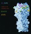
Locations of antigenic regions of the HA trimer. A model of the HA trimer of the H3 subtype (38, 39) is shown in side view with one monomer stained in light blue and the other stained in darker blue. Amino acids that were present in mutated form on individual mutants of PR8 (H1 subtype) and used to assign MAb specificity (7) are colored in yellow for the Cb/E, green for the Ca/A,D, red for the Sa, and dark blue for the Sb regions, with the latter making up the Sa,Sb/B region comprising mostly the tip of the trimer (7). MAbs that displayed reduced reaction with mutations belonging to distinct regions were assigned in this analysis to the region that had a dominant effect. In this side view, most of the mutated amino acids are visible only on one of the monomers, but in some cases, analogous mutations are visible on more than one monomer. The location of the sialic acid binding site, which is a shallow depression in the globular region of the HA monomer, is indicated by a yellow circle. Most mutations other than those marking the Cb/E region cluster around the binding site.

References

    1. Beebe, D. P., R. D. Schreiber, and N. R. Cooper. 1983. Neutralization of influenza virus by normal human sera: mechanisms involving antibody and complement. J. Immunol. 130:1317–1322. - PubMed
    1. Bindon, C. I., G. Hale, and H. Waldmann. 1988. Importance of antigen specificity for complement-mediated lysis by monoclonal antibodies. Eur. J. Immunol. 18:1507–1514. - PubMed
    1. Bizebard, T., B. Gigant, P. Rigolet, B. Rasmussen, O. Diat, P. Boeseccke, S. A. Wharton, J. J. Skehel, and M. Knossow. 1995. Structure of influenza virus haemagglutinin complexed with a neutralizing antibody. Nature 376:92–95. - PubMed
    1. Botto, M., C. Dell’Agnola, A. E. Bygrave, E. M. Thompson, H. T. Cook, F. Petry, M. Loos, P. P. Pandolfi, and M. J. Walport. 1998. Homozygous C1q deficiency causes glomerulonephritis associated with multiple apoptotic bodies. Nat. Genet. 19:56–59. - PubMed
    1. Brekke, O. H., T. E. Michaelsen, and I. Sandlie. 1995. The structural requirements for complement activation by IgG: does it hinge on the hinge. Immunol. Today 16:85–89. - PubMed

Publication types

MeSH terms

LinkOut - more resources