. 2002 Feb;76(3):1422-34.
doi: 10.1128/jvi.76.3.1422-1434.2002.

Heterologous gene expression from transmissible gastroenteritis virus replicon particles

Affiliations

Heterologous gene expression from transmissible gastroenteritis virus replicon particles

Kristopher M Curtis et al. J Virol. 2002 Feb.

Abstract

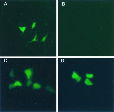
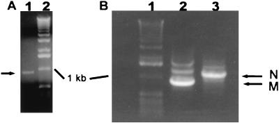
We have recently isolated a transmissible gastroenteritis virus (TGEV) infectious construct designated TGEV 1000 (B. Yount, K. M. Curtis, and R. S. Baric, J. Virol. 74:10600-10611, 2000). Using this construct, a recombinant TGEV was constructed that replaced open reading frame (ORF) 3A with a heterologous gene encoding green fluorescent protein (GFP). Following transfection of baby hamster kidney (BHK) cells, a recombinant TGEV (TGEV-GFP2) was isolated that replicated efficiently and expressed GFP. Replicon constructs were constructed that lacked either the ORF 3B and E genes or the ORF 3B, E, and M genes [TGEV-Rep(AvrII) and TGEV-Rep(EcoNI), respectively]. As the E and M proteins are essential for TGEV virion budding, these replicon RNAs should replicate but not result in the production of infectious virus. Following cotransfection of BHK cells with the replicon RNAs carrying gfp, GFP expression was evident by fluorescent microscopy and leader-containing transcripts carrying gfp were detected by reverse transcription-PCR (RT-PCR). Subsequent passage of cell culture supernatants onto permissive swine testicular (ST) cells did not result in the virus, GFP expression, or the presence of leader-containing subgenomic transcripts, demonstrating the single-hit nature of the TGEV replicon RNAs. To prepare a packaging system to assemble TGEV replicon particles (TGEV VRP), the TGEV E gene was cloned into a Venezuelan equine encephalitis (VEE) replicon expression vector and VEE replicon particles encoding the TGEV E protein were isolated [VEE-TGEV(E)]. BHK cells were either cotransfected with TGEV-Rep(AvrII) (E gene deletion) and VEE-TGEV(E) RNA transcripts or transfected with TGEV-Rep(AvrII) RNA transcripts and subsequently infected with VEE VRPs carrying the TGEV E gene. In both cases, GFP expression and leader-containing GFP transcripts were detected in transfected cells. Cell culture supernatants, collected approximately 36 h posttransfection, were passed onto fresh ST cells where GFP expression was evident approximately 18 h postinfection. Leader-containing GFP transcripts containing the ORF 3B and E gene deletions were detected by RT-PCR. Recombinant TGEV was not released from these cultures. Under identical conditions, TGEV-GFP2 spread throughout ST cell cultures, expressed GFP, and formed viral plaques. The development of infectious TGEV replicon particles should assist studies of TGEV replication and assembly as well as facilitate the production of novel swine candidate vaccines.


Figures

FIG. 1.
FIG. 1.
Construction of TGEV replicon cDNAs. (a) TGEV F fragments. Structural genes are contained in the TGEV F fragment. FiGFP2(PflMI) (∼5.6 kb) was constructed from the wild-type TGEV F fragment (∼5.1 kb) by the deletion of ORF 3A (nt 24828 to 25073) and the insertion of GFP with a 5′ 20-nt N gene IS. Using this construct, we introduced deletions extending from the unique PflMI site at the very 3′ end of GFP to the unique AvrII (nucleotide position 25866) and EcoNI (nucleotide position 26935) sites present within the TGEV E and M genes, respectively. (b) Sequence organization of GFP in FiGFP2(PflMI). GFP was inserted just downstream of the ORF 3A IS. The TGEV sequence originating at the 3′ end of the S gene (nt 24693) through the start of the GFP gene is shown, and the important IS and restriction sites are labeled. (c) Strategy for assembling recombinant TGEV and replicon cDNAs. The six cDNA subclones (TGEV A, B1, B2, C, DE1, and F deletion fragments) spanning the genome are flanked by unique interconnecting BglI and BstXI sites, allowing for directional assembly into a full-length replicon cDNA by in vitro ligation (80). TGEV A contained a unique T7 start site at its 5′ end, and the F deletion fragments (FiGFP2-AvrII and FiGFP2-EcoNI) contain GFP and a 25-nt T tail, allowing for the synthesis of capped T7, polyadenylated transcripts in vitro.
FIG. 2.
FIG. 2.
Recombinant TGEV virus expressing GFP. (a) Cultures of ST cells were infected with either recombinant TGEV-GFP2 virus or the TGEV infectious construct (icTGEV) (80) at an MOI of 5 for 1 h at room temperature. The inocula were removed, and the cultures were incubated in complete medium at 37°C. Samples were harvested at the indicated times and assayed by plaque assay in ST cells. (b) Cultures of ST cells were infected as described for panel a. At ∼12 h postinfection, GFP expression was observed by fluorescent microscopy in TGEV-GFP2-infected cultures (B) but not in icTGEV-infected cultures (A).
FIG. 2.
FIG. 2.
Recombinant TGEV virus expressing GFP. (a) Cultures of ST cells were infected with either recombinant TGEV-GFP2 virus or the TGEV infectious construct (icTGEV) (80) at an MOI of 5 for 1 h at room temperature. The inocula were removed, and the cultures were incubated in complete medium at 37°C. Samples were harvested at the indicated times and assayed by plaque assay in ST cells. (b) Cultures of ST cells were infected as described for panel a. At ∼12 h postinfection, GFP expression was observed by fluorescent microscopy in TGEV-GFP2-infected cultures (B) but not in icTGEV-infected cultures (A).
FIG. 3.
FIG. 3.
Heterologous gene expression from TGEV replicon RNAs. (a) Cultures of BHK cells were transfected with TGEV replicon RNAs encoding GFP that contained ORF 3B and E (A) [TGEV-Rep(AvrII) transcripts] or ORF 3B, E, and M gene deletions (C). Alternatively, transcripts were treated with RNase prior to transfection into BHK cells (B and D) [TGEV-Rep(AvrII) DNA and TGEV-Rep(EcoNI) DNA only, respectively]. At ∼18 h posttransfection, GFP expression was observed by fluorescent microscopy. (b) Intracellular RNA was isolated from transfected cell cultures and used as a template for RT-PCR. Leader-containing GFP subgenomic transcripts were detected using a 5′ leader primer (TGEV-L) and 3′ primers located just downstream of the AvrII [(−)E5546] or EcoNI [M6400(−)] sites. Appropriately sized amplicons of ∼850 bp were generated, corresponding to transcripts encoding GFP. Cells transfected with TGEV-Rep(AvrII) (A) or TGEV-Rep(EcoNI) (B) RNA are shown. A 1-kb ladder is shown in both panels (lanes 1). Arrows indicate leader-containing GFP amplicons (lanes 2).
FIG. 3.
FIG. 3.
Heterologous gene expression from TGEV replicon RNAs. (a) Cultures of BHK cells were transfected with TGEV replicon RNAs encoding GFP that contained ORF 3B and E (A) [TGEV-Rep(AvrII) transcripts] or ORF 3B, E, and M gene deletions (C). Alternatively, transcripts were treated with RNase prior to transfection into BHK cells (B and D) [TGEV-Rep(AvrII) DNA and TGEV-Rep(EcoNI) DNA only, respectively]. At ∼18 h posttransfection, GFP expression was observed by fluorescent microscopy. (b) Intracellular RNA was isolated from transfected cell cultures and used as a template for RT-PCR. Leader-containing GFP subgenomic transcripts were detected using a 5′ leader primer (TGEV-L) and 3′ primers located just downstream of the AvrII [(−)E5546] or EcoNI [M6400(−)] sites. Appropriately sized amplicons of ∼850 bp were generated, corresponding to transcripts encoding GFP. Cells transfected with TGEV-Rep(AvrII) (A) or TGEV-Rep(EcoNI) (B) RNA are shown. A 1-kb ladder is shown in both panels (lanes 1). Arrows indicate leader-containing GFP amplicons (lanes 2).
FIG. 4.
FIG. 4.
Strategy to assemble TGEV-Rep(AvrII) VRPs. (a) In the full-length TGEV-Rep(AvrII) cDNA construct, ORF 3A has been replaced with GFP, and ORF 3B and the 5′ end of the E gene have been deleted. To produce packaged replicon particles, replicon RNA-transfected cells were infected with VEE VRPs expressing the TGEV E protein [VEE-TGEV(E)]. Alternatively, TGEV-Rep(AvrII) replicon RNAs can be coelectroporated with pVR21-E1-derived transcripts. TGEV VRPs should be released from cells that can be used as single-hit expression vectors. (b) Cultures of ST cells were infected with either wild-type (wt) TGEV alone or with TGEV and VEE VRPs expressing a G1 Norwalk-like virus capsid (wt + VEE) at an MOI of 5 for 1 h at room temperature. The inocula were removed, and the cultures were incubated in complete medium at 37°C. Samples were harvested at the indicated times and assayed by plaque assay in ST cells.
FIG. 4.
FIG. 4.
Strategy to assemble TGEV-Rep(AvrII) VRPs. (a) In the full-length TGEV-Rep(AvrII) cDNA construct, ORF 3A has been replaced with GFP, and ORF 3B and the 5′ end of the E gene have been deleted. To produce packaged replicon particles, replicon RNA-transfected cells were infected with VEE VRPs expressing the TGEV E protein [VEE-TGEV(E)]. Alternatively, TGEV-Rep(AvrII) replicon RNAs can be coelectroporated with pVR21-E1-derived transcripts. TGEV VRPs should be released from cells that can be used as single-hit expression vectors. (b) Cultures of ST cells were infected with either wild-type (wt) TGEV alone or with TGEV and VEE VRPs expressing a G1 Norwalk-like virus capsid (wt + VEE) at an MOI of 5 for 1 h at room temperature. The inocula were removed, and the cultures were incubated in complete medium at 37°C. Samples were harvested at the indicated times and assayed by plaque assay in ST cells.
FIG. 5.
FIG. 5.
TGEV-Rep(AvrII) VRPs. Transfected cultures displayed GFP expression at ∼18 h posttransfection (A) [TGEV-Rep(AvrII) pass 0]. Supernatants were harvested ∼36 h posttransfection and used to infect fresh cultures of ST cells for 1 h at room temperature. At ∼18 h postinfection, GFP expression was observed by fluorescent microscopy in these pass 1 cultures (C and D). (C) TGEV VRP production following coelectroporation with VEE-E RNAs [TGEV-Rep(AvrII) plus E protein transcripts pass 1]; (D) TGEV VRP production following infection with VEE VRPs encoding the E protein [TGEV-Rep(AvrII) plus VEE-TGEV(E) pass 1]. Passage of supernatants from cells transfected with TGEV-Rep(AvrII) transcripts without expression of the E protein in trans did not result in detectable GFP expression (B) [TGEV-Rep(AvrII) without E protein pass 1].
FIG. 6.
FIG. 6.
Leader-containing subgenomic transcripts are present in cultures infected with TGEV VRPs. Intracellular RNA was isolated from cell cultures infected with pass 0 supernatants and used as a template for RT-PCR to detect leader-containing GFP, M, and N subgenomic transcripts using a 5′ leader primer (TGEV-L) and 3′ primers located just downstream of each respective gene. (A) An appropriately sized amplicon of ∼850 bp was generated (indicated by an arrow), corresponding to leader-containing transcripts encoding GFP (lane 1). Lane 2 shows a 1-kb ladder. (B) In addition, amplicons of ∼900 bp and ∼1.2 kb were generated, corresponding to leader-containing M and N gene mRNA transcripts, respectively. The arrows correspond to leader-containing M (lane 2) or N (lane 3) transcripts. A 1-kb ladder is shown in lane 1.
FIG. 7.
FIG. 7.
Sequence analysis of leader-containing amplicons encoding GFP. TGEV-Rep(AvrII) and TGEV-Rep(EcoNI) leader-containing amplicons were isolated from agarose gels and subcloned into Topo II TA cloning vectors. Inserts were sequenced using universal primers and an automated sequencer. (A) Sequence of the 5′ end of GFP amplicons generated from TGEV-Rep(EcoNI)-transfected cells, indicating that the leader-containing transcripts initiated from the TGEV ORF 3A IS. (B) Leader-containing GFP transcripts with the PflMI-AvrII deletion derived from TGEV-Rep(AvrII) VRP-infected cells. (C) Leader-containing GFP transcripts with the PflMI-EcoNI deletion derived from TGEV-Rep(EcoNI)-transfected cells. Underlined bases correspond to the GFP stop codon, and the shaded bases correspond to the deletion and blunt-end ligation sites.
FIG. 8.
FIG. 8.
TGEV replicon particles function as single-hit virus vectors. Cultures of ST cells were infected for 1 h at room temperature with either concentrated TGEV VRPs or supernatant from TGEV-GFP2-transfected cells. (a) Cultures were then examined over a 72-h time period for GFP expression by fluorescent microscopy. Expansion of GFP expression was observed in TGEV-GFP2-infected cells (panels A to E) while no expansion was noted in TGEV VRP-infected cells (panels F to J). (b) In addition, supernatants were harvested from the TGEV-GFP2- and TGEV VRP-infected cultures and assayed by plaque assay in ST cells. Symbols: ⧫, TGEV-GFP2; ▪, TGEV-VRP.
FIG. 8.
FIG. 8.
TGEV replicon particles function as single-hit virus vectors. Cultures of ST cells were infected for 1 h at room temperature with either concentrated TGEV VRPs or supernatant from TGEV-GFP2-transfected cells. (a) Cultures were then examined over a 72-h time period for GFP expression by fluorescent microscopy. Expansion of GFP expression was observed in TGEV-GFP2-infected cells (panels A to E) while no expansion was noted in TGEV VRP-infected cells (panels F to J). (b) In addition, supernatants were harvested from the TGEV-GFP2- and TGEV VRP-infected cultures and assayed by plaque assay in ST cells. Symbols: ⧫, TGEV-GFP2; ▪, TGEV-VRP.

References

    1. Agapov, E. V., I. Frolov, B. D. Lindenbach, B. M. Pragai, S. Schlesinger, and C. M. Rice. 1998. Noncytopathic Sindbis virus RNA vectors for heterologous gene expression. Proc. Natl. Acad. Sci. USA 95:12989–12994. - PMC - PubMed
    1. Almazan, F., J. M. Gonzalez, Z. Penzes, A. Izeta, E. Calvo, J. Plana-Duran, and L. Enjuanes. 2000. Engineering the largest RNA virus genome as an infectious bacterial artificial chromosome. Proc. Natl. Acad. Sci. USA 97:5516–5521. - PMC - PubMed
    1. Balasuriya, U. B., H. W. Heidner, J. F. Hedges, J. C. Williams, N. L. Davis, R. E. Johnston, and N. J. MacLachlan. 2000. Expression of the two major envelope proteins of equine arteritis virus as a heterodimer is necessary for induction of neutralizing antibodies in mice immunized with recombinant Venezuelan equine encephalitis virus replicon particles. J. Virol. 74:10623–10630. - PMC - PubMed
    1. Banchereau, J., and R. M. Steinman. 1998. Dendritic cells and the control of immunity. Nature 392:245–252. - PubMed
    1. Baric, R. S., K. Fu, M. C. Schaad, and S. A. Stohlman. 1990. Establishing a genetic recombination map for murine coronavirus strain A59 complementation groups. Virology 177:646–656. - PMC - PubMed

Publication types

MeSH terms