. 2002 Feb;76(3):1461-74.
doi: 10.1128/jvi.76.3.1461-1474.2002.

Adenovirus E1A N-terminal amino acid sequence requirements for repression of transcription in vitro and in vivo correlate with those required for E1A interference with TBP-TATA complex formation

Affiliations

Adenovirus E1A N-terminal amino acid sequence requirements for repression of transcription in vitro and in vivo correlate with those required for E1A interference with TBP-TATA complex formation

Janice M Boyd et al. J Virol. 2002 Feb.

Abstract

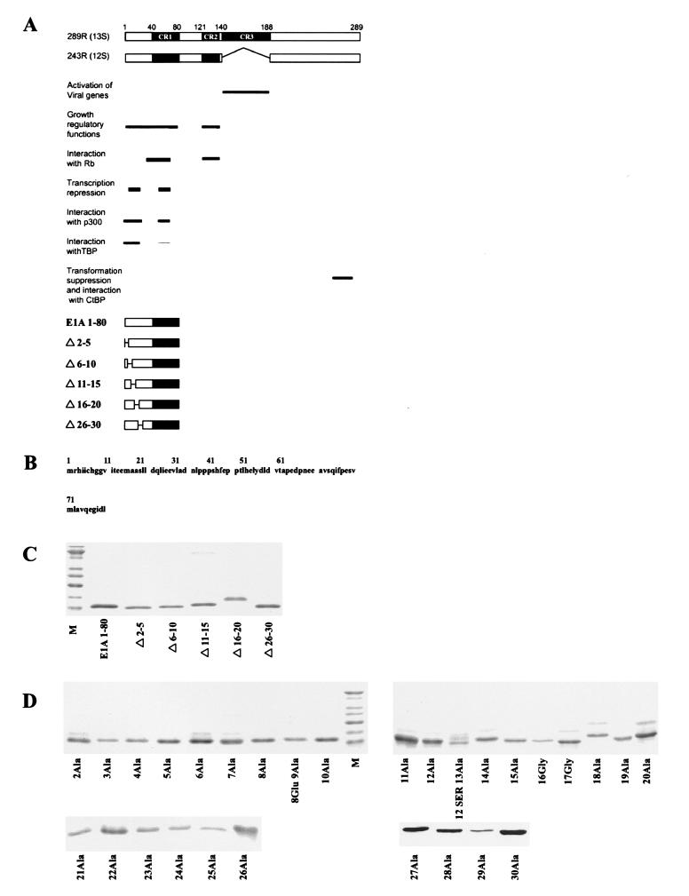
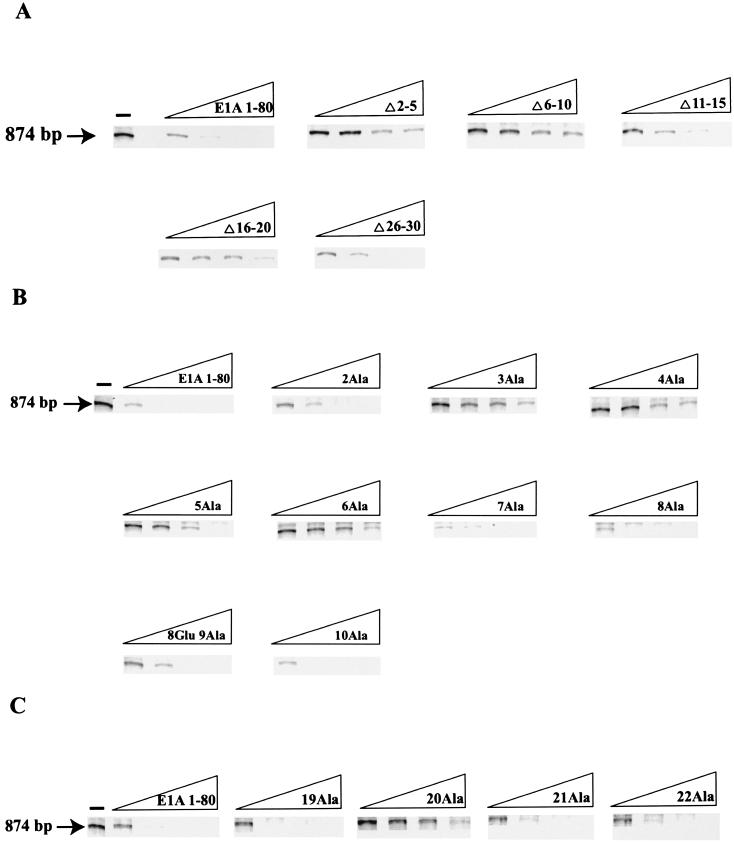
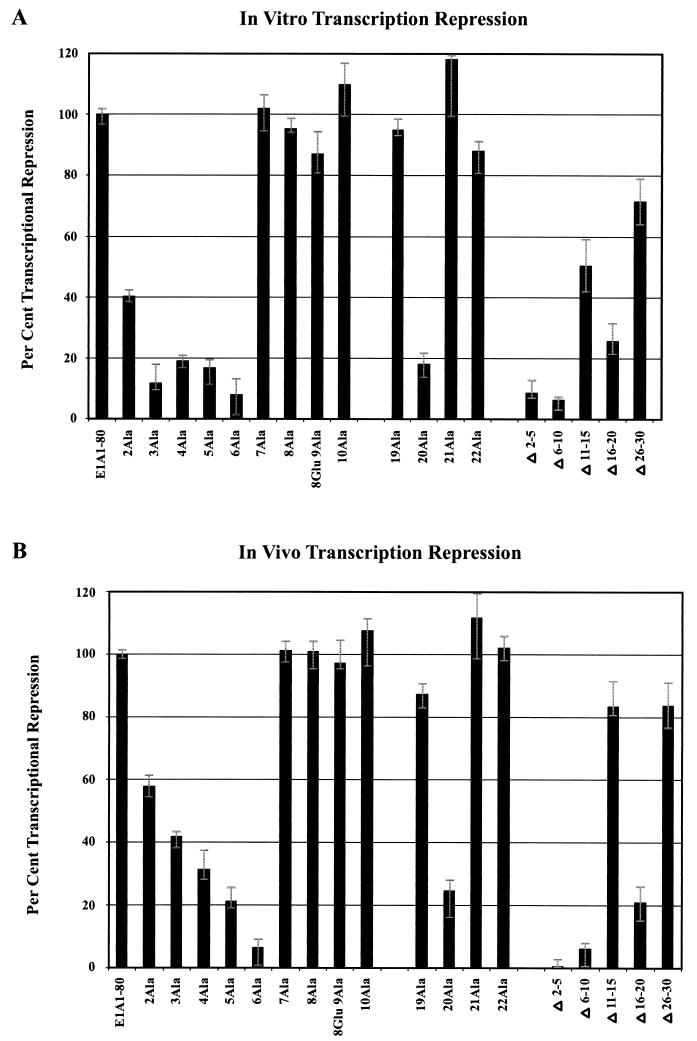
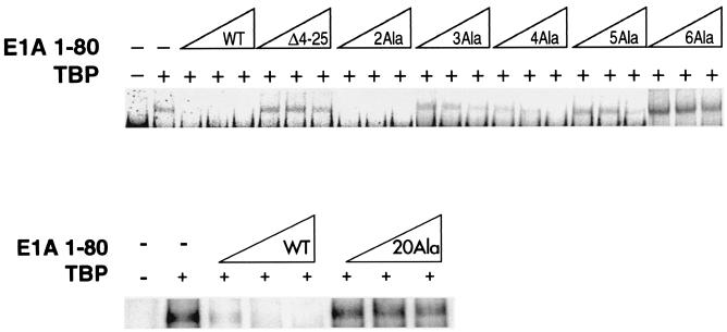
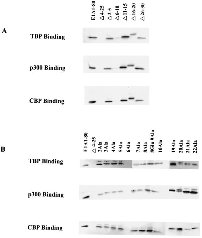
The adenovirus (Ad) E1A 243R oncoprotein encodes an N-terminal transcription repression domain that is essential for early viral functions, cell immortalization, and cell transformation. The transcription repression function requires sequences within amino acids 1 to 30 and 48 to 60. To elucidate the roles of the TATA-binding protein (TBP), p300, and the CREB-binding protein (CBP) in the mechanism(s) of E1A repression, we have constructed 29 amino acid substitution mutants and 5 deletion mutants spanning the first 30 amino acids within the E1A 1-80 polypeptide backbone. These mutant E1A polypeptides were characterized with regard to six parameters: the ability to repress transcription in vitro and in vivo, to disrupt TBP-TATA box interaction, and to bind TBP, p300, and CBP. Two regions within E1A residues 1 to 30, amino acids 2 to 6 and amino acid 20, are critical for E1A transcription repression in vitro and in vivo and for the ability to interfere with TBP-TATA interaction. Replacement of 6Cys with Ala in the first region yields the most defective mutant. Replacement of 20Leu with Ala, but not substitutions in flanking residues, yields a substantially defective phenotype. Protein binding assays demonstrate that replacement of 6Cys with Ala yields a mutant completely defective in interaction with TBP, p300, and CBP. Our findings are consistent with a model in which the E1A repression function involves interaction of E1A with p300/CBP and interference with the formation of a TBP-TATA box complex.


Figures

FIG. 1.
FIG. 1.
(A) Schematic of E1A 289R and 243R proteins, their domain structures, and associated biological activities. Below are schematics of E1A 1-80 and E1A 1-80 deletion polypeptides. (B) Sequence of the first 80 amino acids of E1A (E1A 1-80). (C) Purity of E1A 1-80 deletion mutants. One-microgram amounts of polypeptide were resolved on 15% polyacrylamide gels, stained with SYPRO orange (Molecular Probes), and visualized by blue-green fluorescence on a STORM 840 PhosphorImager. The lowest band of the marker lane (M) is 10 kDa. (D) Purity of E1A 1-80 amino acid substitution mutants. One-microgram amounts of the E1A 1-80 mutants were analyzed as described for panel C above.
FIG. 2.
FIG. 2.
Two regions of E1A 1-80, amino acids 2 to 6 and amino acid 20, are critical for E1A repression in vitro. Each E1A polypeptide was assayed for in vitro transcription repression activity at 62.5, 125, 250, and 500 ng per reaction. Each assay was repeated four to six times. At least two independent preparations of each defective mutant were assayed. (A) Representative dose-response measurements of in vitro repression activity of E1A 1-80 and E1A 1-80 deletion polypeptides. (B) Representative dose-response measurements of in vitro repression activity of E1A 1-80 and E1A 1-80 with Ala substituted for residues 2 to 10 (the polypeptide with Ala substituted for 9Gly also contains a Glu substituted for 8Gly). (C) Representative dose-response measurements of in vitro repression activity of E1A 1-80 and E1A 1-80 19Ala, E1A 1-80 20Ala, E1A 1-80 21Ala, and E1A 1-80 22 Ala.
FIG. 3.
FIG. 3.
Two regions of E1A 1-80, amino acids 2 to 6 and amino acid 20, are critical for E1A repression in vivo. Each individual experiment involved the microinjection of about 100 cells, but only a portion of each microinjected area is seen in the representative photographic fields in these figures. (A) Representative cell microinjection assays demonstrating the transcriptional-repression function of E1A 243R and E1A 1-80. The photomicrographs in each row are of the same field of cells visualized under different conditions. The left column (Phase) is visualized under phase microscopy. The middle column (GFP) is visualized under fluorescence microscopy through an FITC transparent filter and shows cells expressing green fluorescent protein (GFP), a marker for successfully injected cells. The right column (SV40 T-Ag) is visualized under fluorescence microscopy through a rhodamine transparent filter and shows cells expressing SV40 T antigen. Cells in the first row were coinjected with 8 ng of pSV40/μl and 10 ng of pEGFPN-1/μl. About 75% of successfully injected cells were positive for SV40 T antigen. Cells in the second row were coinjected with pSV40, pEGFPN-1, and 10 ng of the E1A 243R-expressing plasmid pcDNA3-E1A 243R/μl. Cells in the third row were coinjected with pSV40, pEGFPN-1, and 25 ng of E1A 1-80 polypeptide/μl. (B) Representative microinjection assays demonstrating that E1A 1-80Δ2-5, E1A 1-80Δ6-10, and E1A 1-80Δ16-20 are defective in transcription repression function in vivo. Cells were coinjected with 8 ng of pSV40/μl, 10 ng of pEGFPN-1/μl, and 25 ng of the indicated E1A 1-80 deletion mutant/μl. (C) Representative assays demonstrating that E1A 1-80 2Ala, 3Ala, 4Ala, 5Ala, and 6Ala are defective to various degrees in transcription repression function in vivo. Cells were injected as described for panel B above except that 25 ng of the indicated Ala substitution mutant/μl was injected. (D) Representative assays demonstrating that E1A 1-80 20Ala is defective in transcription repression function in vivo. Cells were injected as described for panel B above. It should be noted that for E1A 1-80 20Ala, the field shown has fewer cells than fields coinjected with other mutants. E1A 1-80 20 Ala is nonetheless clearly defective: 17 cells were positive for T antigen of 21 cells injected, representing 80% of successfully injected cells visualized in this field.
FIG. 3.
FIG. 3.
Two regions of E1A 1-80, amino acids 2 to 6 and amino acid 20, are critical for E1A repression in vivo. Each individual experiment involved the microinjection of about 100 cells, but only a portion of each microinjected area is seen in the representative photographic fields in these figures. (A) Representative cell microinjection assays demonstrating the transcriptional-repression function of E1A 243R and E1A 1-80. The photomicrographs in each row are of the same field of cells visualized under different conditions. The left column (Phase) is visualized under phase microscopy. The middle column (GFP) is visualized under fluorescence microscopy through an FITC transparent filter and shows cells expressing green fluorescent protein (GFP), a marker for successfully injected cells. The right column (SV40 T-Ag) is visualized under fluorescence microscopy through a rhodamine transparent filter and shows cells expressing SV40 T antigen. Cells in the first row were coinjected with 8 ng of pSV40/μl and 10 ng of pEGFPN-1/μl. About 75% of successfully injected cells were positive for SV40 T antigen. Cells in the second row were coinjected with pSV40, pEGFPN-1, and 10 ng of the E1A 243R-expressing plasmid pcDNA3-E1A 243R/μl. Cells in the third row were coinjected with pSV40, pEGFPN-1, and 25 ng of E1A 1-80 polypeptide/μl. (B) Representative microinjection assays demonstrating that E1A 1-80Δ2-5, E1A 1-80Δ6-10, and E1A 1-80Δ16-20 are defective in transcription repression function in vivo. Cells were coinjected with 8 ng of pSV40/μl, 10 ng of pEGFPN-1/μl, and 25 ng of the indicated E1A 1-80 deletion mutant/μl. (C) Representative assays demonstrating that E1A 1-80 2Ala, 3Ala, 4Ala, 5Ala, and 6Ala are defective to various degrees in transcription repression function in vivo. Cells were injected as described for panel B above except that 25 ng of the indicated Ala substitution mutant/μl was injected. (D) Representative assays demonstrating that E1A 1-80 20Ala is defective in transcription repression function in vivo. Cells were injected as described for panel B above. It should be noted that for E1A 1-80 20Ala, the field shown has fewer cells than fields coinjected with other mutants. E1A 1-80 20 Ala is nonetheless clearly defective: 17 cells were positive for T antigen of 21 cells injected, representing 80% of successfully injected cells visualized in this field.
FIG. 4.
FIG. 4.
Quantitative comparison of in vitro and in vivo transcription repression activity of mutant E1A 1-80 polypeptides. (A) Quantitative PhosphorImage results of four to six independent in vitro experiments at 62.5 ng of polypeptide per reaction were averaged and normalized to the amount of repression exhibited by wild-type E1A 1-80 polypeptide. (B) Quantitative results of two to three independent cell microinjection experiments were averaged and normalized to repression exhibited by wild-type E1A 1-80 polypeptide. The error bars indicate the high and low values of the averaged data.
FIG. 5.
FIG. 5.
Ability of E1A 1-80 polypeptide mutants to interfere with complex formation between TBP and TATA DNA. EMSA was performed using as the probe a 32P-labeled oligonucleotide containing a TATA element. hTBP (TBP) was added (+) as indicated (GST-TBP was used in the lower lanes). E1A 1-80 or mutant E1A 1-80 polypeptide was added as indicated at three concentrations, 50, 100, and 200 ng. The reaction products were analyzed by native polyacrylamide gel electrophoresis and visualized by PhosphorImage analysis as described in Materials and Methods. These analyses were repeated two to three times. Two independent preparations of E1A 1-80 polypeptides exhibiting a mutant phenotype were analyzed with essentially the same results.
FIG. 6.
FIG. 6.
In vitro binding of E1A 1-80 mutant polypeptides to GST-TBP, GST-p300-segment B, and GST-CBP-segment B. E1A 1-80 or mutant E1A 1-80 polypeptide was incubated with an equimolar amount of GSH-agarose immobilized ligand (TBP, p300, or CBP). Bound E1A polypeptide was analyzed by Western blotting and visualized by blue-green fluorescence on a STORM 840 PhosphorImager as described in Materials and Methods. (A) Analysis of E1A 1-80 deletion polypeptides. (B) Analysis of E1A 1-80 substitution polypeptides.

References

    1. Arany, Z., D. Newsome, E. Oldread, D. M. Livingston, and R. Eckner. 1995. A family of transcriptional adaptor proteins targeted by the E1A oncoprotein. Nature 374:81–84. - PubMed
    1. Boulukos, K. E., and E. B. Ziff. 1993. Adenovirus 5 E1A proteins disrupt the neuronal phenotype and growth factor responsiveness of PC12 cells by a conserved region 1-dependent mechanism. Oncogene 8:237–248. - PubMed
    1. Boyd, J. M., T. Subramanian, U. Schaeper, M. LaRegina, and G. Chinnadurai. 1993. A region in the C-terminus of adenovirus 2/5 E1a protein is required for association with a cellular phosphoprotein and important for the negative modulation of T24 ras mediated transformation, tumorigenesis and metastasis. EMBO J. 12:469–478. - PMC - PubMed
    1. Bryant, G. O., L. S. Martel, S. K. Burley, and A. J. Berk. 1996. Radical mutations reveal TATA-box binding protein surfaces required for activated transcription in vivo. Genes Dev. 10:2491–2504. - PubMed
    1. Chakravarti, D., V. LaMorte, M. Nelson, T. Nelson, T. Nakahima, I. Schulman, H. Juguilon, M. Montminy, and R. Evans. 1996. Role of CBP/P300 in nuclear receptor signalling. Nature 365:99–103. - PubMed

Publication types

MeSH terms

LinkOut - more resources