. 2002 Jan 8;99(1):449-54.
doi: 10.1073/pnas.012604899. Epub 2002 Jan 2.

Temporal binding via cortical coincidence detection of specific and nonspecific thalamocortical inputs: a voltage-dependent dye-imaging study in mouse brain slices

Affiliations

Temporal binding via cortical coincidence detection of specific and nonspecific thalamocortical inputs: a voltage-dependent dye-imaging study in mouse brain slices

Rodolfo R Llinas et al. Proc Natl Acad Sci U S A. .

Abstract

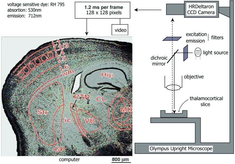
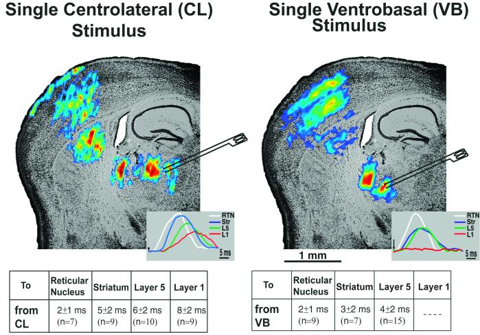
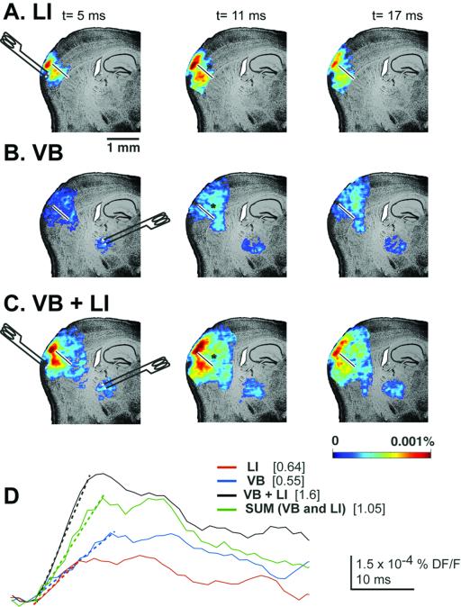
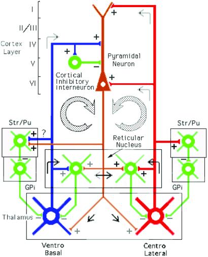
Voltage-sensitive dye imaging of mouse thalamocortical slices demonstrated that electrical stimulation of the centrolateral intralaminar thalamic nucleus (CL) resulted in the specific activation of thalamic reticular nucleus, striatum/putamen, and cortical layers 5, 6, and 1. By contrast, ventrobasal (VB) thalamic stimulation, while activating the reticular and basal ganglia nuclei, also activated directly layers 4 and deep 5 of the cortex. Conjoined stimulation of the VB and CL nuclei resulted in supralinear summation of the two inputs at cortical output layer 5, demonstrating coincidence detection along the apical dendrites. This supralinear summation was also noticed at gamma band stimulus frequency ( approximately 40 Hz). Direct stimulation of cortical layer 1, after a radial section of the cortex that spared only that layer, was shown to sum supralinearly with the cortical activation triggered by VB stimulation, providing a second demonstration for coincidence detection. Coincidence detection by coactivation of the specific (VB) and nonspecific (CL) thalamic nuclei has been proposed as the basis for the temporal conjunction that supports cognitive binding in the brain.


Figures

Figure 1
Figure 1
Schematic diagram of the imaging set-up. Light from a 12-V halogen source was passed through an excitation filter (515 ± 35 nm), dichroic mirror, and microscope objective (×5) before reaching the slice stained with the voltage-sensitive dye RH-795. Emitted fluorescent light was projected onto a charge-coupled device (CCD) camera after passing through the objective, dichroic mirror, and cut-off filter (>590 nm). The CCD camera (HR Deltaron 1700; FUJIX) consisted of 128 × 128 pixels, and each pixel collected light from a surface of about 39 μm × 39 μm. Images were sampled every 1.2 ms. The optical data were analyzed off-line with matlab-based software. A low-magnification, Nissl-stained image of a thalamocortical slice (50-μm thickness) is shown at left. Nuclei and layer subdivisions were demarcated in green lines. Dorsal is up. Hip, hippocampus; ic, internal capsule; Str, neostriatum; VB, ventrobasal nucleus; CL, centrolateral; MD, mediodorsal; wm, white matter; L1, 2/3, 4, 5, 6, different cortical layers. Borders of the cortical “barrels” are demarcated.
Figure 2
Figure 2
Single-pulse stimulation of CL and VB thalamic nuclei. The spread of activity after a single CL (Left) and VB (Right) stimulus is shown superimposed on a Nissl-stained slice. Both CL and VB stimulation activated the reticular nucleus, followed by the striatum. However, at the cortex, different patterns of activation were observed for both stimulations. VB stimulation activated layer 4 followed by layers 2/3 and layer 5; CL stimulation activated layer 6, layer 4–5, and, finally, layer 1. Insets Left and Right correspond to individual pixel profiles at RTN (white lines), striatum/putamen (blue lines), layer 5 (green lines), and layer 1 (red lines) after CL and VB stimulation, respectively. The average delays to different areas of the slice (measured as time between stimulus and the beginning of the individual pixel responses) for both stimulation conditions are shown as a table under each slice.
Figure 3
Figure 3
Stimulation of the CL and VB thalamic nuclei at gamma band frequency. (A) Propagation of optically recorded responses in the somatosensory cortex in response to first and fifth stimuli from CL, VB, and both VB and CL thalamic nuclei. Both thalamic nuclei were stimulated at 40 Hz. Optical signals were superimposed on top of a Nissl-stained slice for a better spatial reference. (Right) Profiles of a single pixel taken from layer 5 are shown for the three different stimulation conditions. Pixel profiles are the averages of 16 trains of 10 stimuli at 40 Hz. Numbers in brackets point at the pixel amplitude corresponding to both first and fifth stimuli. (B) Slope estimation of the rising phase of the first and fifth shock optical responses from CL (red line), VB (blue line), and both CL and VB (black line) is shown with discontinuous lines. Values of the slopes are shown on the right.
Figure 4
Figure 4
Single-pulse stimulation of layer 1 and VB at radially sectioned cortex. Optically recorded responses are shown in the somatosensory cortex 5, 11, and 17 ms after single stimulation of layer 1 (A), VB (B), and both layer 1 and VB (C). Note how layer 1 stimulation on the other side of the cut allowed the spread of activity only through the upper layers of the cortex. (D) Layer 5 pixel profile for the three different stimulation conditions. The slopes of all of them are shown overlapping the pixel profiles (discontinuous lines).
Figure 5
Figure 5
Thalamocortical circuits proposed to subserve temporal binding; diagram of the two thalamocortical systems. The first loop shows the specific ventrobasal nucleus projecting to layer IV of the cortex and to inhibitory interneurons and collaterally to reticular nucleus and striatum/putamen. The second loop shows the nonspecific centrolateral intralaminar nucleus projecting to layers I and VI and also giving collaterals to reticular nucleus and striatum/putamen. Collaterals of these two thalamocortical projections also produce thalamic feedback inhibition via the reticular nucleus and globus pallidus (GPi). The return pathway (in brown) from deep layers V–VI returns the oscillation to the thalamic reticular, ventrobasal, and centrolateral nuclei.

References

    1. Crick F, Koch C. Cold Spring Harbor Symp Quant Biol. 1990;55:953–962. - PubMed
    1. Eckhorn R, Bauer R, Jordan W, Brosch M, Kruse W, Munk M, Reitbock H J. Biol Cybern. 1988;60:121–130. - PubMed
    1. Gray C M, Konig P, Singer W. Nature (London) 1989;338:334–337. - PubMed
    1. Gray C M, Singer W. Proc Natl Acad Sci USA. 1989;86:1698–1702. - PMC - PubMed
    1. Llinás R. In: Fidia Research Foundation Neuroscience Award Lectures. Changeux J-P, Llinás R R, Purves D, Bloom F, editors. Vol. 4. New York: Raven; 1990. pp. 1–10.

Publication types

LinkOut - more resources