. 2002 Jan 22;99(2):815-20.
doi: 10.1073/pnas.022473899. Epub 2002 Jan 2.

Molecular analysis of mutations in DNA polymerase eta in xeroderma pigmentosum-variant patients

Affiliations

Molecular analysis of mutations in DNA polymerase eta in xeroderma pigmentosum-variant patients

Bernard C Broughton et al. Proc Natl Acad Sci U S A. .

Abstract

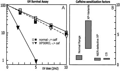
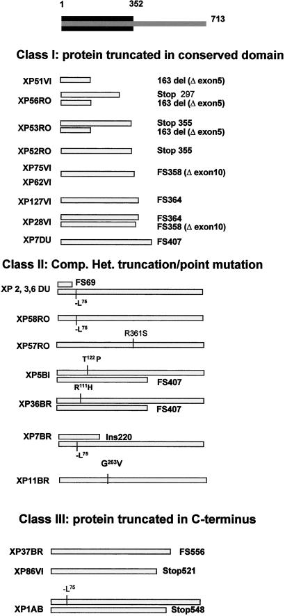
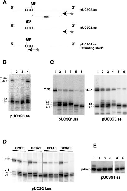
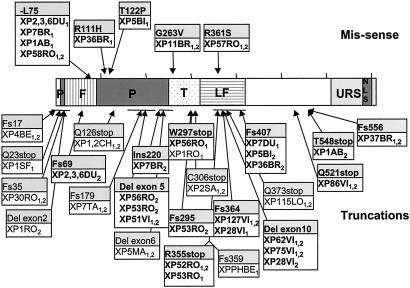
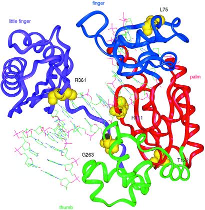
Xeroderma pigmentosum variant (XP-V) cells are deficient in their ability to synthesize intact daughter DNA strands after UV irradiation. This deficiency results from mutations in the gene encoding DNA polymerase eta, which is required for effecting translesion synthesis (TLS) past UV photoproducts. We have developed a simple cellular procedure to identify XP-V cell strains, and have subsequently analyzed the mutations in 21 patients with XP-V. The 16 mutations that we have identified fall into three categories. Many of them result in severe truncations of the protein and are effectively null alleles. However, we have also identified five missense mutations located in the conserved catalytic domain of the protein. Extracts of cells falling into these two categories are defective in the ability to carry out TLS past sites of DNA damage. Three mutations cause truncations at the C terminus such that the catalytic domains are intact, and extracts from these cells are able to carry out TLS. From our previous work, however, we anticipate that protein in these cells will not be localized in the nucleus nor will it be relocalized into replication foci during DNA replication. The spectrum of both missense and truncating mutations is markedly skewed toward the N-terminal half of the protein. Two of the missense mutations are predicted to affect the interaction with DNA, the others are likely to disrupt the three-dimensional structure of the protein. There is a wide variability in clinical features among patients, which is not obviously related to the site or type of mutation.


Figures

Figure 1
Figure 1
Sensitization to UV by caffeine. (A) Typical thymidine incorporation assay on known XP-V strain (triangles) compared with normal fibroblasts (squares), in absence (open symbols) or presence (closed symbols) of 1.5 mM caffeine. (B) Ranges of caffeine-sensitization factors, calculated from survival curves as indicated in Table 1, specified for various categories of UV-sensitive cell strains.
Figure 2
Figure 2
Three classes of polη mutations in patients with XP-V. The conserved N-terminal region of the polη protein is indicated in black at the top of the diagram. The gray bars indicate the mutated protein. Two bars are shown for compound heterozygotes, one for homozygotes.
Figure 3
Figure 3
TLS in XP-V extracts. (A) Different template-primer substrates. (B) Cell extracts were incubated with the pUC3G3.ss template and a 32P end-labeled primer, which annealed 91 nucleotides from the AAF lesion. The products of the reactions were electrophoresed on a sequencing gel. Lane 1, XP1BR; lane 2, XP28VI; lane 3, XP127VI; lane 4, XP86VI. (C) TLS was compared in normal and XP-V extracts, using the pUC3G3.ss and pUC3G1.ss templates. Lane 1, 1BR3; lane 2, XP37BR; lane 3, XP1AB; lane 4, XP86VI; lane 5, XP11BR; lane 6, CTAg. (D) Effect of concentration of various extracts on TLS by using the pUC3G1.ss template. For each strain the amounts of proteins used were 36, 24, and 12 μg. (E) TLS activity of various XP-V extracts using the pUC3G1.ss template and a primer, which anneals immediately upstream of the AAF lesion. Lane 1, XP86VI; lane 2, XP75VI; lane 3, XP62VI; lane 4, XP51VI; lane 5, XP127VI; lane 6, XP28VI.
Figure 4
Figure 4
Sites of mutations identified in polη. Cell strain designations are indicated in boxes, with those used in this study shown in bold. Others are from Masutani et al. (9), Johnson et al. (10), and Yuasa et al. (26). Subscripts 1 and 2 denote different alleles. Horizontal lines indicate deletions. Single amino acid changes are shown in the upper part of the diagram, and truncations are shown in the lower part. The central part of the diagram shows the different domains in the protein (see Fig. 5), with the finger (F), palm (P), thumb (T), and little finger (LF) domains deduced from ref. delineated by different shading. Also shown are the nuclear localization signal (NLS) and the domain involved in localizing polη into replication foci (URS) (16).
Figure 5
Figure 5
Sites of missense mutations superimposed on crystal structure of Y-family DNA polymerase. The figure shows the structure of Dpo4 from Sulfolobus solfataricus bound to DNA (25). The predicted sites of the five missense mutations in polη, based on the sequence alignment in ref. , are indicated on this structure. The different colors indicate the finger, palm, thumb, and little finger domains.

References

    1. Kraemer K H, Lee M M, Scotto J. Arch Dermatol. 1987;123:241–250. - PubMed
    1. Bootsma D, Kraemer K H, Cleaver J E, Hoeijmakers J H J. In: The Genetic Basis of Human Cancer. Vogelstein B, Kinzler K W, editors. New York: McGraw–Hill; 1998. pp. 245–274.
    1. Berneburg M, Lehmann A R. Advances in Genetics. Vol. 43. San Diego: Academic; 2001. pp. 71–102. - PubMed
    1. Maher V M, Ouellette L M, Curren R D, McCormick J J. Nature (London) 1976;261:593–595. - PubMed
    1. Myhr B C, Turnbull D, DiPaolo J A. Mutat Res. 1979;62:341–353. - PubMed

Publication types