Case Reports
. 2002 Jan 8;99(1):473-8.
doi: 10.1073/pnas.022533099. Epub 2002 Jan 2.

The nuclear receptor NR2E3 plays a role in human retinal photoreceptor differentiation and degeneration

Affiliations
Case Reports

The nuclear receptor NR2E3 plays a role in human retinal photoreceptor differentiation and degeneration

Ann H Milam et al. Proc Natl Acad Sci U S A. .

Abstract

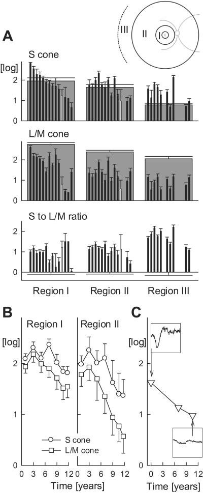
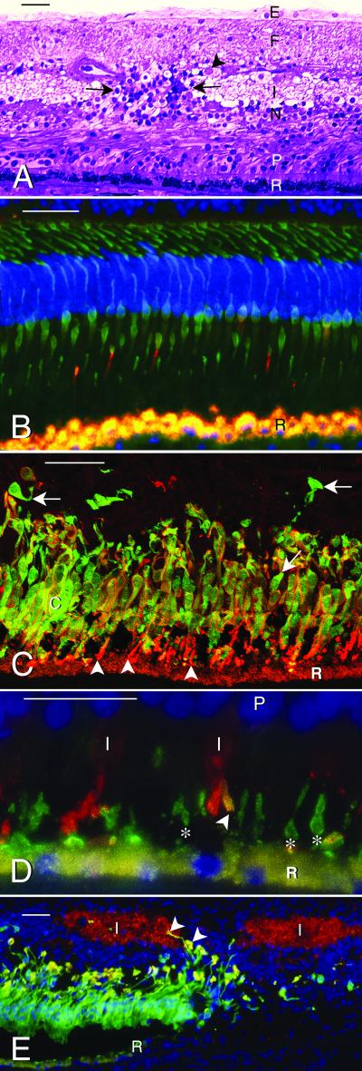
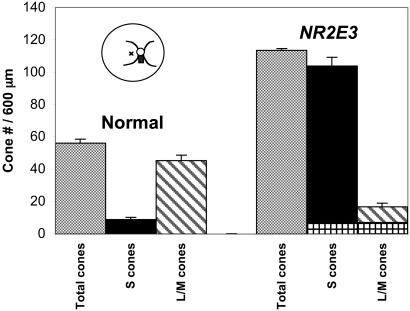
Normal human retinal development involves orderly generation of rods and cones by complex mechanisms. Cell-fate specification involves progenitor cell lineage and external signals such as soluble factors and cell-cell interactions. In most inherited human retinal degenerations, including retinitis pigmentosa, a mutant gene causes loss of visual function, death of mature rods, and eventually death of all cone subtypes. Only one inherited retinal disorder, the enhanced S cone syndrome (ESCS), shows increased visual function, involving the minority S (blue) cones, and decreased rod and L/M (red/green) cone function. This autosomal recessive disease is caused by mutations in NR2E3, a photoreceptor nuclear receptor transcription factor, and may result from abnormal cell-fate determination, leading to excess S cones at the expense of other photoreceptor subtypes. In 16 ESCS patients with the most common NR2E3 mutation, R311Q, we documented an abnormal ratio of S to L/M cone function and progressive retinal degeneration. We studied the postmortem retina of an ESCS patient homozygous for NR2E3 R311Q. No rods were identified, but cones were increased approximately 2-fold, and 92% were S cones. Only 15% of the cones expressed L/M cone opsin, and some coexpressed S cone opsin. The retina was disorganized, with densely packed cones intermixed with inner retinal neurons. The retina was also degenerate, retaining photoreceptors in only the central and far peripheral regions. These observations suggest a key role for NR2E3 in regulation of human photoreceptor development. Degeneration of the NR2E3 retina may result from defective development, known S cone fragility, or abnormal maintenance of mature photoreceptors.


Figures

Figure 1
Figure 1
S versus L/M cone function and disease progression in patients with NR2E3 R311Q mutations. (A) Psychophysically measured S and L/M cone sensitivities and their ratio summarized over three annular regions corresponding to eccentricities of 3–10°, 10–34°, and >34°, respectively (I, II, III shown in Inset). Patient data (thin black bars) are ordered (left to right) in declining S cone sensitivity in region I. S to L/M cone sensitivity ratios are normalized by the mean normal ratio in each region. Data from the eye donor at age 70 years are shown as white. Normal data are gray except in the bottom row, where mean normal is 0 log. Error bars, 2 SEM. (B) Serial data over 11 years in a patient between the ages of 40 and 51 showing S and L/M cone sensitivities in regions I and II. (C) Serial dark-adapted ERG a-wave amplitudes in the same patient. Insets (ordinate range 100 μV; abscissa −10 to 160 ms) show two representative waveforms.
Figure 2
Figure 2
Microscopy of normal and NR2E3 mutant human retinas. The retinal pigment epithelium is indicated by (R). Bar indicates 50 μm in A–C and E and 25 μm in D. (A) Central macula of retina with NR2E3 R311Q mutation. Glycol methacrylate section stained with Richardson's methylene blue/azure II. Photoreceptors (P) are reduced to 2–3 layers of nuclei (normal is 6–8 layers). Outer segments are short to absent. The inner nuclear layer (N) varies in thickness, and the inner plexiform layer (I) is interrupted by a cluster of cells and radial fibers (between arrows). Ganglion cells (arrowhead) are reduced to a discontinuous layer (normal is 6–8 layers), the nerve fiber layer (F) is gliotic, and an epiretinal membrane (E) is present. (B–E) Immunofluorescence images. The RPE (R) contains gold autofluorescent lipofuscin granules. The nuclei have been stained (blue) with 4′,6′-diamidino-2-phenylindole in B, D, and E. (B) Central part of normal human retina processed for immunofluorescence with mAb 7G6 (green), which labels the cytoplasm of all cones, and anti-S cone opsin (red), which labels four S cone outer segments. (C) Central part of ESCS retina labeled with mAb 7G6 (green), specific for cone cytoplasm, and anti-S cone opsin (red). Note multilayered cones (c) with outer segments (arrowheads) positive for S cone opsin. Most cones are double-labeled (gold) and have S cone opsin reactivity (red) in the surface membranes of the inner segments and cell bodies. A few 7G6 positive cones (arrows) are S cone opsin negative. (D) The NR2E3 mutant retina labeled with anti-S cone opsin (green) (mAb OS-2, which labels S cone outer segments, indicated by *) and anti-L/M cone opsin (red). Note opsin delocalization to the inner segments (I) of the L/M cones and one cone outer segment (gold, arrowhead) that is positive for both L/M and S cone opsin. P, photoreceptor nuclei. (E) Low magnification micrograph showing loss of autofluorescent RPE (gold) and S-cones (green) from the periphery (right side) of the macula. The inner plexiform layer (I), labeled (red) with anti-SV2, is interrupted by S cones and their axons (arrowheads). R, edge of RPE.
Figure 3
Figure 3
Cone cell types in a normal and NR2E3 mutant retina, identified by immunocytochemistry with mAb 7G6 (to label all cones), anti-S cone opsin (to label blue cones), and anti-L/M cone opsin (to label red/green cones). The cone counts are expressed per 600-μm length of retina. Compared with the normal retina, the NR2E3 mutant shows an approximately 2-fold increase in total cones, the majority of which are S cones. The L/M cones are decreased in the NR2E3 mutant retina, and some cones (crosshatched bars) coexpress L/M and S cone opsin. The retinal sections were taken from a region (black rectangle in cartoon of fundus) just inferior to the optic nerve head (small circle in cartoon with major retinal blood vessels emerging from the superior and inferior edges). The small × in the cartoon indicates the fovea.

References

    1. Livesey F J, Cepko C L. Nat Rev. 2001;2:109–118. - PubMed
    1. Rothermel A, Layer P G. Eur J Neurosci. 2001;13:949–958. - PubMed
    1. Johnson P T, Williams R R, Reese B E. Visual Neurosci. 2001;18:157–168. - PubMed
    1. Egea P F, Klaholz P, Moras D. FEBS Lett. 2000;476:62–67. - PubMed
    1. Kumar R, Thompson E B. Steroids. 1999;64:310–319. - PubMed

Publication types

MeSH terms

Substances

LinkOut - more resources