

Decreased pulmonary vascular permeability in aquaporin-1-null humans
- PMID: 11773634
- PMCID: PMC117429
- DOI: 10.1073/pnas.022626499
Decreased pulmonary vascular permeability in aquaporin-1-null humans
Abstract
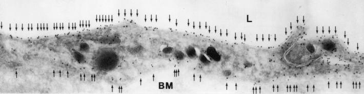
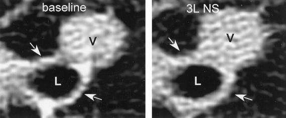
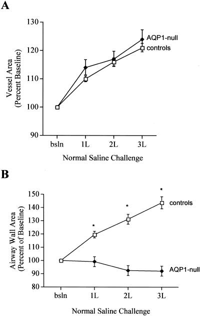
The molecular determinants of water permeability in the human lung are incompletely defined. Aquaporins (AQP) are water-specific membrane channel proteins. AQP1 is present in endothelial cells in the lung, including those in the vascular plexus around the airways. Rare individuals have been identified who are deficient in AQP1. High-resolution computed tomography scans of the lung were used to evaluate the response to i.v. fluid challenge in two unrelated AQP1-null individuals and five normal controls. The airways and pulmonary vessels were measured at baseline and after i.v. administration of 3 liters of saline. Increases in airway wall thickness after fluid administration reflect peribronchiolar edema formation. Both control and AQP1 null subjects had approximately a 20% increase in pulmonary vessel area in response to saline infusion, suggesting similar degrees of volume loading. Control subjects had a 44% increase in the thickness of the airway wall, consistent with peribronchiolar edema formation. In marked contrast, airway wall thickness did not change in AQP1-null subjects in response to saline infusion. These studies indicate that AQP1 is a determinant of vascular permeability in the lung, and demonstrate a role for aquaporins in human pulmonary physiology.
Figures
References
-
- Denker B M, Smith B L, Kuhajda F P, Agre P. J Biol Chem. 1988;263:15634–15642. - PubMed
-
- Joshi S R, Wagner F F, Vasantha K, Panjwani S R, Flegel W A. Immunohematology. 2001;41:1273–1278. - PubMed
-
- Preston G M, Smith B L, Zeidel M L, Moulds J J, Agre P. Science. 1994;265:1585–1587. - PubMed
-
- King L S, Choi M, Fernandez P C, Cartron J P, Agre P. N Engl J Med. 2001;345:175–179. - PubMed
Publication types
MeSH terms
Substances
Grants and funding
LinkOut - more resources
Full Text Sources
