. 2001 Nov-Dec;3(6):469-79.
doi: 10.1038/sj.neo.7900196.

Bromelain reversibly inhibits invasive properties of glioma cells

Affiliations

Bromelain reversibly inhibits invasive properties of glioma cells

B B Tysnes et al. Neoplasia. 2001 Nov-Dec.

Abstract

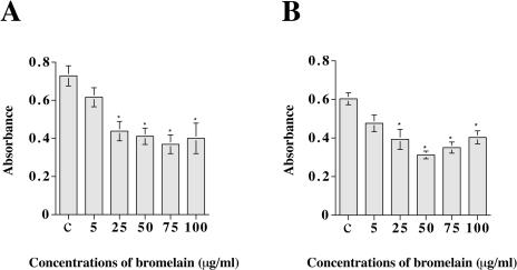
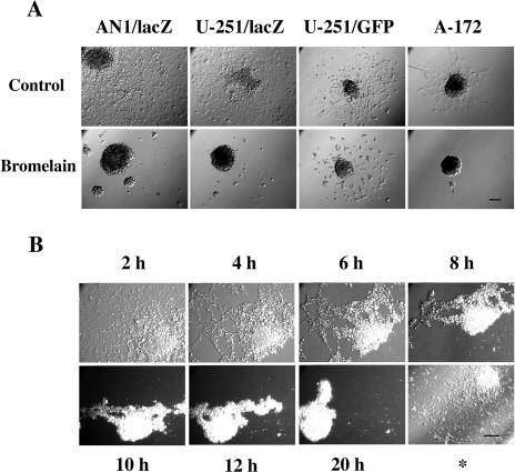
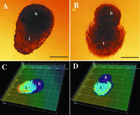
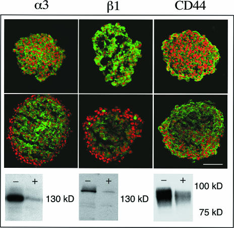
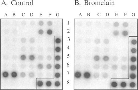
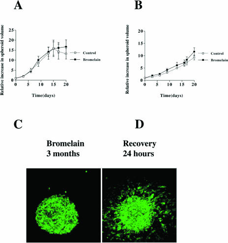
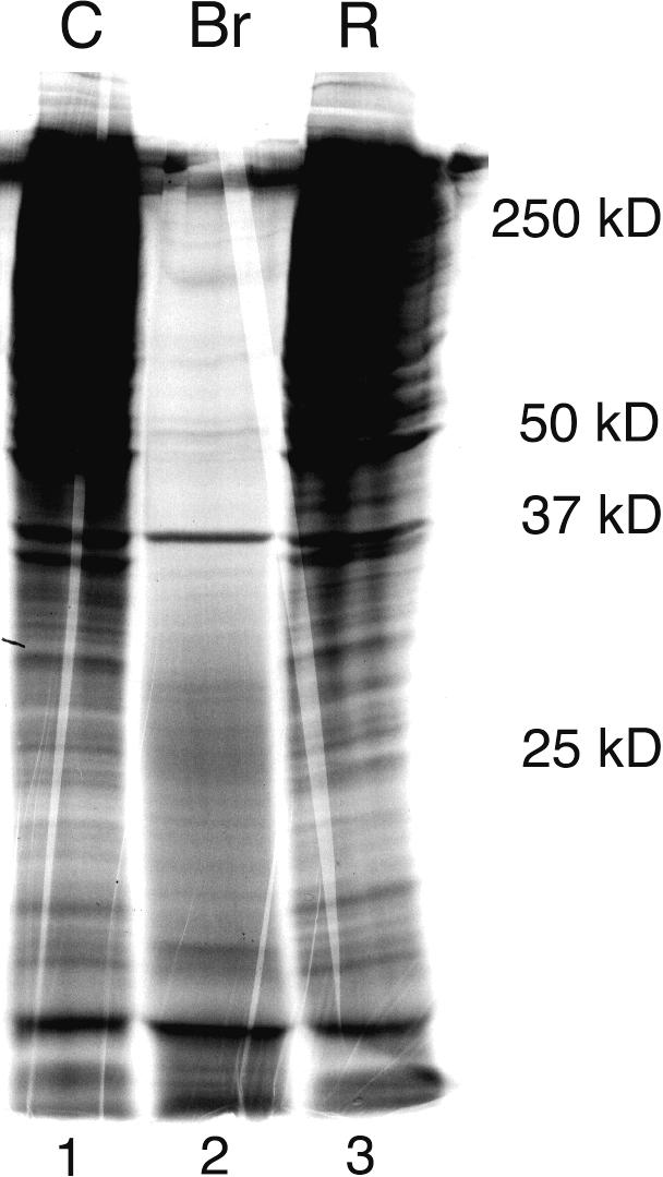
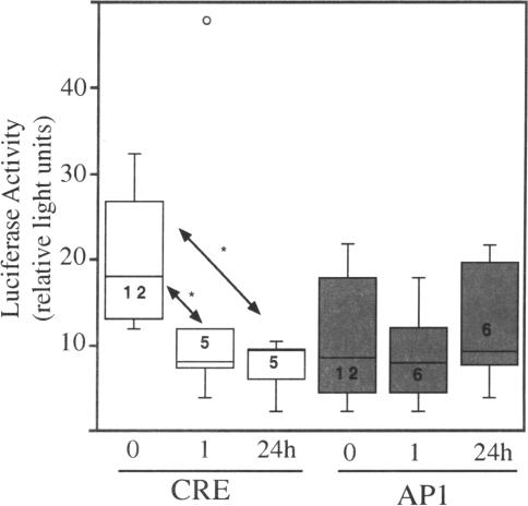
Bromelain is an aqueous extract from pineapple stem that contains proteinases and exhibits pleiotropic therapeutic effects, i.e., antiedematous, antiinflammatory, antimetastatic, antithrombotic, and fibrinolytic activities. In this study, we tested bromelain's effects on glioma cells to assess whether bromelain could be a potential contributor to new antiinvasive strategies for gliomas. Several complementary assays demonstrated that bromelain significantly and reversibly reduced glioma cell adhesion, migration, and invasion without affecting cell viability, even after treatment periods extending over several months. Immunohistochemistry and immunoblotting experiments demonstrated that alpha3 and beta1 integrin subunits and hyaluronan receptor CD44 protein levels were reduced within 24 hours of bromelain treatment. These effects were not reflected at the RNA level because RNA profiling did not show any significant effects on gene expression. Interestingly, metabolic labelling with 35-S methionine demonstrated that de novo protein synthesis was greatly attenuated by bromelain, in a reversible manner. By using a transactivating signaling assay, we found that CRE-mediated signaling processes were suppressed. These results indicate that bromelain exerts its antiinvasive effects by proteolysis, signaling cascades, and translational attenuation.


Figures

Figure 1
Figure 1
Bromelain inhibits glioma cell adhesion. The diagrams show adhesion of AN1/lacZ (A) and U-251/lacZ (B) human glioma cells exposed to increasing concentrations of bromelain. The number of cells adhering to the plastic was quantified using photometry (y-axis). Data expressed as mean±SEM; n=8 in each group. Asterisks indicate significant differences from control (ANOVA and Sheffé's test). c=control (complete growth medium).
Figure 2
Figure 2
Bromelain inhibits glioma cell migration. (A) Cell migration (48 hours) from multicellular spheroids from different human glioma cell lines in 96-wells with and without bromelain (50 µg/ml). Bar, 100 µm. (B) Time lapse study of cells from an AN1/lacZ spheroid, which had been allowed 48 hours of migration in DMEM supplemented with 10% serum, before the medium was changed to a medium containing 50 µg/ml bromelain. The time points refer to the time after bromelain was added to the medium. *This picture was taken 96 hours after the bromelain medium was replaced with ordinary serum supplemented medium. Bar, 100 µm.
Figure 3
Figure 3
Bromelain reduces glioma cell invasion into normal brain cell aggregates. Tumor cell invasion in 72-hour co-cultures of spheroids prepared from the U-87/GFP human glioma cell line and normal fetal rat brain cell aggregates. Transmission light microscopy pictures of a control co-culture (A) and a bromelain-treated co-culture (B). Scale bars, 100 µm. t=tumor, b=brain. Three-dimensional reconstructions of confocal microscopy sections of the same tumor spheroids combined with transmission pictures of the brain aggregate (C, control; D, bromelain-treated). A linear color map ranging from zero (dark blue, no transmission) to 255 (red, highest fluorescence intensity) was used. The distance between the lines is 200 µm along the x-and y-axes, and 27 µm along the z-axis.
Figure 4
Figure 4
Bromelain affects cell surface protein expression. Scanning confocal microscopy pictures of α3 and β1 integrin subunits and CD44 immunostained spheroid sections (green signal) from the AN1/lacZ human glioma cell line. Nuclei are stained with propidium iodide (red). Upper panel: control spheroids; middle panel: bromelain-treated spheroids. Scale bar, 100 µm. Lower panel: corresponding Western blot of monolayer cultures; control cells marked with (-) and bromelain-treated cells marked with (+).
Figure 5
Figure 5
Transcriptional profiling following bromelain treatment. Gene expression profiles of control (A) and bromelain-treated (24 hours) AN1/lacZ human glioma cells (B) in a cancer/metastasis GEArray. Probes were prepared using RT-PCR techniques and hybridized to membrane genes involved in tumor cell adhesion and genes for proteolysis of ECM during tumor cell invasion. Following extensive washing, membranes were exposed to a phosphoimager to capture an image. No large variations in gene expression were detected following the bromelain treatment. The α3 integrin and CD44 are located, respectively, at 5C, 5D and 1E, 1F. The two housekeeping genes actin and GAPDH are located, respectively, at 3G, 4G and 5G, 6G, 7G, 8E, 8F, 8G, whereas the negative control (pUC18, bacterial plasmid) is located at 1G, 2G. The signals were found to be within the linear range of exposure.
Figure 6
Figure 6
Glioma spheroid growth and reversal of inhibited migration from spheroids. Cell growth of AN1/lacZ (A) and U-87/GFP (B) human glioma spheroids with and without bromelain (50 µg/ml). The relative increase in volume is expressed as mean±SEM; control, n=4 (AN1/lacZ), n=5 (U-87/GFP); bromelain-treated group, n=5. (C) U-87/GFP human glioma spheroid grown in bromelain-containing medium for 3 months. The spheroid is floating in the medium. (D) Twenty-four hours after bromelain removal. The spheroid is now attached to the culture dish and cells are migrating out from the spheroid.
Figure 7
Figure 7
Bromelain attenuates de novo protein synthesis in glioma cells. SDS-PAGE showing newly synthesized proteins from 35-S methionine-labelled U-87/GFP- transfected human glioma control cells (Lane 1), cells treated with bromelain for 7 days (Lane 2), and cells treated with bromelain for 4 days and then allowed a 3-day recovery period in complete growth medium (Lane 3). Molecular size markers are indicated to the right.
Figure 8
Figure 8
Bromelain affects nuclear signaling. AN1/lacZ cells were transfected with luciferase reporter constructs containing CRE or AP-1 cis-acting elements prior to bromelain treatment for 0, 1, or 24 hours. Cells were extracted and luciferase was measured. The data, shown in the form of a box plot, indicate that CRE signaling was significantly affected following bromelain treatment. The rectangle represents 50% of the data, and the mean is indicated by the horizontal line dissecting the box. The lines above and below the box establish the range of the data; a small open circle represents an outlier. Numbers in the rectangle represent trial numbers. Trials were done in duplicates. Asterisks denote significance [CRE 1 hour: P<.002; CRE 24 hours: P<.01, Kaleidagraph (V3.5, Synergy)].

References

    1. Russell DS, Rubinstein LJ. Pathology of Tumours of the Nervous System. 5th ed. London: Williams and Wilkins; 1989. Tumours of Central Neuroepithelial Origin; pp. 83–350.
    1. Giese A, Westphal M. Glioma invasion in the central nervous system. Neurosurgery. 1996;39:235–250. - PubMed
    1. Rutka JT, Apodaca G, Stern R, Rosenblum M. The extracellular matrix of the central and peripheral nervous systems: structure and function. J Neurosurg. 1988;69:155–170. - PubMed
    1. Uhm JH, Gladson CL, Rao JS. The role of integrins in the malignant phenotype of gliomas. Front Biosci. 1999;4:D188–D199. - PubMed
    1. Paulus W, Baur I, Schuppan D, Roggendorf W. Characterization of integrin receptors in normal and neoplastic human brain. Am J Pathol. 1993;143:154–163. - PMC - PubMed

Publication types

MeSH terms