Review
. 2001;28(4):265-75.

Gender and cardiac arrhythmias

Affiliations
Review

Gender and cardiac arrhythmias

R P Villareal et al. Tex Heart Inst J. 2001.

Abstract

The incidence of certain clinical arrhythmias varies between and women. Clinical and experimental observations suggest the existence of true differences in electrophysiologic properties between the sexes. We review these differences, possible mechanisms, clinical implications, and therapeutic considerations in the treatment of various arrhythmias in women.


Figures

None
Fig. 1 Sinus cycle length before and after double autonomic blockade. In both instances, men had a significantly longer cycle length (slower heart rate) than did women. *P <0.05 (Reprinted from American Journal of Medicine, Vol 100. Burke JH, Goldberger JJ, Ehlert FA, Kruse JT, Parker MA, Kadish AH. Gender differences in heart rate before and after autonomic blockade: evidence against an intrinsic gender effect. p. 537–43, copyright 1996, with permission from Excerpta Medica Inc., and from the author.)
None
Fig. 2 Heart rate variability between women with and without hormone (estrogen) replacement therapy. HF = high frequency; HRT = hormone replacement therapy; LF = low frequency; pNN50 = percentage difference between successive RR intervals >50 msec *P <0.05 (Data adapted with permission from Huikuri HV, et al. 17)
None
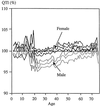
Fig. 3 Corrected QTI interval according to age (birth to 75 yr) in 14,379 children and adults. Note a decrease in the men's QT interval after puberty, which then increases over time but never reaches that of women. (From Rautaharju PM, Zhou SH, Wong S, Calhoun HP, Berenson GS, Prineas R, Davignon A. Sex differences in the evolution of the electrocardiographic QT interval with age. Can J Cardiol 1992;8:690–5. Reproduced with permission of Can J Cardiol.)
None
Fig. 4 Sinus cycle lengths before and after double autonomic blockade during the various menstrual phases. The sinus cycle length was significantly longer (*P <0.03) during the menstrual phase of the cycle at baseline. This difference was no longer evident after autonomic blockade. (Reprinted from American Journal of Medicine, Vol 100. Burke JH, Goldberger JJ, Ehlert FA, Kruse JT, Parker MA, Kadish AH. Gender differences in heart rate before and after autonomic blockade: evidence against an intrinsic gender effect. p. 537–43, copyright 1996, with permission from Excerpta Medica Inc., and from the author.)
None
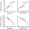
Fig. 5 Incidence of supraventricular tachycardia (SVT) plotted against serum levels of plasma ovarian hormones on day 28 of the menstrual cycle. There was significant positive correlation between progesterone and SVT episodes and an inverse correlation between estradiol-17β and SVT. (Reprinted with permission from Elsevier Science [The Lancet 1996;347:786–8 23] and from the author.)
None
Fig. 6 The effects of the IKR blocker, E4031, on placebo-, estrogen-, and dihydrotestosterone-treated rabbit papillary muscles. DHT = dihydrotestosterone; EAD = early afterdepolarization; EST = estrogen (From Hara M, Danilo P Jr, Rosen MR. Effects of gonadal steroids on ventricular repolarization and on the response of E4031. J Pharmacol Exper Ther 1998;285:1068–72. Reproduced with permission from the publisher and from the author.)
None
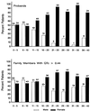
Fig. 7 Percentage distribution of probands (top) and family members with corrected QT (QTc) interval >440 ms (bottom) by sex and age at baseline electrocardiography. Up until the age of 15 years, the proportion of males:females is approximately 1:1. After 15 years, there is a marked female predominance. (From Locati HE, Zareba W, Moss AJ, Schwartz PJ, Vincent M, Lehmann MH, et al. Age- and sex-related differences in clinical manifestations in patients with congenital long-QT syndrome: findings from the International LQTS Registry. Circulation 1998;97:2237-44. Reproduced with permission from Lippincott Williams & Wilkins and from the author.)
None
Fig. 8 Sex- and age-specific rates for sudden death, morbidity, and total coronary deaths in subjects aged 35 to 84 years: 26-year follow-up, Framingham study. morb. = morbidity; mort. = mortality (From Lerner DJ, Kannel WB. Patterns of coronary heart disease morbidity and mortality in the sexes: a 26-year follow-up of the Framingham population. Am Heart J 1986;111:383-90. Reproduced with permission from Mosby, Inc., A Harcourt Health Sciences Company.)
None
Fig. 9 Relative proportions of underlying heart disease in survivors of cardiac arrest: A) female and B) male. CAD = coronary artery disease; DCM = dilated cardiomy-opathy; RV = right ventricular; VHD = valvular heart disease (From Albert CM, McGovern BA, Newell JB, Ruskin JN. Sex differences in cardiac arrest survivors. Circulation 1996;93:1170–6. Reproduced with permission from Lippincott Williams & Wilkins and from the author.)

References

    1. Linde C. Women and arrhythmias. Pacing Clin Electrophysiol 2000;23:1550–60. - PubMed
    1. Benjamin EJ, Levy D, Vaziri SM, D'Agostino RB, Belanger AJ, Wolf PA. Independent risk factors for atrial fibrillation in a population-based cohort. The Framingham Heart Study. JAMA 1994;271:840–4. - PubMed
    1. Rodriguez LM, de Chillou C, Schlapfer J, Metzger J, Baiyan X, van den Dool A, et al. Age at onset and gender of patients with different types of supraventricular tachycardias. Am J Cardiol 1992;70:1213–5. - PubMed
    1. Timmermans C, Smeets JL, Rodriguez LM, Vrouchos G, van den Dool A, Wellens HJ. Aborted sudden death in the Wolff-Parkinson-White syndrome. Am J Cardiol 1995;76:492–4. - PubMed
    1. Bazett HC. An analysis of the time-relations of electrocardiograms. Heart 1920;7:353–70.