. 2002 Jan 7;156(1):199-209.
doi: 10.1083/jcb.200109021. Epub 2002 Jan 3.

Conditional disruption of beta 1 integrin in Schwann cells impedes interactions with axons

Affiliations

Conditional disruption of beta 1 integrin in Schwann cells impedes interactions with axons

M Laura Feltri et al. J Cell Biol. .

Abstract

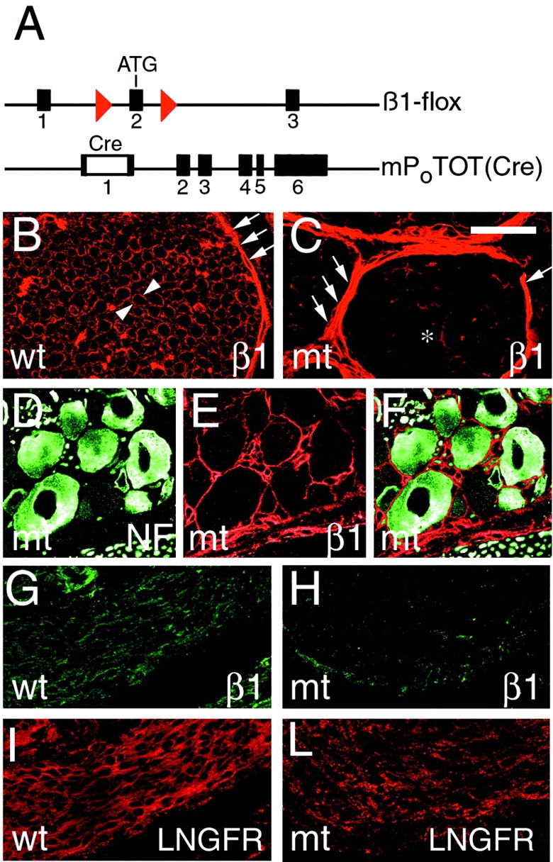
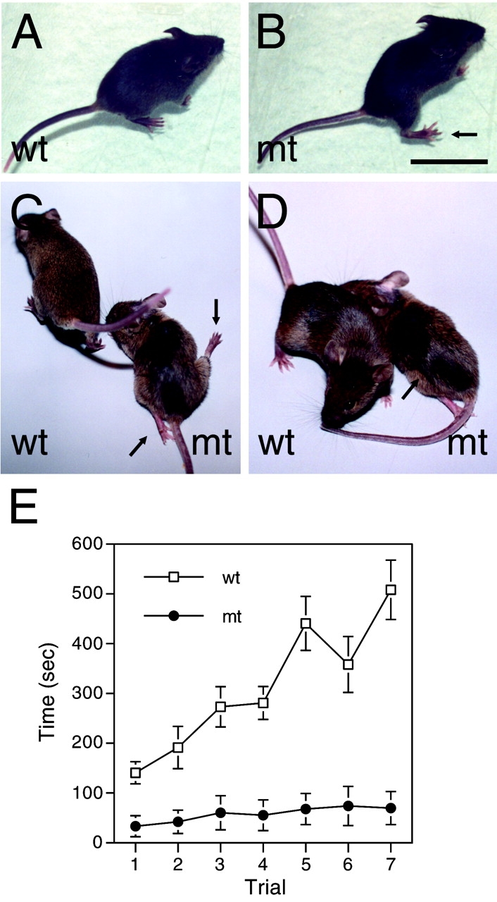
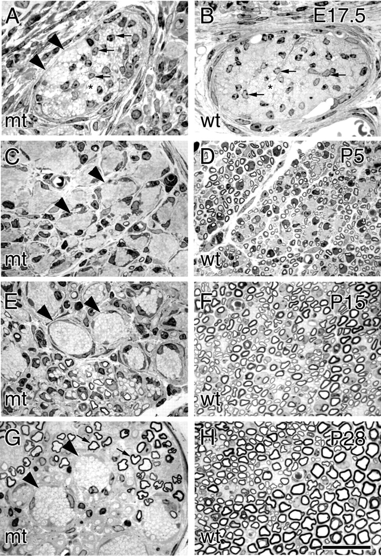
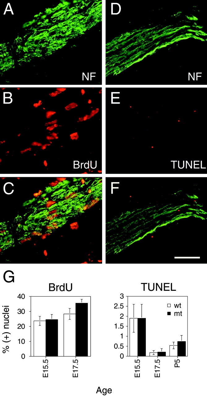
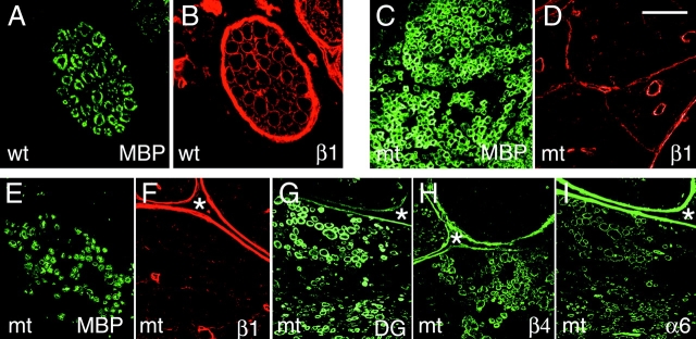
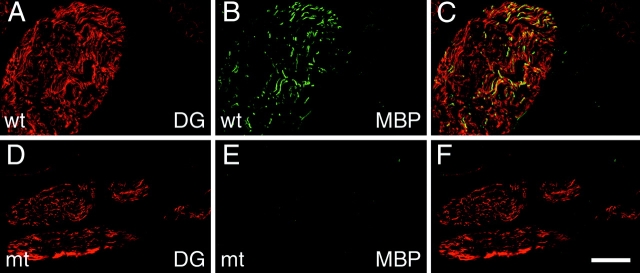
In dystrophic mice, a model of merosin-deficient congenital muscular dystrophy, laminin-2 mutations produce peripheral nerve dysmyelination and render Schwann cells unable to sort bundles of axons. The laminin receptor and the mechanism through which dysmyelination and impaired sorting occur are unknown. We describe mice in which Schwann cell-specific disruption of beta1 integrin, a component of laminin receptors, causes a severe neuropathy with impaired radial sorting of axons. beta 1-null Schwann cells populate nerves, proliferate, and survive normally, but do not extend or maintain normal processes around axons. Interestingly, some Schwann cells surpass this problem to form normal myelin, possibly due to the presence of other laminin receptors such as dystroglycan and alpha 6 beta 4 integrin. These data suggest that beta 1 integrin links laminin in the basal lamina to the cytoskeleton in order for Schwann cells to ensheath axons, and alteration of this linkage contributes to the peripheral neuropathy of congenital muscular dystrophy.


Figures

Figure 1.
Figure 1.
Disruption of β1 integrin specifically in Schwann cells in peripheral nerves. A schematic representation of the “floxed” Itgβ1 allele shows that two loxP sites (red) flank exon 2, containing the ATG start site of translation and the P0Cre-mP0TOT(Cre) transgene, containing the Cre gene inserted into exon 1 of the Mpz gene. Transverse sections of control (B; wt) or mutant (C; mt) sciatic nerves at P28 show that β1 is normally detected in the endoneurium at the outer surface of each myelinating Schwann cell (arrowheads) and in the perineurium (arrows), whereas in mutant nerves, β1 is absent in Schwann cells (asterisk), but is preserved in the perineurium (arrows). Low level endoneurial staining for β1 is associated with vessels and perineurial cells abnormally present in the endoneurium (C; see text). (D–F) Double staining of DRG for neurofilament (D; green) and β1 integrin (E; red) shows that β1 expression in mutant sensory neurons is preserved; (F) merge. Longitudinal sections of control (G and I) and mutant (H and L) nerves at E17.5 are double stained for β1 (G and H; green) and LNGFR (I and L; red). Note that the β1 integrin staining in Schwann cells is markedly reduced in mutant nerves by E17.5. Bar: (B and C) 30 μm; (D and F) 50 μm; (G and I) 60 μm; (H and L) 90 μm.
Figure 2.
Figure 2.
Mutant mice manifest severe motor impairment. (A–D) Mutant (mt) and control (wt) mice at P28 (A and B) or P90 (C and D) were observed sitting on an inclined surface (A and B) or walking on a flat surface (C and D). Mutant mice at P28 sit with their hind limbs displaced laterally (arrow in B) and walk with a wider base, swinging their hind limb laterally (unpublished data). By P90, many mutant mice manifest nearly paralyzed hind limbs (arrows in C) and more obvious muscle atrophy (arrow in D). (E) Rotarod test was performed with 2–3-mo-old mutant and control mice. In a series of seven consecutive trials the time (seconds; mean ± SEM) for which animals remained on a rod rotating at increasing speed is plotted. Hold time is significantly reduced in mutant mice (n = 10) as compared with control mice (n = 14). 0.01 < P < 0.001 in the first trial, and P < 0.001 in subsequent trials, by two-tailed Student t test. Bar: (A–C) 3 cm; (D) 2.7 cm.
Figure 3.
Figure 3.
Mutant mice develop a severe dysmyelinating neuropathy. Transverse semithin sections of comparable nerves from mutant (mt; A, C, E, and G) and control (wt; B, D, F, and H) mice at E17.5 (A and B), P5 (C and D), P15 (E and F), and P28 (G and H). At P28, myelination is complete in normal nerves (H), whereas mutant nerves show large bundles of unsorted axons (G, arrowheads) and few myelinated fibers (arrows). At E17.5 (A and B) both control and mutant nerves contain groups of yet unsorted axons (asterisks) and numerous Schwann cells between them (arrows); groups of tightly packed axons begin to be distinguishable in mutant nerves (A, arrowheads), but not in controls (B). In P5 control nerves (D), most large caliber axons have reached the proper 1:1 relationship with a Schwann cell, and many thin myelin sheaths appear. In mutant nerves (C), in contrast, large groups of unsegregated axons are present (arrowheads), and only rare, very thin, myelin sheaths have formed. Unsorted groups of axons become progressively more obvious during nerve maturation (E and G, arrowheads), and are often grouped sectorially in the nerve. More thin myelin sheaths appear with delay in mutant nerves at P15 (E) and become thicker at P28 (G). Bar: (A and B) 27 μm; (C–H) 29 μm.
Figure 4.
Figure 4.
Ultrastructural analysis of mutant nerves reveals perturbed relationships with axons. Transverse sections of mutant sciatic nerves at P28 (A and B) reveal bundles containing unsorted, mixed caliber axons (asterisks) that are not invested by Schwann cell cytoplasm (“naked”). Several Schwann cells have attained the proper 1:1 relationship with an axon and occasionally are forming a myelin sheath (A). At high magnification (C), the periodicity of compact myelin appears normal. Axonal bundles are always surrounded by a layer of perineurial cells, abnormally present in the endoneurium (arrowheads). Some of the perineurial cells contain lipid inclusions (B) and at high magnification (inset in B–G) display a basal lamina (arrowhead in G), tight junctions (arrow in G), and caveolae (arrows in insets indicated by asterisks in G; magnified 1.6×). In D–F several axons are shown surrounded by a loose, undulated basal lamina (arrowheads), which in some cases contain Schwann cell processes variably ensheathing axons (arrows), whereas in C, a myelinated fiber has a tightly apposed basal lamina (arrowheads). Transverse sections of control (H) and mutant (I) nerves at P1 show differences in radial sorting and cytoplasmic processes. In control nerves (H), most axons >1 μm are either segregated at the periphery of a bundle (black asterisks), in groups of 1–3 (arrows), or clearly in a 1:1 relationship with a Schwann cell. In contrast, in mutant nerves (I), most large axons are either unsegregated from bundles of smaller axons (black asterisks), or in groups of greater than three (thick arrows). Whereas control Schwann cells (H) send thin processes around axons, mutant Schwann cells (I) send abnormally shaped, thick cytoplasmic processes (white asterisks) in every direction (thin arrows). Bars: (A) 4.4 μm; (B) 3.7 μm; (C and G) 1.2 μm; (D and E) 1.6 μm; (F) 1 μm; (H and I) 7.6 μm.
Figure 5.
Figure 5.
β1-null Schwann cells proliferate and survive normally. (A–C) Nuclei of mutant Schwann cells associated with longitudinal sections of nerves at E17.5 that have been stained for neurofilament (A, green) and BrdU (B, red) after a 1-h pulse; C, merge. (D–F) Apoptotic nuclei associated with mutant nerves at E17.5 that have been stained for neurofilament (NF) (D, green) were identified by TUNEL staining (E, red); F, merge. (G) The percentages (mean ± SEM) of BrdU- or TUNEL-positive nuclei in mutant (mt) and control (wt) nerves. See also Table I. By a paired, two-tailed t test the percentages in mutant and controls were not significantly different. Bar, (A–F) 60 μm.
Figure 6.
Figure 6.
Synthesis of other laminin receptors and myelin in β1-null Schwann cells. Transverse sections of sciatic nerves from control (wt) (A and B) and mutant (mt) (E and F) mice at P28 were double stained for MBP (A and E; green) and β1 integrin (B and F; red). In the control nerve, every MBP-positive myelin sheath is encircled by a β1-positive Schwann cell cytoplasm, whereas in the mutant nerve, the MBP-positive myelin sheaths are not. Staining of serial sections from the same mutant nerve for dystroglycan (DG; G), β4 integrin (H), and α6 integrin (I) detects multiple myelinated fibers in the same field (next to an asterisk as a point of reference) positive for α6, β4 integrin, and dystroglycan. Transverse sections of spinal roots of mutant mice at P28 double stained for MBP (C) and β1 integrin (D) reveal numerous MBP-positive fibers with no associated β1 integrin. Bar: (A and B) 20 μm; (C and D) 40 μm; (E–I) 70 μm.
Figure 7.
Figure 7.
Myelination is delayed despite normal onset of dystroglycan synthesis in mutant nerves. Longitudinal sections of control (wt) (A–C) and mutant (mt) (D–F) nerves at P1 were double stained for dystroglycan (A and D; red) and MBP (B and E; green). In all nerves, dystroglycan is detected in multiple fibers (A, C, D, and F), but only in control nerves do a subset of these fibers stain for MBP (compare B to E). Bar: (A–F) 60 μm.

Comment in

  • Myelination: some receptors required.
    Scherer SS. Scherer SS. J Cell Biol. 2002 Jan 7;156(1):13-5. doi: 10.1083/jcb.200112017. Epub 2002 Jan 7. J Cell Biol. 2002. PMID: 11781330 Free PMC article.

References

    1. Akagi, K., V. Sandig, M. Vooijs, M. Van der Valk, M. Giovannini, M. Strauss, and A. Berns. 1997. Cre-mediated somatic site-specific recombination in mice. Nucleic Acids Res. 25:1766–1773. - PMC - PubMed
    1. Anderson, D.J. 1997. Cellular and molecular biology of neural crest cell lineage determination. Trends Genet. 13:276–280. - PubMed
    1. Arroyo, E.J., J.R. Bermingham, Jr., M.G. Rosenfeld, and S.S. Scherer. 1998. Promyelinating Schwann cells express Tst-1/SCIP/Oct-6. J. Neurosci. 18:7891–7902. - PMC - PubMed
    1. Bradley, W.G., and M. Jenkison. 1973. Abnormalities of peripheral nerves in murine muscular dystrophy. J. Neurol. Sci. 18:227–247. - PubMed
    1. Bronnerfraser, M., M. Artinger, J. Muschler, and A.F. Horwitz. 1992. Developmentally regulated expression of α6 integrin in avian embryos. Development. 115:197–211. - PubMed

Publication types

MeSH terms