

doi: 10.1126/science.1066274.
Role of cell-specific SpoIIIE assembly in polarity of DNA transfer
Affiliations
- PMID: 11778051
- PMCID: PMC2885158
- DOI: 10.1126/science.1066274
Item in Clipboard
Role of cell-specific SpoIIIE assembly in polarity of DNA transfer
Science.
.
Abstract
SpoIIIE mediates postseptational chromosome partitioning in Bacillus subtilis, but the mechanism controlling the direction of DNA transfer remains obscure. Here, we demonstrated that SpoIIIE acts as a DNA exporter: When SpoIIIE was synthesized in the larger of the two cells necessary for sporulation, the mother cell, DNA was translocated into the smaller forespore; however, when it was synthesized in the forespore, DNA was translocated into the mother cell. Furthermore, the DNA-tracking domain of SpoIIIE inhibited SpoIIIE complex assembly in the forespore. Thus, during sporulation, chromosome partitioning is controlled by the preferential assembly of SpoIIIE in one daughter cell.
Figures
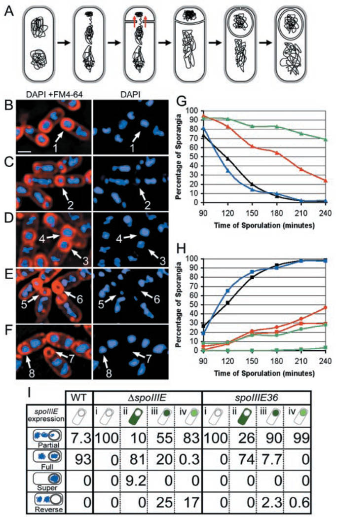
Effect of cell-specific SpoIIIE expression on DNA translocation. (A) Engulfment diagram. (B to F) Samples from t3 were processed as described (20). (B) Wild-type sporangia, with fully translocated forespore chromosomes (arrow 1). (C) ΔspoIIIE sporangia, with partially translocated forespore chromosomes (arrow 2). (D) Sporangia expressing spoIIIE in the mother cell (PspoIID-spoIIIE-gfp) showing a fully translocated chromosome (arrow 3) and a sporangium with two chromosomes in the forespore (arrow 4), which might reflect a defect in chromosome decatenation. (E) Sporangia expressing spoIIIE in the forespore (PspoIIR-spoIIIE-gfp) showing both partial (arrow 5) and reverse (arrow 6) chromosome translocation (13). (F) Sporangia expressing spoIIIE from the spoIIQ promoter (PspoIIQ-spoIIIE-gfp) showing both reverse (arrow 7) and forward (arrow 8) chromosome translocation. Scale bar, 1 µm. (G and H) Chromosome translocation phenotypes in wild type (black), when SpoIIIE was produced in the mother cell (PspoIID-spoIIIE-gfp, blue) and in the forespore at low (PspoIIR-spoIIIE-gfp, green) or high (PspoIIQ-spoIIIE-gfp, red) levels. (G) Disappearance of partially translocated chromosomes (triangles). (H) Appearance of chromosomes that have been translocated into (squares) or out of (circles) the forespore. (I) Chromosome translocation at t3, when SpoIIIE is expressed in the mother cell (PspoIID-spoIIIE-gfp, ii) or the forespore at high (PspoIIQ-spoIIIE-gfp, iii) or low levels (PspoIIR-spoIIIE-gfp, iv) in the ΔspoIIIE null mutant versus the spoIIIE36 missense mutant. The WT column shows chromosome translocation in wild type; column i shows translocation in spoIIIE36 and ΔspoIIIE. The numbers refer to the percentage of sporangia in each strain showing the chromosome translocation phenotypes at left.
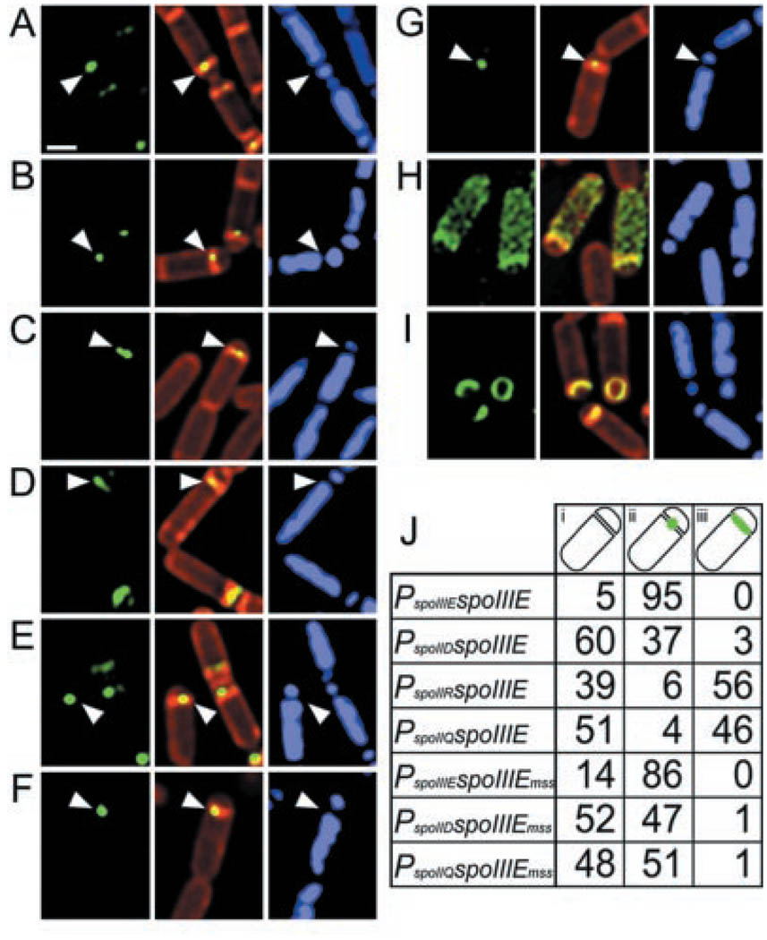
Cell-specific assembly of SpoIIIE-GFP in a ΔspoIIIE mutant background. The full-length protein (spoIIIE), the membrane domain (spoIIIEmss), and the DNA-tracking domain (spoIIIEcyto) were expressed from the natural spoIIIE promoter (PspoIIIE), in the mother cell (PspoIID) or in the forespore at low (PspoIIR) or high (PspoIIQ) levels. Samples from t1.5 (A to G) or t2.0 (H and I) were processed as described (20). Arrowheads indicate the predominant localization phenotype, either a focus at the polar septum (A, B, E, F, and G) or a line across the septum (C and D). (A) PspoIIIE-spoIIIE-gfp. (B) PspoIID-spoIIIE-gfp. (C) PspoIIR-spoIIIE-gfp. (D) PspoIIQ-spoIIIE-gfp. (E) PspoIIIE-spoIIIEmss-gfp. (F) PspoIID-spoIIIEmss-gfp. (G) PspoIIQ-spoIIIEmss-gfp. (H) PspoIID-spoIIIEcyto-gfp: the DNA-tracking domain fills the mother cell cytoplasm. (I) PspoIIQ-spoIIIEcyto-gfp: the DNA-tracking domain localizes to the septal membranes of both early and late sporangia. We were unable to detect the DNA-tracking domain of SpoIIIE expressed from its own promoter (PspoIIIE-spoIIIEcyto-gfp), probably because GFP fusions expressed at such low levels are only detectable if they localize. Scale bar, 1 µm. (J) Percentage of sporangia at t1.5 that have complete sporulation septa and the following localization patterns for the GFP fusions indicated in the first column: none apparent (i), a focus at the polar septum (ii), and a line along the septum (iii). Between 70 and 145 sporangia were scored for each strain.
References
-
- Stragier P, Losick R. Annu. Rev. Genetics. 1996;30:297. - PubMed
-
- Errington J, Bath J, Wu L-J. Nature Rev. Mol. Cell Biol. 2001;2:538. - PubMed
-
- Pogliano J, Sharp MD, Pogliano K. J. Bacteriol. in press.
-
- Wu LJ, Lewis PJ, Allmansberger R, Hauser PM, Errington J. Genes Dev. 1995;9:1316. - PubMed
-
- Wu LJ, Errington J. Science. 1994;264:572. - PubMed
Publication types
MeSH terms
Substances
Grants and funding
LinkOut - more resources
Full Text Sources
Molecular Biology Databases
