

Molecular mechanism of transforming growth factor beta-mediated cell-cycle modulation in primary human CD34(+) progenitors
- PMID: 11781230
- PMCID: PMC4382314
- DOI: 10.1182/blood.v99.2.499
Molecular mechanism of transforming growth factor beta-mediated cell-cycle modulation in primary human CD34(+) progenitors
Abstract
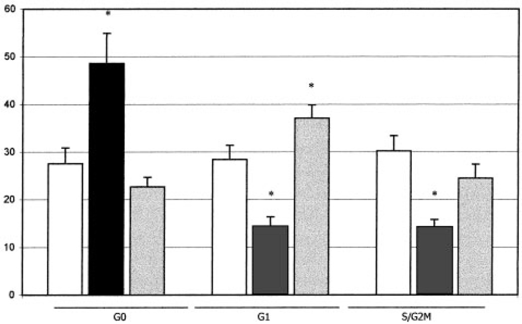
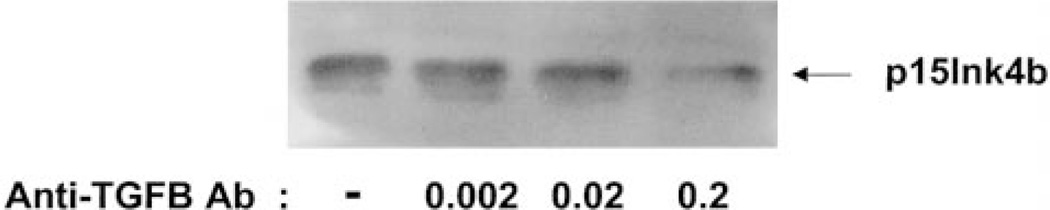
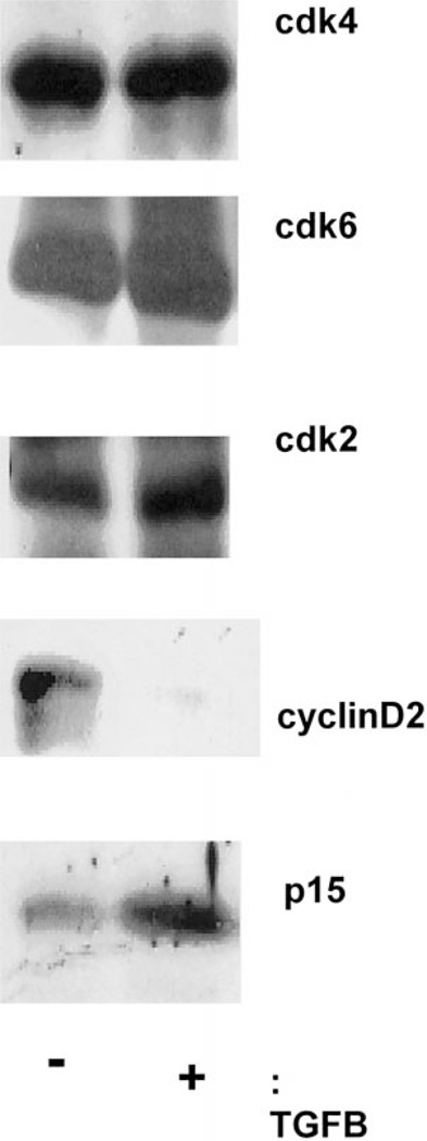
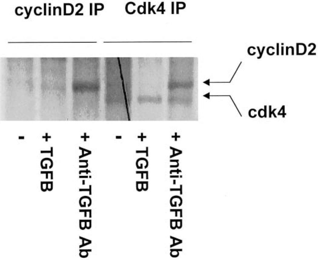
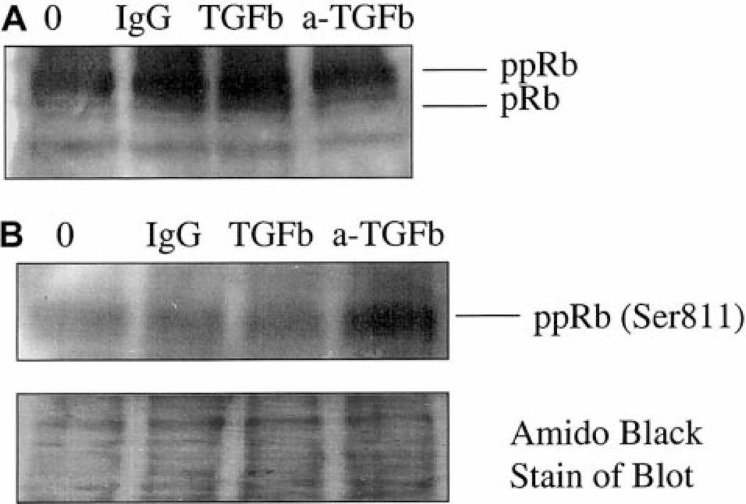
The mechanisms by which transforming growth factor beta (TGF-beta) exerts a negative effect on cell-cycle entry in primary human hematopoietic stem/progenitor cells were examined at the molecular and cellular levels. After treatment of primary human CD34+ progenitors with TGF-beta there was a decrease in the levels of cyclin D2 protein and an increase in levels of the cyclin-dependent kinase inhibitor (CDKI) p15 as compared to the levels in untreated cells. The converse was true after addition of neutralizing anti-TGF-beta antibody. Administration of TGF-beta to CD34+ cells in the presence of cytokines prevented retinoblastoma protein (pRb) phosphorylation, which occurred in the same cells treated with cytokines alone or cytokines and anti-TGF-beta antibody. Neutralization of TGF-beta during 24 to 48 hours of culture with cytokines significantly increased the number of colony-forming progenitors, but did not modulate the human stem cell pool, as measured in 6- to 12-month xenotransplantation assays. Equivalent numbers of human B, T, and myeloid cells were obtained after transplantation of cells treated with or without neutralization of TGF-beta.
Figures
Similar articles
-
Reduction in levels of the cyclin-dependent kinase inhibitor p27(kip-1) coupled with transforming growth factor beta neutralization induces cell-cycle entry and increases retroviral transduction of primitive human hematopoietic cells.Proc Natl Acad Sci U S A. 1998 Oct 27;95(22):13006-11. doi: 10.1073/pnas.95.22.13006. Proc Natl Acad Sci U S A. 1998. PMID: 9789031 Free PMC article.
-
Cdk4 integrates growth stimulatory and inhibitory signals during G1 phase of hematopoietic cells.Oncogene. 1995 Feb 16;10(4):751-5. Oncogene. 1995. PMID: 7862452
-
Latent membrane protein-1 induces cyclin D2 expression, pRb hyperphosphorylation, and loss of TGF-beta 1-mediated growth inhibition in EBV-positive B cells.J Immunol. 1995 Aug 1;155(3):1047-56. J Immunol. 1995. PMID: 7636179
-
Cell cycle and transcriptional control of human myeloid leukemic cells by transforming growth factor beta.Leuk Lymphoma. 2000 Jul;38(3-4):235-46. doi: 10.3109/10428190009087015. Leuk Lymphoma. 2000. PMID: 10830731 Review.
-
Cyclin-dependent kinase regulation during G1 phase and cell cycle regulation by TGF-beta.Adv Cancer Res. 1997;71:165-207. doi: 10.1016/s0065-230x(08)60099-8. Adv Cancer Res. 1997. PMID: 9111866 Review.
Cited by
-
CD93 Marks a Non-Quiescent Human Leukemia Stem Cell Population and Is Required for Development of MLL-Rearranged Acute Myeloid Leukemia.Cell Stem Cell. 2015 Oct 1;17(4):412-21. doi: 10.1016/j.stem.2015.08.008. Epub 2015 Sep 18. Cell Stem Cell. 2015. PMID: 26387756 Free PMC article.
-
Mesenchymal stem cells for trinucleotide repeat disorders.Methods Mol Biol. 2013;1010:79-91. doi: 10.1007/978-1-62703-411-1_6. Methods Mol Biol. 2013. PMID: 23754220 Free PMC article.
-
Cell cycle entry of hematopoietic stem and progenitor cells controlled by distinct cyclin-dependent kinase inhibitors.Int J Hematol. 2002 Jun;75(5):460-5. doi: 10.1007/BF02982107. Int J Hematol. 2002. PMID: 12095144 Review.
-
Hematopoietic stem cell: self-renewal versus differentiation.Wiley Interdiscip Rev Syst Biol Med. 2010 Nov-Dec;2(6):640-53. doi: 10.1002/wsbm.86. Wiley Interdiscip Rev Syst Biol Med. 2010. PMID: 20890962 Free PMC article. Review.
-
Deconstructing the Complexity of TGFβ Signaling in Hematopoietic Stem Cells: Quiescence and Beyond.Curr Stem Cell Rep. 2016 Dec;2(4):388-397. doi: 10.1007/s40778-016-0069-x. Epub 2016 Oct 29. Curr Stem Cell Rep. 2016. PMID: 28529843 Free PMC article.
References
-
- Massague J. TGF-beta signal transduction. Annu Rev Biochem. 1998;67:753–791. - PubMed
-
- Ewen ME, Sluss HK, Whitehouse LL, Livingston DM. TGF beta inhibition of Cdk4 synthesis is linked to cell cycle arrest. Cell. 1993;74:1009–1020. - PubMed
-
- Hannon GJ, Beach D. p15INK4B is a potential effector of TGF-beta-induced cell cycle arrest. Nature. 1994;371:257–261. [see comments]. - PubMed
-
- Satterwhite DJ, Aakre ME, Gorska AE, Moses HL. Inhibition of cell growth by TGF beta 1 is associated with inhibition of B-myb and cyclin A in both BALB/MK and Mv1Lu cells. Cell Growth Differ. 1994;5:789–799. - PubMed
-
- Liu JH, Wei S, Burnette PK, Gamero AM, Hutton M, Djeu JY. Functional association of TGF-beta receptor II with cyclin B. Oncogene. 1999;18:269–275. - PubMed
Publication types
MeSH terms
Substances
Grants and funding
LinkOut - more resources
Full Text Sources
Other Literature Sources
Medical
