. 2002 Jan;109(1):19-27.
doi: 10.1172/JCI12035.

Pneumococcal pneumolysin and H(2)O(2) mediate brain cell apoptosis during meningitis

Affiliations

Pneumococcal pneumolysin and H(2)O(2) mediate brain cell apoptosis during meningitis

Johann S Braun et al. J Clin Invest. 2002 Jan.

Abstract

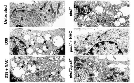
Pneumococcus is the most common and aggressive cause of bacterial meningitis and induces a novel apoptosis-inducing factor-dependent (AIF-dependent) form of brain cell apoptosis. Loss of production of two pneumococcal toxins, pneumolysin and H(2)O(2), eliminated mitochondrial damage and apoptosis. Purified pneumolysin or H(2)O(2) induced microglial and neuronal apoptosis in vitro. Both toxins induced increases of intracellular Ca(2+) and triggered the release of AIF from mitochondria. Chelating Ca(2+) effectively blocked AIF release and cell death. In experimental pneumococcal meningitis, pneumolysin colocalized with apoptotic neurons of the hippocampus, and infection with pneumococci unable to produce pneumolysin and H(2)O(2) significantly reduced damage. Two bacterial toxins, pneumolysin and, to a lesser extent, H(2)O(2), induce apoptosis by translocation of AIF, suggesting new neuroprotective strategies for pneumococcal meningitis.


Figures

Figure 1
Figure 1
Inactivation of pneumolysin and blocking H2O2 inhibits apoptosis induced by live pneumococci. (a and b) Human microglia incubated with wild-type pneumococci D39 (107 CFU/ml, 6 hours) in the absence or presence of NAC (10 mM) underwent massive shrinkage and condensation of their nuclei by AO/EB staining, whereas NAC effectively impaired changes in nuclear morphology and cell membrane damage in cells exposed to plnA (107 CFU/ml, 6 hours). Bars = 10 μm. (b) Quantification of apoptotic microglia after exposure to wild-type (D39) or mutant pneumococci defective in pneumolysin (plnA) or H2O2 production (spxB) or both (plnA/spxB). Shown are the results (means + SD) of three independent experiments after 6 hours of incubation. Addition of the antioxidant NAC prevented apoptosis induced by plnA, but not that induced by D39 or spxB (b). plnA in the presence of NAC or catalase (Cat) (1,250 U/ml) and plnA/spxBwere significantly different compared with all others (P < 0.05). plnA combined with NAC or catalase was more efficient to prevent apoptosis than was the double mutant plnA/spxB (P < 0.05). ANOVA and Student-Newman-Keuls test were used. WT, wild-type.
Figure 2
Figure 2
Inactivation of pneumolysin and blocking H2O2 inhibits mitochondrial damage induced by live pneumococci. Changes of ultrastructure of microglial cells exposed to pneumococci (107 CFU/ml) for 3 hours were monitored by transmission electron microscopy. D39 and plnA caused massive swelling of the mitochondria (arrows) and endoplasmic reticulum (ER). Treatment of D39-exposed cells with NAC (10 mM) failed to prevent that damage. By contrast, when cells exposed to plnA were treated with NAC (10 mM), swelling of mitochondria and the endoplasmic reticulum was markedly attenuated. The double mutant plnA/spxB also showed attenuated mitochondrial damage. ×7,500. N, nucleus.
Figure 3
Figure 3
Pneumolysin and H2O2 are each sufficient to induce apoptosis in microglia and neurons. (a) Microglia cells were incubated for 8 hours with purified pneumolysin (0.1 μg/ml) or H2O2 (0.2 mM) and stained with AO and EB. Either treatment resulted in shrinkage and condensation typical of apoptosis induced by wild-type pneumococci. The apoptosis-inducing activity of pneumolysin correlates with its cytolytic (Lys) but not its complement-activating (Compl) function. Microglial cells were incubated with wild-type or mutant pneumolysin (0.1 μg/ml) for 12 hours and stained with AO and EB. A point mutant in the domain required for cytolytic activity of pneumolysin (W433F) failed to induce apoptosis, whereas a point mutant in the domain required for complement activation (D385N) retained full apoptosis-inducing capacity. Bars = 10 μm. (b) Quantification of the effects of wild-type or mutant pneumolysin on primary rat hippocampal neurons. Results shown (means + SD) are representative for three independent experiments performed in triplicate. (c) Neurotoxicity (primary rat hippocampal neurons) of various concentrations of H2O2.
Figure 4
Figure 4
Pneumococcus triggers early increases of intracellular ROS and Ca2+. Microglial cells were untreated (Control) or incubated with pneumococcus D39 (107 CFU/ml) for 45 minutes to 3 hours. ROS (a) and Ca2+ (b) levels were visualized by fluorescence of the dyes DHR123 (10 μM) and Fluo-4 (10 μM), respectively, and quantified with a multiwell fluorescence plate reader. Results shown (mean + SD) are representative for three independent experiments performed in triplicate. (b and c) The effects of the Ca2+ chelator BAPTA-AM (5 μM) were investigated with the AO/EB assay after exposure of cells to D39 in the presence and absence of BAPTA-AM. Bars = 10 μm.
Figure 5
Figure 5
Pneumolysin and H2O2 induce increase of intracellular Ca2+ and release of mitochondrial AIF. Primary rat neurons were exposed to either pneumolysin (0.1 μg/ml) or H2O2 (0.2 mM). Intracellular Ca2+ was visualized by Fluo-4 (10 μM) after 1.5 hours. Release of mitochondrial AIF, its translocation to the nucleus, and the effects of BAPTA-AM (5 μM) were assessed immuncytochemically after 6 hours’ incubation. Insets show colocalization of HOECHST 33258 (blue fluorescence) and AIF (red fluorescence). Bar = 10 μm.
Figure 6
Figure 6
Pneumolysin and H2O2 contribute to neuronal damage in the rabbit model of pneumococcal meningitis. (a) Neurotoxicity in the dentate gyrus was quantified by neuronal loss or cell shrinkage assessed in H&E-stained sections. Bars represent means + SD of five brain sections per rabbit. Compared with control animals challenged with PBS, wild-type pneumococci D39 induced significant neurotoxicity (*P < 0.05). Damage in D39-challenged animals was also statistically significant compared with all other tested groups except D39 + NAC (P < 0.05; ANOVA, Student-Newman-Keuls test). #Significant difference between plnA/spxB and plnA, P = 0.04. ##Significant difference between plnA/spxB with and without catalase, P = 0.004; t test. (b) Immunohistochemical staining with anti-pneumolysin antibody followed by peroxidase-conjugated secondary antibody and diaminobenzidine (dark color) detected pneumolysin in dentate gyrus neurons of D39- but not plnA- or PBS-treated animals. Bar = 10 μm.
Figure 7
Figure 7
Schematic model of pneumococcal-induced apoptosis of neurons. Neuronal apoptosis is triggered in part by the host inflammation and is mediated in part by caspase activation. Pneumolysin and H2O2 are direct triggers of S. pneumoniae, inducing apoptosis by increasing intracellular Ca2+, damaging mitochondria, and causing the release and translocation of mitochondrial AIF.

References

    1. Zychlinsky A, Sansonetti P. Apoptosis in bacterial pathogenesis. J Clin Invest. 1997; 100:493–496. - PMC - PubMed
    1. Bohr V, Paulson OB, Rasmussen N. Pneumococcal meningitis. Late neurologic sequelae and features of prognostic impact. Arch Neurol. 1984; 41:1045–1049. - PubMed
    1. Durand ML, et al. Acute bacterial meningitis in adults. A review of 493 episodes. N Engl J Med. 1993; 328:21–28. - PubMed
    1. Pfister HW, Feiden W, Einhäupl KM. Spectrum of complications during bacterial meningitis in adults. Results of a prospective clinical study. Arch Neurol. 1993; 50:575–581. - PubMed
    1. Zysk G, et al. Anti-inflammatory treatment influences neuronal apoptotic cell death in the dentate gyrus in experimental pneumococcal meningitis. J Neuropathol Exp Neurol. 1996; 55:722–728. - PubMed

Publication types

MeSH terms