. 2002 Jan;109(1):29-39.
doi: 10.1172/JCI13696.

Pulmonary overexpression of IL-9 induces Th2 cytokine expression, leading to immune pathology

Affiliations

Pulmonary overexpression of IL-9 induces Th2 cytokine expression, leading to immune pathology

Ulla-Angela Temann et al. J Clin Invest. 2002 Jan.

Abstract

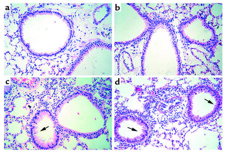
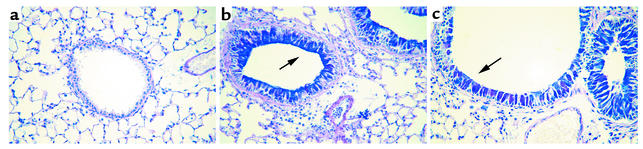
IL-9 is a pleiotropic cytokine with multiple functions on many cell types involved in the pathology of human asthma. The constitutive overexpression of IL-9 in the lungs of transgenic mice resulted in an asthma-like phenotype. To define the contribution of IL-9 to lung inflammation we generated transgenic mice in which lung-specific expression of the IL-9 transgene is inducible by doxycycline. Transgene induction resulted in lymphocytic and eosinophilic infiltration of the lung, airway epithelial cell hypertrophy with mucus production, and mast cell hyperplasia, similar to that seen in mice that constitutively expressed IL-9 in their lungs. Various cytokines, including IL-4, IL-5, and IL-13, were expressed in the lung in response to IL-9. Blockade of IL-4 or IL-5 following IL-9 induction reduced airway eosinophilia without affecting mucus production. In contrast, neutralization of IL-13 completely abolished both lung inflammation and mucus production. These findings suggest that pathologic changes in the lung require additional signals beyond IL-9, provided by IL-4, IL-5, and IL-13, to develop fully.


Figures

Figure 1
Figure 1
Genetic constructs used for the generation of transgenic mice with regulated overexpression of IL-9 by doxycycline. The inducible, lung-specific expression system consists of two separate constructs. (a) The first construct contains the rtTA cDNA under the control of the lung epithelial cell–specific, rat CC10 promoter and the hGH intronic and polyadenylation sequences. (b) In the second construct the IL-9 cDNA is linked to the tet-O-CMV promoter and the SV 40 small t intron and polyadenylation sequence.
Figure 2
Figure 2
Total cell counts from lung lavage fluid. IL-9 transgene-negative (TG) and -positive (TG+) mice, 6 weeks of age, were housed with or without doxycycline-supplemented food over a period of 14 days. At three different time points mice were sacrificed, subjected to lung lavage, and total cell numbers assessed. Data are expressed as mean ± SD of counts from four to five animals per group. Significant differences in cell numbers compared with that of transgene-negative and -positive mice that did not receive doxycycline are indicated by an asterisk.
Figure 3
Figure 3
Characterization of inflammatory cells in lung lavage fluid. Differential cell counts were performed on lung lavage cells retrieved from transgene-negative (TG) and -positive (TG+) mice that had been housed with or without doxycycline-supplemented food over a period of 14 days. At day 1, 7, and 14, mice were sacrificed and lungs lavaged. Differential cell counts were derived from at least 200 cell counts and are expressed as total cell numbers. Data represent the mean ± SD of counts from four to five animals per group. Significant differences in cell numbers compared with that of transgene-negative or –positive mice at day 1 that did not receive doxycycline are indicated with an asterisk. Notice the differences in the scale of the y axis between the graphs.
Figure 4
Figure 4
Lung histology of conducting airways and parenchyma. Lung sections from IL-9 transgene-negative (a) and -positive (bd) mice were stained with H&E for examination using light microscopy. Induction of the IL-9 transgene by doxycycline over a period of 7 days (c) or 14 days (d) resulted in infiltration of lung tissue with inflammatory cells. Furthermore, airway epithelial cells were hypertrophic (arrows). Inflammatory cells were not observed in lung sections from six-week-old transgene-negative (a) or -positive (b) mice without doxycycline treatment. Original magnification ×300.
Figure 5
Figure 5
Histologic staining for mucin and mast cells. Lung sections from IL-9 transgene-negative (a) or -positive (bd) mice were stained with AB/PAS for light microscopic identification of mucin-containing cells. Intense, positive (purple) staining for mucin was observed in hypertrophied airway epithelial cells from IL-9 transgene-positive mice after 7 days (c) (arrow) and 14 days (d) (arrow) of transgene induction by doxycycline. In lung sections from transgene-positive mice that did not receive doxycycline some mucin-positive epithelial cells were also detected (b) (arrow). Airway epithelium from transgene-negative mice did not stain for mucin (a). For the identification of mast cells, lung sections from transgene-negative (e) or -positive (f) mice were stained with toluidine blue. Dark blue–stained mast cell granules (arrow) were present in the airway epithelium from transgene-positive mice that were housed with doxycycline-supplemented food over a period of 14 days (f), but not in transgene-negative mice (e). Original magnification: ad, ×300; ef, ×600.
Figure 6
Figure 6
Analysis of cytokine expression in total lung tissue. (a) Levels of various mRNAs encoding predominantly Th2 cytokines from individual IL-9 transgene-negative (lane 1, 2) and -positive (lane 3, 4) mice after transgene induction by doxycycline were compared by RPA. The P32-labeled multiprobe template set (T) was used as a size marker. Levels of L32 and GAPDH mRNAs encoding housekeeping genes were used to ensure equal loading of samples. (b) Levels of mRNA encoding IL-4 were evaluated by RT-PCR using total lung RNA isolated from transgene-negative (lane 1, 2) and -positive (lane 3, 4) mice 14 days after IL-9 transgene induction by doxycycline. Levels of mRNA encoding HPRT, a housekeeping gene, were used to demonstrate equal loading of samples. Fragment sizes of a molecular-weight marker are indicated in base pairs.
Figure 7
Figure 7
Characterization of inflammatory cells in lung lavage fluid after cytokine blockade. IL-9 transgene-negative (TG) or -positive (TG+) mice were housed with (+Dox) or without (–Dox) doxycycline-supplemented food for 14 days. During the entire time of IL-9 transgene induction, selected groups of animals received anti–IL-4 Ab’s, sIL-13Rα-Fc, or isotype control Ab’s (hIgG) as described in Methods. At day 14, mice were sacrificed and the lung lavaged. (a) Total lung lavage cell counts. (b) Differential cell counts on lung lavage cells. Results are expressed as total cell numbers and were obtained from at least 200 cell counts. Data are expressed as mean ± SD. Significant differences in total cell number (a) or number of eosinophils (b) compared with that of transgene-positive mice that received doxycycline but no Ab’s are indicated with an asterisk.
Figure 8
Figure 8
Histologic staining for mucin after cytokine blockade. Light micrographs of lung sections stained with AB/PAS from IL-9 transgene-negative (a) or –positive (bf) mice that were housed for 14 days with doxycycline-supplemented food. During the entire time of IL-9 transgene induction selected groups of animals received Ab’s: (c) control Ab’s, hIgG; (d) anti–IL-4; (e) sIL-13Rα-Fc; (f) anti–IL-4 and sIL-13Rα-Fc. Lung sections from transgene-positive mice that received anti–IL-4 Ab’s, control Ab’s, or no Ab’s showed strong positive (magenta) staining for mucin in airway epithelial cells (arrows) and accumulation of inflammatory cells near blood vessels and airways (bd). Infiltration of lung tissue with inflammatory cells or positive staining for mucin was not observed in sections from transgene-positive mice that received sIL-13Rα-Fc alone (e), or in combination with anti–IL-4 (f), or in transgene-negative mice (a). Original magnification ×300.
Figure 9
Figure 9
Characterization of inflammatory cells in lung lavage fluid after IL-5 blockade. IL-9 transgene-negative (TG) or -positive (TG+) mice were kept with (+Dox) or without (–Dox) doxycycline-supplemented food. After 14 days, mice were sacrificed, lungs lavaged, and cell numbers in lavage fluid evaluated. During the entire time of transgene induction, selected groups of animals received intraperitoneal anti–IL-5 Ab’s or isotype control Ab’s (GL113). (a) Total cell numbers retrieved by lung lavage. (b) Differential cell counts. Results are expressed as total cell numbers and were derived from at least 200 cell counts. Data are presented as mean ± SD. Significant differences compared with counts from transgene-positive mice that did not receive Ab’s during IL-9 transgene expression are indicated with an asterisk.
Figure 10
Figure 10
Histologic staining for mucin after IL-5 blockade. Lung sections from IL-9 transgene-negative (a) and -positive (b and c) mice that were kept 14 days on doxycycline-supplemented food, were stained with AB/PAS. Intense, positive (purple) staining for mucin was observed in airway epithelial cells in transgene-positive mice (arrow) (b) and in mice that additionally received anti–IL-5 Ab’s during IL-9 transgene expression (arrow) (c). Lung sections from IL-9 transgene-negative mice did not stain for mucin (a). Original magnification ×300.

References

    1. Holgate ST. The inflammatory basis of asthma and its implications for drug treatment. Clin Exp Allergy. 1996; 4(Suppl.):1–4. - PubMed
    1. Lukacs NW, Strieter RM, Kunkel SL. Leukocyte infiltration in allergic airway inflammation. Am J Respir Cell Mol Biol. 1995; 13:1–6. - PubMed
    1. Robinson DS, et al. Predominant TH2-like bronchoalveolar T-lymphocyte population in atopic asthma. N Engl J Med. 1992; 326:298–304. - PubMed
    1. Riffo-Vasquez Y, Pitchford S, Spina D. Cytokines in airway inflammation. Int J Biochem Cell Biol. 2000; 32:833–853. - PubMed
    1. Gessner A, Blum H, Rollinghoff M. Differential regulation of IL-9-expression after infection with Leishmania major in susceptible and resistant mice. Immunobiology. 1993; 189:419–435. - PubMed

Publication types

MeSH terms