. 2002 Jan;109(1):121-30.
doi: 10.1172/JCI14080.

The cardiac phenotype induced by PPARalpha overexpression mimics that caused by diabetes mellitus

Affiliations

The cardiac phenotype induced by PPARalpha overexpression mimics that caused by diabetes mellitus

Brian N Finck et al. J Clin Invest. 2002 Jan.

Abstract

Recent evidence has defined an important role for PPARalpha in the transcriptional control of cardiac energy metabolism. To investigate the role of PPARalpha in the genesis of the metabolic and functional derangements of diabetic cardiomyopathy, mice with cardiac-restricted overexpression of PPARalpha (MHC-PPAR) were produced and characterized. The expression of PPARalpha target genes involved in cardiac fatty acid uptake and oxidation pathways was increased in MHC-PPAR mice. Surprisingly, the expression of genes involved in glucose transport and utilization was reciprocally repressed in MHC-PPAR hearts. Consistent with the gene expression profile, myocardial fatty acid oxidation rates were increased and glucose uptake and oxidation decreased in MHC-PPAR mice, a metabolic phenotype strikingly similar to that of the diabetic heart. MHC-PPAR hearts exhibited signatures of diabetic cardiomyopathy including ventricular hypertrophy, activation of gene markers of pathologic hypertrophic growth, and transgene expression-dependent alteration in systolic ventricular dysfunction. These results demonstrate that (a) PPARalpha is a critical regulator of myocardial fatty acid uptake and utilization, (b) activation of cardiac PPARalpha regulatory pathways results in a reciprocal repression of glucose uptake and utilization pathways, and (c) derangements in myocardial energy metabolism typical of the diabetic heart can become maladaptive, leading to cardiomyopathy.


Figures

Figure 1
Figure 1
Increased expression of PPARα target genes in diabetic mouse myocardium. (a) Bars represent mean (± SEM) mRNA levels as determined by phosphorimager analysis of Northern blots performed with RNA isolated from mouse ventricle, shown as arbitrary units (AU) corrected for GAPDH signal intensity and normalized to the value of vehicle-injected controls (= 1.0). *P < 0.05 versus vehicle-injected mice (n = 4 for each group). A representative autoradiograph of the Northern blot studies is shown at the right. Each lane contained 15 μg total RNA isolated from mice 6 weeks after an injection of saline (vehicle) or a single dose of STZ (180 mg/kg). The blot was sequentially hybridized with the radiolabeled cDNA probes shown on the left. (b) Increased expression of PPARα target genes in db/db diabetic mice. Bars represent mRNA levels shown as arbitrary units (AU) corrected for GAPDH signal intensity and normalized to the value of db/+ littermate controls (= 1.0). *P < 0.05 versus db/+ littermate controls; n = 5 for each group. A representative autoradiograph is shown at the right. Each lane contained 15 μg of ventricular total RNA isolated from 10-week-old heterozygote control (db/+) or littermate diabetic (db/db) mice.
Figure 2
Figure 2
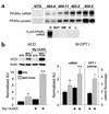
Increased expression of PPARα target genes in hearts of transgenic mouse lines (MHC-PPAR mice) that express PPARα in a cardiac-restricted manner. (a) Representative autoradiographs of Northern (top) and Western (middle) blot studies performed with heart samples from 6-week-old mice from four independent MHC-PPAR lines. At the exposure shown, endogenous PPARα could not be detected in NTG samples. FLAG-PPARα protein was detected in the transgenic mice using an antibody directed against either PPARα (shown here) or FLAG (data not shown). Cardiac-specific expression of FLAG-PPARα mRNA was confirmed by Northern blot analysis performed with multiple tissues (bottom panel represents the 402-2 line). H, heart; BAT, brown adipose tissue; SM, skeletal muscle; K, kidney; L, liver. (b) The expression of PPARα target genes is increased and inducible by PPARα activator in the hearts of MHC-PPAR mice. The graphs represent the results of Northern blot analyses performed with the probes indicated using RNA isolated from ventricle of male and female NTG or MHC-PPAR littermate mice fed control chow or a 7-day course of chow containing Wy-14,643 (0.1% wt/wt). Bars represent mean mRNA levels (arbitrary units, AU) quantified as described in Methods (n ≥ 7 mice for each group). The inset shows representative autoradiographs of Western blot analyses. *P < 0.05 versus NTG littermate mice. **P < 0.05 versus MHC-PPAR mice fed control chow and NTG mice. Far right: Bars represent mean malonyl-CoA−inhibited CPT enzyme activity (n = 3 for each group). P < 0.05 versus NTG littermate mice.
Figure 3
Figure 3
Myocardial lipid accumulation in fasted MHC-PPAR mice. (a) Photomicrographs depicting the histologic appearance of ventricular tissue from NTG and MHC-PPAR mice following a 24-hour fast at low (upper panels) and high (lower panels) magnification. Frozen tissue sections were stained with oil red O. Red droplets indicate neutral lipid staining. (b) Lipid profile of mouse ventricle samples prepared from MHC-PPAR or NTG mice given ad libitum access to food or fasted for 24 hours. Lipid species were separated and analyzed using ESIMS (see Methods). Mass/charge (m/z) ratios of 814, 840, and 864 denote TAGs containing fatty acyl groups containing chain lengths of 16:0/16:0/16:0, 16:0/16:0/18:1, and 16:0/18:1/18:2, as shown at the top. (c) Representative autoradiographs of Northern blot analyses performed with total RNA isolated from cardiac ventricle of mice fed ad libitum or after a 24-hour fast using cDNA probes for diacylglyceride acyltransferase (DGAT), glycerol-3-phosphate acyltransferase (GPAT), and adipophilin. Ethidium bromide−stained 28S rRNA is shown as a control for loading.
Figure 4
Figure 4
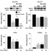
Altered expression of genes involved in glucose utilization pathways in MHC-PPAR heart. The graphs represent the results of Northern blot analyses performed with total RNA isolated from the cardiac ventricles of MHC-PPAR or littermate NTG mouse hearts. The blots were sequentially hybridized with radiolabeled cDNA probes for GLUT4, GLUT1, PFK, and pyruvate dehydrogenase kinase 4 (PDK4). Bars represent mean mRNA levels as determined by phosphorimager analysis (n ≥ 7 for each group) corrected to the GAPDH signal and normalized to that of the NTG mice treated with control chow (= 100 or 1.0). Groups receiving a 7-day course of Wy-14,643 are denoted at the bottom of each graph. Top: Representative autoradiographs of Western blot analyses using anti-GLUT4 or anti-GLUT1 antibodies with protein extracts containing total cell membrane−enriched fractions of cardiac ventricle from MHC-PPAR mice and NTG littermates. *P < 0.05 versus NTG littermate mice fed control chow. **P < 0.05 versus MHC-PPAR mice fed control chow and NTG mice. Bars represent the mean of at least seven individual animals.
Figure 5
Figure 5
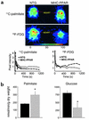
Myocardial palmitate utilization is increased and FDG reduced in MHC-PPAR mice. (a) The top two panels contain representative images of 11C-palmitate and 18F-FDG uptake into myocardium as assessed by microPET in NTG and MHC-PPAR mice following a 6-hour fast. The relative amount of tracer uptake into the mouse heart 15 seconds after bolus injection of 11C-palmitate or 18F-FDG into the jugular vein is indicated by the color scale (0−100). The arrows indicate the cardiac field. As shown in the upper panel, the color field is increased into the red scale in the hearts of MHC-PPAR mice infused with 11C-palmitate, which is indicative of enhanced myocardial uptake of fatty acid. Conversely, uptake of 18F-FDG is substantially lower in hearts of MHC-PPAR mice than in those of NTG littermates. The graphs below the images contain time-activity curves for the cardiac fields. (b) Myocardial palmitate oxidation is increased and glucose oxidation reduced in MHC-PPAR mice. The oxidation of [9,10-3H]palmitate and [U-14C]glucose was assessed in isolated working hearts (as described in Methods) of MHC-PPAR (line 402-2; n = 6) or NTG littermate (n = 4) mice fed control chow. Bars represent mean (± SEM) oxidation rates expressed as nanomoles of substrate oxidized per minute per gram dry mass. *P < 0.05 versus NTG littermate mice.
Figure 6
Figure 6
Induction of the cardiac hypertrophic growth gene regulatory program and left ventricular dysfunction in MHC-PPAR mice. (a) Bars represent mean biventricular-to-body-weight (BV/BW) ratios (n ≥ 20 for each group) for male and female MHC-PPAR and NTG littermate mice. All mice were between 2 and 4 months of age. *P < 0.05 versus NTG littermate mice. (b) Representative autoradiographs of Northern blot analyses performed with total RNA isolated from cardiac ventricle from 6-week-old NTG or MHC-PPAR (404-3 line) mice using cDNA probes for skeletal (Sk.) α-actin, brain-type natriuretic peptide (BNP), atrial natriuretic factor (ANF), phospholamban (PLB), and sarcoplasmic/endoplasmic reticulum Ca2+ ATPase 2a (SERCA2a). (c) Ventricular dysfunction in MHC-PPAR mice is related to transgene expression level. Representative two-dimensional guided M-mode echocardiographic images of the left ventricle obtained from the parasternal view at the midventricular level of NTG and four different transgenic lines of female mice at 2 months of age.

References

    1. Kannel WB, Hjortland M, Castelli WP. Role of diabetes in congestive heart failure: the Framingham study. Am J Cardiol. 1974; 34:29–34. - PubMed
    1. Rubler S, et al. New type of cardiomyopathy associated with glomerulosclerosis. Am J Cardiol. 1972; 30:595–602. - PubMed
    1. Stanley WC, Lopaschuk GD, McCormack JG. Regulation of energy substrate metabolism in the diabetic heart. Cardiovasc Res. 1997; 34:25–33. - PubMed
    1. Rodrigues B, Cam MC, McNeill JH. Myocardial substrate metabolism: implications for diabetic cardiomyopathy. J Mol Cell Cardiol. 1995; 27:169–179. - PubMed
    1. Neely JR, Rovetto MJ, Oram JF. Myocardial utilization of carbohydrate and lipids. Prog Cardiovasc Dis. 1972; 15:289–329. - PubMed

Publication types

MeSH terms