. 2002 Jan 7;195(1):125-33.
doi: 10.1084/jem.20011097.

Antitumor monoclonal antibodies enhance cross-presentation ofcCellular antigens and the generation of myeloma-specific killer T cells by dendritic cells

Affiliations

Antitumor monoclonal antibodies enhance cross-presentation ofcCellular antigens and the generation of myeloma-specific killer T cells by dendritic cells

Kavita M Dhodapkar et al. J Exp Med. .

Abstract

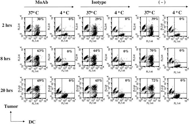
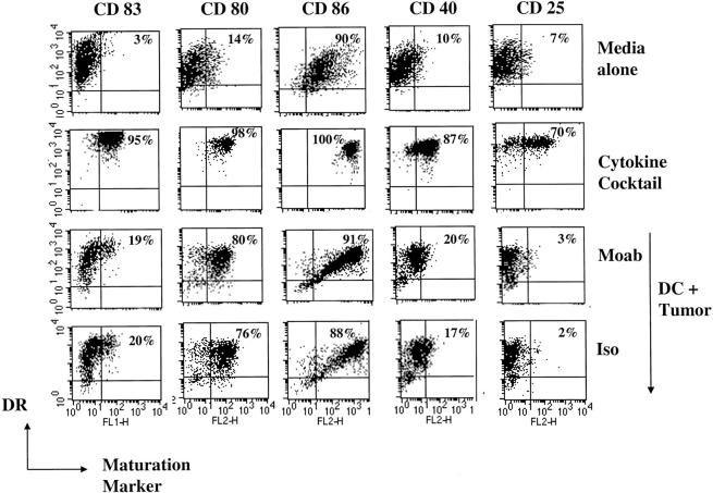
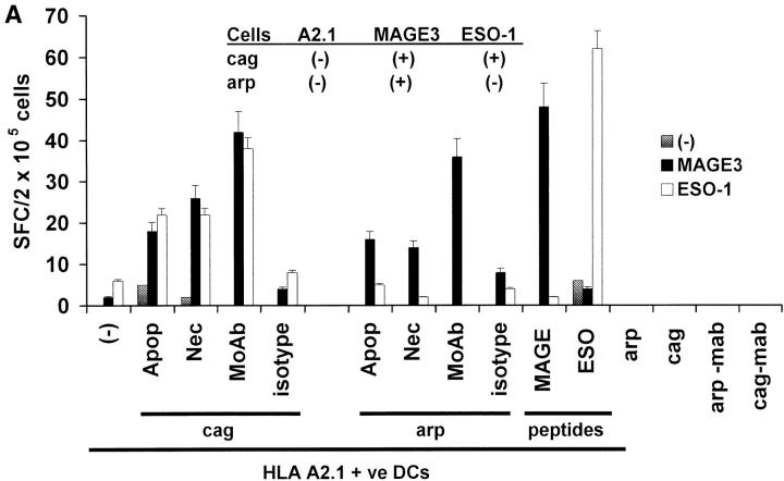
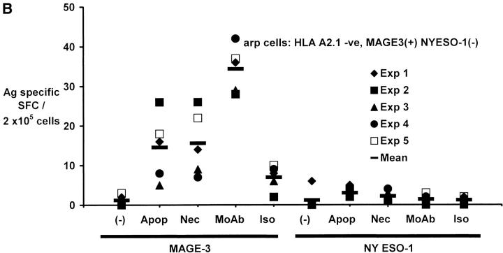
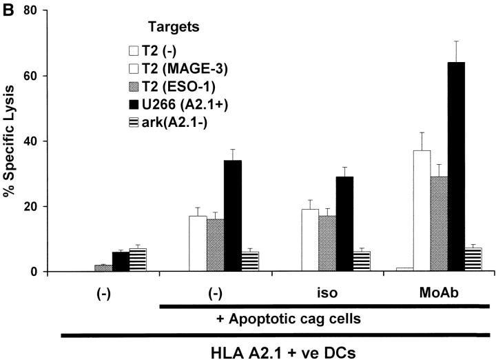
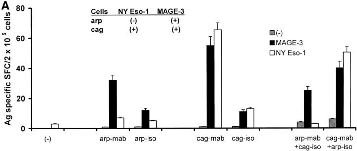
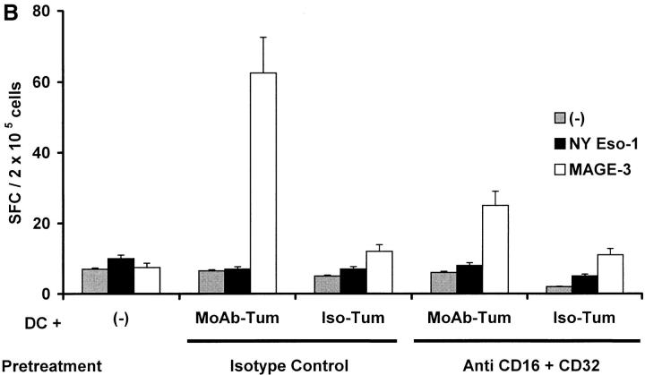
The mechanism of antitumor effect of monoclonal antibodies (mAbs) is not fully understood. Here we show that coating myeloma cells with anti-syndecan-1 antibody promotes cross-presentation of cellular antigens by dendritic cells (DCs) to autologous T cells from healthy donors. The tumor cells treated with anti-syndecan-1 or isotype-matched control antibody were fed to HLA-mismatched monocyte-derived immature DCs. Tumor cell-loaded mature DCs induced a strong CD8(+) T cell response that was specific for the cancer-testis (C-T) antigens expressed in the tumor. The CD8(+) T cells killed peptide-pulsed targets, as well as myeloma tumor cells. Importantly, mAbs-coated tumor-loaded DCs were consistently superior to DCs loaded with peptides or dying cells for eliciting tumor-specific killer T cells. This enhanced cross-presentation was not due to enhanced tumor cell uptake or to DC maturation. When mixtures of NY-Eso-1-positive and -negative myeloma cells were captured by DCs, the anti-syndecan-1 antibody had to be on the NY-Eso-1-positive cells to elicit NY-Eso-1-specific response. Cross-presentation was inhibited by pretreatment of DCs with Fc gamma receptor blocking antibodies. Targeting of mAb-coated tumors to DCs may contribute to the efficacy of tumor-reactive mAb and offers a new strategy for immunotherapy.


Figures

Figure 1.
Figure 1.
Uptake of tumor cells by DCs. Myeloma cells were labeled with dye (PKH26) and either stained with anti–syndecan-1 antibody (mAb), isotype control antibody, or left unstained. Tumor cells were irradiated (3 Gy) just before coculture with dye (PKH67) labeled DCs at 4°C or 37°C, and the percent of double-positive DCs (top right of each panel) analyzed by flow cytometry.
Figure 2.
Figure 2.
Phagocytosis of mAb-coated tumor does not trigger extensive DC maturation. Monocyte-derived DCs at day 6 of culture were cultured in media alone, with inflammatory cytokine cocktail, or with tumor cells pretreated with anti–syndecan-1 or isotype control antibody. After 1 d of culture, the cells were stained with anti-HLA DR (FITC/PE) and one of the following antibodies (anti-CD80, CD86, CD83, CD40, and CD25). Percentage of HLA-DR +ve DCs expressing CD83, CD80, CD86, CD40, or CD25 (top right of each panel) were quantified by flow cytometry.
Figure 3.
Figure 3.
Generation of cancer testis antigen-specific IFN-γ–producing T cells by DCs. (A) Generation of MAGE-3 or NY-Eso-1 specific T cells by peptide-pulsed or tumor cell–loaded DCs. HLA A2.1 −ve myeloma cells (cag, arp) were killed either by γ-irradiation (30 Gy) (apoptosis), freeze thaw cycles (necrosis), or coated with anti–syndecan-1 antibody (mAb)/isotype control, irradiated (3 Gy), and added to HLA A2.1+ ve immature DCs as live (annexin V-negative) cells. DCs were matured using cytokine cocktail. Autologous T cells were stimulated with mature peptide-pulsed or tumor cell–loaded DCs, or tumor cells alone. After two stimulations, the number of HLA A*0201-restricted MAGE3/NY-Eso-1 peptide specific IFN-γ–producing cells were quantified with an ELISPOT assay, using peptide pulsed autologous DCs as APCs. (B) Summary of MAGE3/NY-Eso-1–specific T cells elicited in five experiments, using DCs loaded with dying (apoptotic/necrotic) or antibody-treated (anti–syndecan-1 mAb/isotype) myeloma cells (arp cells, MAGE3+, NY-Eso-1 –ve). (C) Stimulation using CD11c+ DCs from DC-tumor cocultures. CD11c+ DCs were purified using magnetic beads from 4-h cocultures of HLA A2.1 +ve DCs with dying HLA A2.1−ve myeloma (cag) cells (apoptosis/necrosis, as described earlier); or coated with anti–syndecan-1 antibody/isotype. These DCs were then matured using cytokine cocktail and used as APCs to either directly stimulate autologous T cells, or in transwell cultures separated from T cells and unpulsed DCs. After two stimulations, HLA A*0201-restricted MAGE-3 or NY-Eso-1 peptide-specific T cells were quantified by ELISPOT using autologous peptide-pulsed DCs as APCs. Data are representative of two similar experiments.
Figure 3.
Figure 3.
Generation of cancer testis antigen-specific IFN-γ–producing T cells by DCs. (A) Generation of MAGE-3 or NY-Eso-1 specific T cells by peptide-pulsed or tumor cell–loaded DCs. HLA A2.1 −ve myeloma cells (cag, arp) were killed either by γ-irradiation (30 Gy) (apoptosis), freeze thaw cycles (necrosis), or coated with anti–syndecan-1 antibody (mAb)/isotype control, irradiated (3 Gy), and added to HLA A2.1+ ve immature DCs as live (annexin V-negative) cells. DCs were matured using cytokine cocktail. Autologous T cells were stimulated with mature peptide-pulsed or tumor cell–loaded DCs, or tumor cells alone. After two stimulations, the number of HLA A*0201-restricted MAGE3/NY-Eso-1 peptide specific IFN-γ–producing cells were quantified with an ELISPOT assay, using peptide pulsed autologous DCs as APCs. (B) Summary of MAGE3/NY-Eso-1–specific T cells elicited in five experiments, using DCs loaded with dying (apoptotic/necrotic) or antibody-treated (anti–syndecan-1 mAb/isotype) myeloma cells (arp cells, MAGE3+, NY-Eso-1 –ve). (C) Stimulation using CD11c+ DCs from DC-tumor cocultures. CD11c+ DCs were purified using magnetic beads from 4-h cocultures of HLA A2.1 +ve DCs with dying HLA A2.1−ve myeloma (cag) cells (apoptosis/necrosis, as described earlier); or coated with anti–syndecan-1 antibody/isotype. These DCs were then matured using cytokine cocktail and used as APCs to either directly stimulate autologous T cells, or in transwell cultures separated from T cells and unpulsed DCs. After two stimulations, HLA A*0201-restricted MAGE-3 or NY-Eso-1 peptide-specific T cells were quantified by ELISPOT using autologous peptide-pulsed DCs as APCs. Data are representative of two similar experiments.
Figure 4.
Figure 4.
Generation of tumor-specific killer T cells. (A) Generation of killer T cells using peptide-pulsed or tumor cell–loaded DCs. T cells from experiment in Fig. 3 A were tested for killing of T2 cells pulsed with 10 μM HLA A*0201-restricted MAGE3/ NY-Eso-1 peptide or HLA A*0201-positive (U266) or -negative (ark) myeloma cells, at a E/T ratio of 20:1, with a 5 h 51Cr release assay. Lysis of K562 cells (as control) was <10% (data not shown). (B) Summary of experiments on three donors using DCs loaded with dying (apoptotic/necrotic) or antibody-treated (anti–syndecan-1 mAb/isotype) myeloma cells (arp cells: MAGE-3 +ve, NY-Eso-1 –ve).
Figure 4.
Figure 4.
Generation of tumor-specific killer T cells. (A) Generation of killer T cells using peptide-pulsed or tumor cell–loaded DCs. T cells from experiment in Fig. 3 A were tested for killing of T2 cells pulsed with 10 μM HLA A*0201-restricted MAGE3/ NY-Eso-1 peptide or HLA A*0201-positive (U266) or -negative (ark) myeloma cells, at a E/T ratio of 20:1, with a 5 h 51Cr release assay. Lysis of K562 cells (as control) was <10% (data not shown). (B) Summary of experiments on three donors using DCs loaded with dying (apoptotic/necrotic) or antibody-treated (anti–syndecan-1 mAb/isotype) myeloma cells (arp cells: MAGE-3 +ve, NY-Eso-1 –ve).
Figure 5.
Figure 5.
Effects of antitumor antibodies on cross presentation of cellular antigens from dying tumor cells. (A) HLA A2.1−ve myeloma cells were killed either by γ-irradiation (apoptosis) or freeze thaw (necrosis), and either left untreated, or coated with anti–syndecan-1 antibody (mAb) or isotype. HLA A2.1+ DCs were fed with tumor cells, matured with cytokine cocktail and then used to stimulate autologous T cells. After two stimulations, generation of HLA A*0201-restricted MAGE-3 or NY-Eso-1 peptide-specific T cells was quantified in a 16 h ELISPOT using autologous peptide-pulsed mature DCs as APCs. (B) T cells from experiment in A were tested for killing of T2 cells pulsed with HLA A*0201-restricted MAGE3/NY-Eso-1 peptide or HLA A*0201-positive (U266) or -negative (ark) myeloma cells, at a E/T ratio of 20:1, with a standard 5 h 51Cr release assay.
Figure 5.
Figure 5.
Effects of antitumor antibodies on cross presentation of cellular antigens from dying tumor cells. (A) HLA A2.1−ve myeloma cells were killed either by γ-irradiation (apoptosis) or freeze thaw (necrosis), and either left untreated, or coated with anti–syndecan-1 antibody (mAb) or isotype. HLA A2.1+ DCs were fed with tumor cells, matured with cytokine cocktail and then used to stimulate autologous T cells. After two stimulations, generation of HLA A*0201-restricted MAGE-3 or NY-Eso-1 peptide-specific T cells was quantified in a 16 h ELISPOT using autologous peptide-pulsed mature DCs as APCs. (B) T cells from experiment in A were tested for killing of T2 cells pulsed with HLA A*0201-restricted MAGE3/NY-Eso-1 peptide or HLA A*0201-positive (U266) or -negative (ark) myeloma cells, at a E/T ratio of 20:1, with a standard 5 h 51Cr release assay.
Figure 6.
Figure 6.
Cross-presentation requirements beyond tumor cell uptake. (A) Antibody enhances cross-presentation only when coating the tumor antigen-expressing myeloma cell. arp (NY-Eso-1−ve) and cag (NY-Eso-1 +ve) cells were treated with anti–syndecan-1 or isotype control antibody and fed to HLA A2.1 +ve DCs, either alone, or together, at DC/tumor ratio of 1:1. Tumor cell–loaded DCs (after maturation with cytokine cocktail), were used to stimulate autologous T cells. After two stimulations, the number of peptide-specific T cells was quantified using peptide-pulsed DCs as APCs in an ELISPOT assay. (B) FcγR blocking antibodies decrease cross-presentation. DCs were pretreated with anti-FcγR blocking antibodies (CD16+CD32) or with isotype controls, before feeding tumor cells treated with anti-syndecan (mAb-Tum) or isotype (Iso-Tum) control antibody, as described in the legend to Fig. 3 A. Antigen-specific T cells were quantified after two stimulations, in an ELISPOT assay using peptide-pulsed DCs as APCs.
Figure 6.
Figure 6.
Cross-presentation requirements beyond tumor cell uptake. (A) Antibody enhances cross-presentation only when coating the tumor antigen-expressing myeloma cell. arp (NY-Eso-1−ve) and cag (NY-Eso-1 +ve) cells were treated with anti–syndecan-1 or isotype control antibody and fed to HLA A2.1 +ve DCs, either alone, or together, at DC/tumor ratio of 1:1. Tumor cell–loaded DCs (after maturation with cytokine cocktail), were used to stimulate autologous T cells. After two stimulations, the number of peptide-specific T cells was quantified using peptide-pulsed DCs as APCs in an ELISPOT assay. (B) FcγR blocking antibodies decrease cross-presentation. DCs were pretreated with anti-FcγR blocking antibodies (CD16+CD32) or with isotype controls, before feeding tumor cells treated with anti-syndecan (mAb-Tum) or isotype (Iso-Tum) control antibody, as described in the legend to Fig. 3 A. Antigen-specific T cells were quantified after two stimulations, in an ELISPOT assay using peptide-pulsed DCs as APCs.

Comment in

References

    1. Schubert, U., L.C. Anton, J. Gibbs, C.C. Norbury, J.W. Yewdell, and J.R. Bennink. 2000. Rapid degradation of a large fraction of newly synthesized proteins by proteasomes. Nature. 404:770–774. - PubMed
    1. Heath, W.R., and F.R. Carbone. 2001. Cross-presentation, dendritic cells, tolerance and immunity. Annu. Rev. Immunol. 19:47–64. - PubMed
    1. Berard, F., P. Blanco, J. Davoust, E.M. Neidhart-Berard, M. Nouri-Shirazi, N. Taquet, D. Rimoldi, J.C. Cerottini, J. Banchereau, and A.K. Palucka. 2000. Cross-priming of naive CD8 T cells against melanoma antigens using dendritic cells loaded with killed allogeneic melanoma cells. J. Exp. Med. 192:1535–1544. - PMC - PubMed
    1. Nouri-Shirazi, M., J. Banchereau, D. Bell, S. Burkeholder, E.T. Kraus, J. Davoust, and K.A. Palucka. 2000. Dendritic cells capture killed tumor cells and present their antigens to elicit tumor-specific immune responses. J. Immunol. 165:3797–3803. - PubMed
    1. Subklewe, M., C. Paludan, M. Tsang, K. Mahnke, R. Steinman, and C. Munz. 2001. Dendritic cells cross-present latency gene products from Epstein-Barr virus-transformed B cells and expand tumor-reactive CD8+ killer T cells. J. Exp. Med. 193:405–412. - PMC - PubMed

Publication types

MeSH terms