. 2002 Jan 15;21(1-2):43-52.
doi: 10.1093/emboj/21.1.43.

Shy1p is necessary for full expression of mitochondrial COX1 in the yeast model of Leigh's syndrome

Affiliations

Shy1p is necessary for full expression of mitochondrial COX1 in the yeast model of Leigh's syndrome

Antoni Barrientos et al. EMBO J. .

Abstract

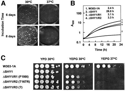
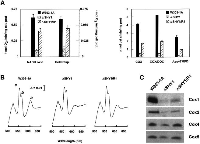
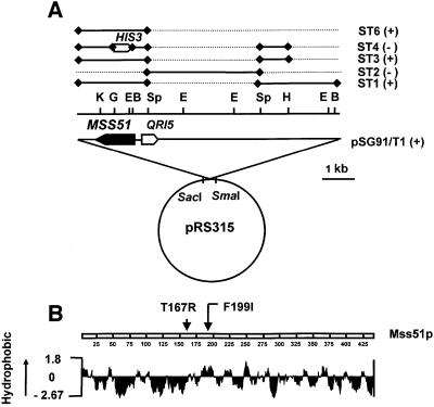
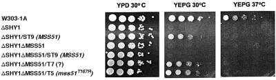
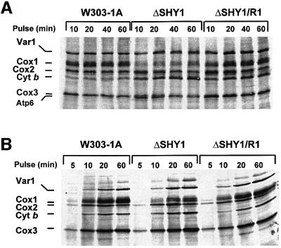
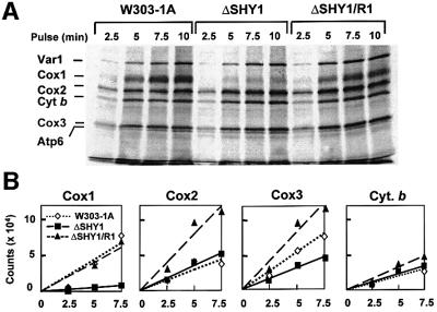
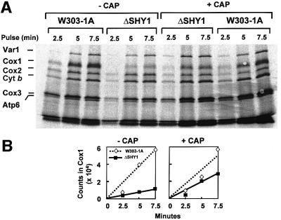
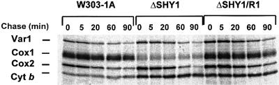
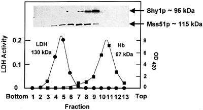
SHY1 codes for a mitochondrial protein required for full expression of cytochrome oxidase (COX) in Saccharomyces cerevisiae. Mutations in the homologous human gene (SURF1) have been reported to cause Leigh's syndrome, a neurological disease associated with COX deficiency. The function of Shy1p/Surf1p is poorly understood. Here we have characterized revertants of shy1 null mutants carrying extragenic nuclear suppressor mutations. The steady-state levels of COX in the revertants is increased by a factor of 4-5, accounting for their ability to respire and grow on non-fermentable carbon sources at nearly wild-type rates. The suppressor mutations are in MSS51, a gene previously implicated in processing and translation of the COX1 transcript for subunit 1 (Cox1) of COX. The function of Shy1p and the mechanism of suppression of shy1 mutants were examined by comparing the rates of synthesis and turnover of the mitochondrial translation products in wild-type, mutant and revertant cells. We propose that Shy1p promotes the formation of an assembly intermediate in which Cox1 is one of the partners.


Figures

None
Fig. 1. Growth properties of shy1 mutants and revertants. (A) The shy1 null mutant ΔSHY1 was plated at a density of 107 cells/plate on rich ethanol/glycerol (YEPG). The plates were photographed after 6 and 8 days of incubation at 30 and 37°C. (B) The respiratory-competent strain W303-1A, the shy1 null mutant ΔSHY1, and two independent revertants ΔSHY/R1 and ΔSHY/R2 were inoculated into liquid YEPG media and incubated with vigorous shaking at 30°C. Growth was monitored by absorbance at 600 nm. The doubling times for the different strains are indicated. (C) Serial dilutions of the respiratory-competent strain W303-1A, the shy1 mutant ΔSHY1, and three revertants ΔSHY/R1, ΔSHY/R2 and ΔSHY/R3 with the mss51 mutations indicated were spotted on YPD and YEPG plates and incubated at 30 and 37°C for 2–3 days.
None
Fig. 2. Functional characterization of the shy1 null mutant and revertant. (A) In the left panel, mitochondria prepared from the wild type (W303-1A), from the shy1 mutant (ΔSHY1) and from the revertant (ΔSHY1/R1) were assayed polarographically for NADH oxidase (NADH oxid.). Respiration was also assayed in whole cells in the presence of glucose (Cell Resp.). The specific activities reported were corrected for AA-insensitive respiration. The bars indicate the mean ± SD from three independent sets of measurements. In the right panel, COX was assayed in frozen–thawed mitochondria (COX) and in mitochondria permeabilized with potassium deoxycholate (COX/DOC) by measuring oxidation of ferrocytochrome c at 550 nm. COX activity was also assayed polarographically by measuring the oxygen consumption rate in the presence of ascorbate plus TMPD. (B) Cytochrome spectra. Mitochondria were extracted at a protein concentration of 5 mg/ml with potassium deoxycholate under conditions that quantitatively solubilize all the cytochromes (Tzagoloff et al., 1975). Difference spectra of the reduced (sodium dithionite) versus oxidized (potassium ferricyanide) extracts were recorded at room temperature. The α absorption bands corresponding to cytochromes a and a3 have maxima at 603 nm (a). The maxima for cytochrome b (b) and for cytochrome c and c1 (c) are 560 and 550 nm, respectively. (C) Steady-state concentrations of COX subunits. Total mitochondrial proteins (30 µg) separated by 16.5% SDS–PAGE were transferred to nitrocellulose and probed with subunit-specific antibodies to COX subunits.
None
Fig. 3. Identification of MSS51 as the suppressor gene. (A) Restriction maps of pSG91/T1 and of subclones. The locations of the restriction sites for BamHI (B), EcoRI (E), BglII (G), HindIII (H), KpnI (K) and SphI (Sp) are shown above the nuclear DNA insert in pSG91/T1. The regions of the nuclear insert in pSG91/T1 subcloned in YIp351 are represented by the solid bars in the upper part of the figure. The discontinuous lines represent the regions of pSG91/T1 deleted in each subclone. The plus and minus signs indicate suppression or lack thereof, respectively, of the shy1 null mutant by the subclones. The MSS51 reading frame and the direction of transcription of the gene are indicated by the solid arrow. The direction of transcription of the adjoining QRI5 gene is shown by the open arrow for orientation purposes. (B) Hydropathy profile of Mss51p (Kyte and Doolittle, 1982). The arrows indicate the location of the suppressor mutations identified.
None
Fig. 4. Suppression of the respiratory defect of shy1 mutants by wild-type and mutant MSS51. MSS51 was cloned in YIp351 and integrated at the chromosomal LEU2 locus of the shy1 null mutant. MSS51 and the two suppressor genes mss51T167R and mss51? were also integrated at the LEU2 locus of ΔSHY1ΔMSS51, a mutant construct with null mutations in MSS51 and SHY1. Serial dilution of the wild-type W303-1A, the single and double mutants and the transformants starting with 105 cells were spotted on rich glucose (YPD) and rich glycerol/ethanol (YEPG) plates and incubated at 30 and 37°C for 2.5 days.
None
Fig. 5. Mitochondrial protein synthesis in shy1 null mutants and revertants. (AIn vivo labeling of mitochondrial DNA products. Wild-type (W303), mutant (ΔSHY1) and revertant (ΔSHY1/R1) cells were labeled with [35S]methionine at 30°C for the times indicated in the presence of cycloheximide. (BIn organello protein synthesis. Mitochondria isolated from the same strains were labeled with [35S]methionine at 30°C in the absence of cycloheximide. Equivalent amounts of total cellular or mitochondrial proteins were separated by PAGE on a 17.5% polyacrylamide gel, transferred to a nitrocellulose membrane and exposed to an X-ray film. The mitochondrial translation products are identified on the left. The migration of Cox2 relative to Cox1 differs from that seen in (A) because a different PAGE buffer system was used for the separation.
None
Fig. 6. Kinetics of in vivo labeling of mitochondrial products during short pulses. (A) Wild-type (W303), mutant (ΔSHY1) and revertant (ΔSHY1/R1) cells were labeled with [35S]methionine. Total cellular proteins were extracted, depolymerized in sample buffer and separated on a 17.5% polyacrylamide gel. The labeling conditions, sample preparation and gel electrophoresis were identical to those described in Figure 5A. The proteins were transferred to nitrocellulose and exposed to X-ray film. (B) The radioactivity associated with Cox1, Cox2 and Cox3, and with cytochrome b was also quantitated in a PhosphorImager. The results were analyzed by linear regression.
None
Fig. 7. Effect of chloramphenicol on the kinetics of in vivo labeling of mitochondrial products. (A) Cells were grown and labeled at 30°C as in Figure 6, except that one half of the culture was incubated in the presence of 2 mg/ml chloramphenicol during the last 2 h of growth (+ CAP). Cells were harvested from both media and washed twice with a solution containing 40 mM potassium phosphate plus 2% galactose prior to labeling. Samples were removed after the labeling times indicated. Total cellular proteins were separated on a 17.5% gel as in Figure 6. (B) The radiolabeled Cox1 was quantitated in a PhosphorImager and the results were analyzed by linear regression.
None
Fig. 8. Turnover of in vivo labeled mitochondrial translation products. Cells were grown and labeled for 20 min at 30°C with [35S]methionine as in Figure 5. The labeling reaction was terminated by addition of excess 80 mM cold methionine and 4 µg/ml puromycin (0 time). Samples of the cultures were collected after the incubation times at 30°C indicated and processed as in Figure 6.
None
Fig. 9. Sedimentation properties of Shy1p and Mss51p. Mitochondria (7 mg of protein) from the wild-type haploid strain W303-1A were solubilized in the presence of 1 M KCl and 1% KDOC. The clarified extract obtained by centrifugation at 200 000 gav for 15 min was mixed with hemoglobin and lactate dehydrogenase, and applied to 5 ml of a linear 10–25% sucrose gradient containing 10 mM Tris–HCl pH 7.5, 0.5 mM EDTA and 0.1% Triton X-100. Following centrifugation for 14 h at 50 000 r.p.m. in a Beckman SW65 rotor, the gradient was collected in 13–14 equal fractions. Each fraction was assayed for hemoglobin (Hb) by absorption at 410 nm, and for lactate dehydrogenase (LDH) activity by measuring NADH-dependent conversion of pyruvate to lactate. The distributions of Shy1p and Mss51p were assayed by western blot analysis. The masses of Shy1p and Mss51p were determined from the positions of the respective peaks relative to those of the markers (Martin and Ames, 1961).

References

    1. Beyer R.E. (1964) A protein factor required for phosphorylation coupled to electron flow between reduced coenzyme Q and cytochrome c in the electron transfer chain. Biochem. Biophys. Res. Commun., 16, 460–465. - PubMed
    1. Choquet Y., Wostrikoff,K., Rimbault,B., Zito,F., Girard-Bascou,J., Drapier,D. and Wollman,F.A. (2001) Assembly-controlled regulation of chloroplast gene translation. Biochem Soc. Trans., 29, 421–426. - PubMed
    1. Coenen M.J., van den Heuvel,L.P., Nijtmans,L.G., Morava,E., Marquardt,I., Girschick,H.J., Trijbels,F.J., Grivell,L.A. and Smeitink, J.A. (1999) SURFEIT-1 gene analysis and two-dimensional blue native gel electrophoresis in cytochrome c oxidase deficiency. Biochem. Biophys. Res. Commun., 265, 339–344. - PubMed
    1. Costanzo M.C., Seaver,E.C. and Fox,T.D. (1989) The PET54 gene of Saccharomyces cerevisiae: characterization of a nuclear gene encoding a mitochondrial translational activator and subcellular localization of its product. Genetics, 122, 297–305. - PMC - PubMed
    1. Decoster E., Simon,M., Hatat,D. and Faye,G. (1990) The MSS51 gene product is required for the translation of the COX1 mRNA in yeast mitochondria. Mol. Gen. Genet., 224, 111–118. - PubMed

Publication types

MeSH terms