. 2002 Jan 15;21(1-2):53-63.
doi: 10.1093/emboj/21.1.53.

CREB activation induced by mitochondrial dysfunction is a new signaling pathway that impairs cell proliferation

Affiliations

CREB activation induced by mitochondrial dysfunction is a new signaling pathway that impairs cell proliferation

T Arnould et al. EMBO J. .

Abstract

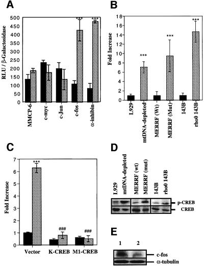
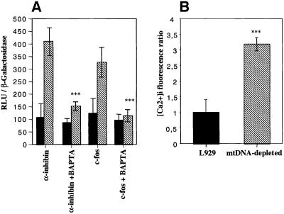
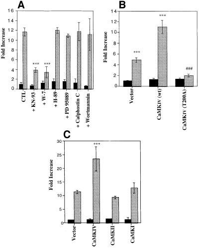
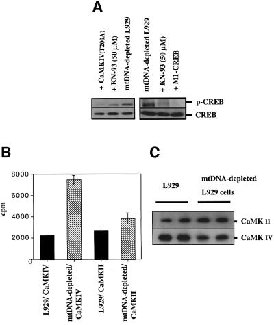
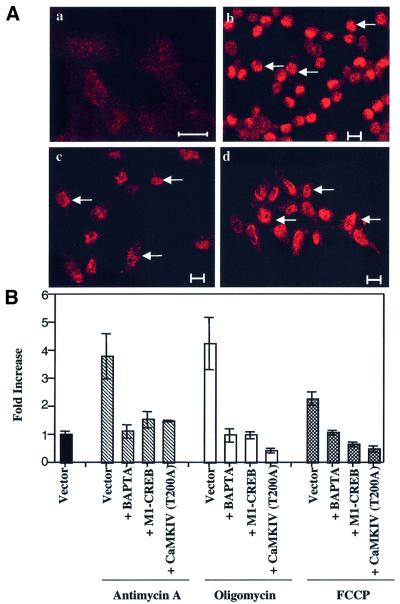
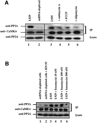
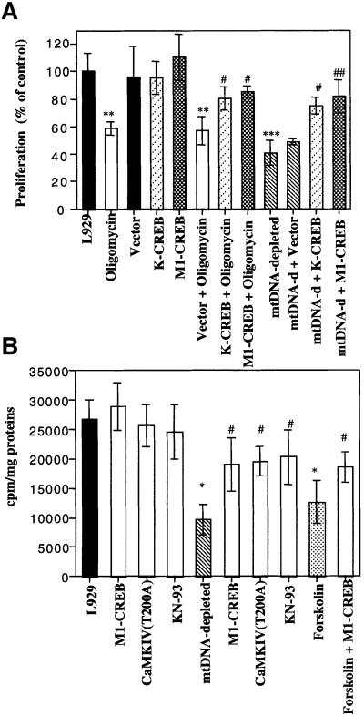
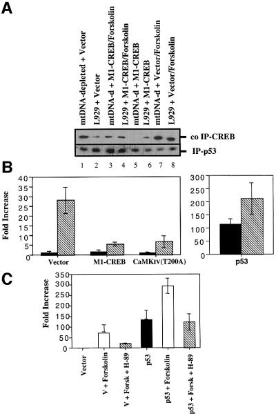
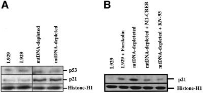
We characterized a new signaling pathway leading to the activation of cAMP-responsive element-binding protein (CREB) in several cell lines affected by mitochondrial dysfunction. In vitro kinase assays, inhibitors of several kinase pathways and overexpression of a dominant-negative mutant for calcium/calmodulin kinase IV (CaMKIV), which blocks the activation of CREB, showed that CaMKIV is activated by a mitochondrial activity impairment. A high calcium concentration leading to the disruption of the protein interaction with protein phosphatase 2A explains CaMKIV activation in these conditions. Transcrip tionally active phosphorylated CREB was also found in a rho0 143B human osteosarcoma cell line and in a MERRF cybrid cell line mutated for tRNA(Lys) (A8344G). We also showed that phosphorylated CREB is involved in the proliferation defect induced by a mitochondrial dysfunction. Indeed, cell proliferation inhibition can be prevented by CaMKIV inhibition and CREB dominant-negative mutants. Finally, our data suggest that phosphorylated CREB recruits p53 tumor suppressor protein, modifies its transcriptional activity and increases the expression of p21(Waf1/Cip1), a p53-regulated cyclin-dependent kinase inhibitor.


Figures

None
Fig. 1. Effects of EtBr-treatment on mitochondria in L929 cells. (A) PCR amplification of a specific mtDNA fragment in the mitochondrial D-loop performed on purified mtDNA (lanes 1 and 2) and nuclear DNA (lanes 4 and 5). (B) Immunofluorescence patterns of cells stained with a monoclonal antibody against the COX I subunit: (a) mtDNA-depleted cells; (b) L929 cells. Asterisks indicate the localization of nuclei. Scale bars = 10 µm.
None
Fig. 2. (A) Luciferase activity in transiently transfected L929 control and mtDNA-depleted cells with luciferase constructs driven by c-myc, c-Jun, authentic MMCP-6, c-fos and α-inhibin promoters. L929 (black) and mtDNA-depleted L929 (hatched) cells, were transiently co-transfected with β-galactosidase and the luciferase constructs. (B) CREB-dependent luciferase activity in L929, mtDNA-depleted L929, wild-type (wt) MERRF cybrid, mutated (mut) MERRF cybrid, 143B and ρ0 143B cells transiently transfected with the luciferase reporter construct driven by the α-inhibin promoter and an expression plasmid encoding β-galactosidase. (C) CREB-dependent luciferase activity in L929 (black) and mtDNA-depleted L929 (hatched) cells transiently transfected with K-CREB or M1-CREB, together with the CREB-sensitive luciferase reporter construct and an expression plasmid encoding β-galactosidase. Luciferase activity was determined 24 h post-transfection. Results are expressed in relative light units (RLU) after normalization for β-galactosidase activity (A) or in fold increase of control cells (B and D) as means ± 1 SD (*** and ###, significantly different from L929 and mtDNA-depleted L929 cells with P <0.001). (D) Western blot analysis of phospho-CREB (p-CREB) and CREB assessed for the different cell lines used in (B). (E) Western blot analysis for c-fos and α-tubulin assessed for L929 (lane 2) and mtDNA-depleted L929 (lane 1).
None
Fig. 3. (A) Effect of a 6 h treatment with 20 µM BAPTA-AM on the CREB-sensitive luciferase activity after transfection of L929 (black) or mtDNA-depleted L929 (hatched) cells with luciferase reporter genes driven by either CREB-regulated c-fos or α-inhibin promoters. Luciferase activity was determined and expressed as RLU after normalization for β-galactosidase activity (n = 6; ***, significantly different from mtDNA-depleted cells with P <0.001). (B) Intracellular calcium concentration ratio in L929 and mtDNA-depleted cells loaded with 10 µM Fluo-3-AM. Results are expressed in fold increase of control cells, and represent the mean ± SD for n = 3 (***, significantly different from L929 with P <0.001).
None
Fig. 4. (A) Effect of kinase pathway inhibitors on CREB-dependent luciferase activity in L929 (black) and mtDNA-depleted (hatched) cells. After transfection, cells were incubated for 16 h before the assay with 50 µM KN-93, 50 µM W-7, 10 µM H-89, 20 µM PD 95089, 1 µM calphostin C or 1 µM wortmannin. (B) L929 (black) and mtDNA-depleted L929 (hatched) cells were transiently co-transfected with a plasmid encoding β-galactosidase, the CREB luciferase reporter construct driven by the α-inhibin promoter and an expression vector encoding wild-type CaMKIV or the dominant-negative form CaMKIV T200A. (C) Effect of overexpression of CaMKI, CaMKII and CaMKIV on the activation of the CREB luciferase reporter construct driven by the α-inhibin promoter. L929 (black) and mtDNA-depleted L929 (hatched) cells were transiently co-transfected as described in (B) with an expression vector encoding wild-type CaMKI, II or IV. Results are normalized for β-galactosidase activity, are expressed in fold increase of luciferase activity of the L929 control cells and represent the mean ± SD for n = 3 (***, significantly different from L929 with P <0.001; ###, significantly different from mtDNA-depleted L929 cells with P <0.001).
None
Fig. 5. (A) Effect of dominant-negative CaMKIV T200A or M1-CREB overexpression and the calmodulin kinase inhibitor KN-93 on the CREB phosphorylation pattern in mtDNA-depleted L929 cells. mtDNA-depleted L929 cells were either transfected with a plasmid encoding CaMKIV T200A or M1-CREB or treated with 50 µM KN-93 for 24 h before phospho-CREB and CREB expression were analyzed by western blotting. (B) Endogenous CaMKIV or CaMKII activity in L929 and mtDNA-depleted cells was measured after immuno precipitation. Representative results of one experiment in duplicate are shown and are expressed as c.p.m. (C) The amounts of immunoprecipitated kinases were controlled by western blotting.
None
Fig. 6. (A) Immunolocalization of phospho-CREB on Ser133 in L929 cells treated or not (a) for 6 h with 10 µM FCCP (b), 8 µM oligomycin (c) and 1 µM antimycin A (d) and analyzed the next day by immunofluorescence and confocal microscopy. Bars = 10 µm. Arrows indicate a nuclear localization. (B) CREB-dependent luciferase activity in 293 cells triggered by treatment with 1 µM antimycin A (hatched bars), 8 µM oligomycin (white bars) or 10 µM FCCP (black bars) is inhibited by 20 µM BAPTA for 6 h, or by expression of a dominant-negative form of CREB (M1-CREB) or a dominant-negative mutant of CaMKIV (CaMKIV T200A). Results are expressed as fold increase of control cells (vector) after normalization. The results are representative of two experiments in triplicate and are expressed as means ± 1 SD.
None
Fig. 7. (A) Co-immunoprecipitation of PP2A catalytic C subunit with CaMKIV. CaMKIV was immunoprecipitated (IP) from L929 cells (lanes 1 and 3) treated or not with mitochondrial inhibitors (8 µM oligomycin, 1 µM antimycin A and 10 µM FCCP) (lanes 4–6) or from mtDNA-depleted L929 cells (lane 2). (B) Co-immunoprecipitation of PP2A catalytic C subunit with CaMKIV. CaMKIV was immuno precipitated from L929 cells (lane 3) treated or not with ionomycin at 10, 100 or 500 nM for 16 h (lanes 4–6) or from mtDNA-depleted L929 cells treated or not with 50 µM KN-93 for 16 h (lanes 1 and 2). Immune complexes were analyzed by immunoblotting with an anti-PP2A C subunit antibody (anti-PP2A) or an anti-CaMKIV antibody (anti-CaMKIV). Expression levels of the PP2A C subunit in the lysates are also shown.
None
Fig. 8. (A) Incorporation of [3H]thymidine into DNA was analyzed in L929 cells (treated or not with oligomycin) and compared with the incorporation for mtDNA-depleted L929 cells. In some conditions, these cells were transiently transfected before treatment with an empty vector or plasmids encoding dominant-negative CREB (K-CREB and M1-CREB). At 16 h post-transfection, oligomycin (8 µM) was added or not for 6 h and, the next day, cells were labeled with 2 µCi of [3H]thymidine for 4 h and processed accordingly. (B) Effect of M1-CREB and CaMKIV T200A overexpressions or KN-93 treatment on [3H]thymidine incorporation. L929 and mtDNA-depleted L929 cells were transiently transfected with an empty vector or plasmids encoding M1-CREB or CaMKIV T200A. The vector-transfected cells were treated or not with 50 µM KN-93 for 24 h before the [3H]thymidine incorporation assay. A control was perfomed with forskolin (10–5 M) for 24 h with or without M1-CREB overexpression. Values were determined and are expressed either as c.p.m./mg of proteins (B, n = 3) or as a percentage of [3H]thymidine incorporation for control L929 cells (A, n = 4). (*, **, *** or #, ##, ###, significantly different from L929 control cells or mtDNA-depleted L929 cells with P <0.05, P <0.01 or P <0.001).
None
Fig. 9. (A) CREB directly interacts with p53 tumor suppressor protein in a phosphorylation-dependent manner. p53 protein was immunoprecipitated (IP) from L929 and mtDNA-depleted L929 cells transiently transfected or not with a plasmid encoding M1-CREB. When indicated, cells were treated with 10 µM forskolin for 24 h. Bound CREB protein was detected by western blot analysis. (B) p53-dependent transcription is increased in mtDNA-depleted L929 cells. The p53-responsive pG13-Luc reporter was co-transfected into L929 (black) or mtDNA-depleted L929 cells (hatched) with the β-galactosidase construct, together with 1 µg of an empty vector, of a plasmid encoding M1-CREB and CaMKIV T200A (left panel) or a p53 expression plasmid (pC53-SN3) (right panel). (C) p53-dependent transcription is stimulated by forskolin treatment in an H-89 inhibitable manner. The p53-responsive pG13-Luc reporter was co-transfected into L929 cells with the β-galactosidase construct, together with 1 µg of an empty vector or a plasmid encoding wild-type p53 (pC53-SN3). Cells were then stimulated with 10 µM forskolin with or without 10 µM H-89 for 16 h. Luciferase activity was measured 48 h post-transfection. Reported values are expressed in fold increase of L929 control cells transfected with an empty vector as means ± SD (n = 3).
None
Fig. 10. (A) p21 is overexpressed in mtDNA-depleted L929 cells. Western blotting analysis for p53 and p21 derived from L929 and mtDNA-depleted L929 cells. (B) p21 expression is stimulated in L929 cells treated with 10 µM forskolin for 16 h (lanes 1 and 2) while its expression is reduced in mtDNA-depleted cells transiently transfected with M1-CREB or incubated with 50 µM KN-93 for 16 h (lanes 3–5). Protein loading was controlled by the expression of histone H1.

Similar articles

Cited by

References

    1. Biswas G., Adebanjo,O.A., Freedman,B.D., Anandatheerthavarada,H.K., Vijayasarathy,C., Zaidi,M., Kotlikoff,M. and Avadhani,N.G. (1999) Retrograde Ca2+ signaling in C2C12 skeletal myocytes in response to mitochondrial genetic and metabolic stress: a novel mode of inter-organelle crosstalk. EMBO J., 18, 522–533. - PMC - PubMed
    1. Bonni A., Brunet,A., West,A.E., Datta,S.R., Takasu,M.A. and Greenberg,M.E. (1999) Cell survival promoted by the Ras–MAPK signaling pathway by transcription-dependent and -independent mechanisms. Science, 286, 1358–1362. - PubMed
    1. Brady P.S., Park,E.A., Liu,J.S., Hanson,R.W. and Brady,L.J. (1992) Isolation and characterization of the promoter for the gene coding for the 68 kDa carnitine palmitoyltransferase from the rat. Biochem. J., 286, 779–783. - PMC - PubMed
    1. Buchet K. and Godinot,C. (1998) Functional F1-ATPase essential in maintaining growth and membrane potential of human mitochondrial DNA-depleted ρ° cells. J. Biol. Chem., 273, 22983–22989. - PubMed
    1. Buck M., Turler,H. and Chojkier,M. (1994) LAP (NF-IL-6), a tissue-specific transcriptional activator, is an inhibitor of hepatoma cell proliferation. EMBO J., 13, 851–860. - PMC - PubMed

Publication types

MeSH terms

Substances