. 2002 Jan 15;21(1-2):64-71.
doi: 10.1093/emboj/21.1.64.

Regulation of Raf-1 activation and signalling by dephosphorylation

Affiliations

Regulation of Raf-1 activation and signalling by dephosphorylation

Amardeep S Dhillon et al. EMBO J. .

Abstract

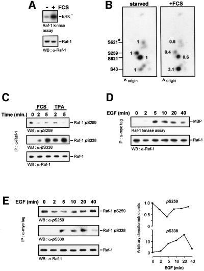
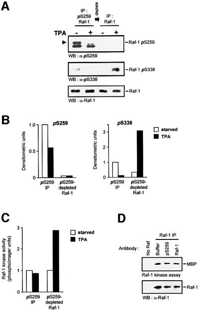
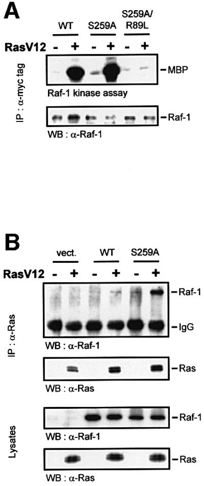
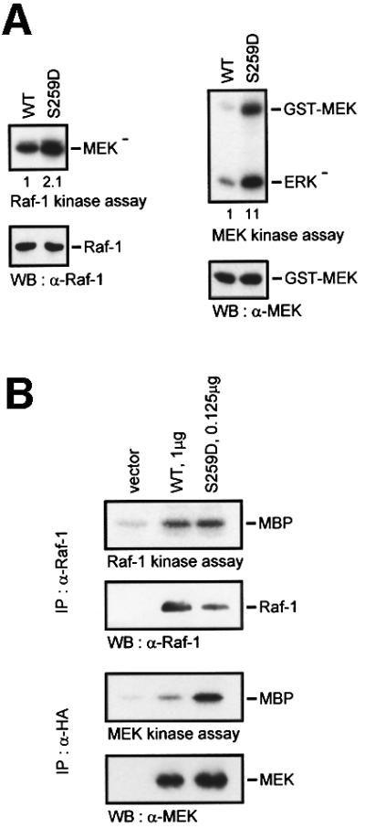
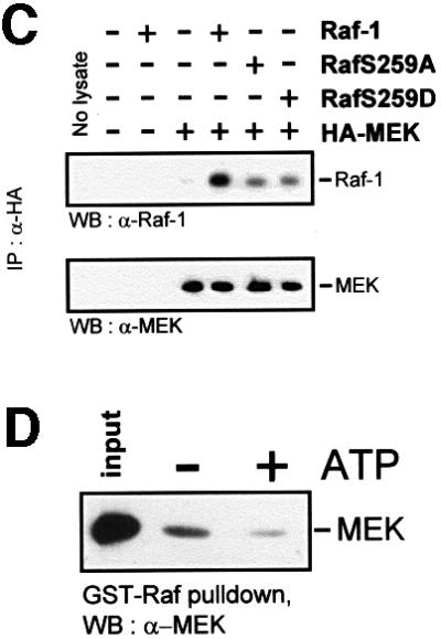
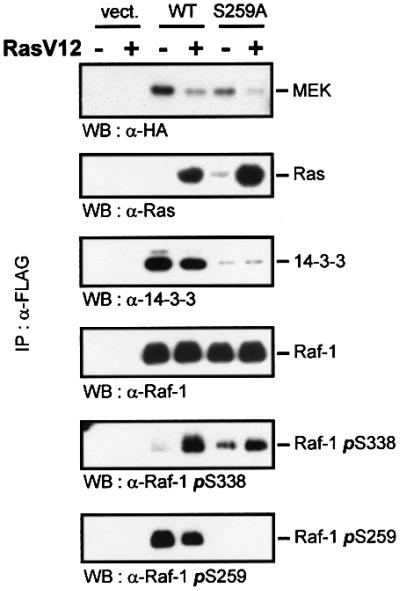
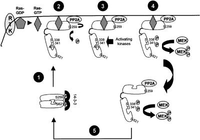
The Raf-1 kinase is regulated by phosphorylation, and Ser259 has been identified as an inhibitory phosphorylation site. Here we show that the dephosphorylation of Ser259 is an essential part of the Raf-1 activation process, and further reveal the molecular role of Ser259. The fraction of Raf-1 that is phosphorylated on Ser259 is refractory to mitogenic stimulation. Mutating Ser259 elevates kinase activity because of enhanced binding to Ras and constitutive membrane recruitment. This facilitates the phosphorylation of an activating site, Ser338. The mutation of Ser259 also increases the functional coupling to MEK, augmenting the efficiency of MEK activation. Our results suggest that Ser259 regulates the coupling of Raf-1 to upstream activators as well as to its downstream substrate MEK, thus determining the pool of Raf-1 that is competent for signalling. They also suggest a new model for Raf-1 activation where the release of repression through Ser259 dephosphorylation is the pivotal step.


Figures

None
Fig. 1. Changes in Raf-1 phosphorylation during activation. Serum-starved NIH 3T3 cells were labelled with 0.5 mCi/ml [32P]ortho phosphoric acid for 6 h and stimulated with 20% FCS for 5 min. Endogenous Raf-1 was immunoprecipitated, and (A) examined for kinase activity. (B) Another Raf-1 aliquot was digested with trypsin and subjected to two-dimensional phosphopeptide mapping. Equal amounts of Raf were loaded, as evident from the western blot shown in (A). Phosphorylation sites were assigned by comparison with the respective Raf-1 point mutants. The peptide containing Ser621 produces two spots due to partial digestion; the minor spot is labelled by an asterisk. Spots were quantified by phosphoimager, and the changes relative to serum-starved cells are shown. (C) Dephosphoryl ation of endogenous Raf-1 during serum and mitogen stimulation. COS cells were treated with 10% FCS or 100 ng/ml TPA for the indicated times. Raf-1 was immunoprecipitated from the cells with an anti-Raf-1 antibody. The immunoprecipitates were then blotted with antibodies against Raf-1 and phosphoserines 259 and 338. (D) Time course of Raf-1 kinase activation. COS cells were transfected with myc-Raf, serum starved and stimulated with 20 ng/ml EGF for the indicated times. myc-Raf-1 was immunoprecipitated with the 9E10 monoclonal antibody, and catalytic activity was measured using a two-step kinase assay. (E) Myc-Raf immunoprecipitates were blotted with phospho specific antisera against phosphoserines 259 and 338. The blots were quantified by laser densitometry shown in the right hand panels.
None
Fig. 2. Raf-1 phosphorylated on Ser259 is refractory to mitogen stimulation. Cells were lysed in RIPA buffer and Raf-1 was immunodepleted from serum-starved or TPA-treated (100 ng/ml for 20 min) cells using the affinity-purified anti-pSer259 antibody. The supernatant subsequently was immunoprecipiated with anti-Raf antiserum. Both fractions of Raf-1 were examined for phosphorylation of Ser259 and Ser338, using phosphospecific antisera (A and B), and for kinase activity (C). Raf-1 phosphorylated on Ser259 is indicated by the arrowhead; the lower of the two bands is non-specific. Similar results were obtained with EGF stimulation. For (B), the immunoblots of (A) were quantified by laser densitometry. The pSer259 and pSer338 signals, respectively, were normalized to the signals obtained with a non-discriminatory Raf-1 antibody (lowest panel in A). (D) The Raf-1 Ser259-phosphospecific antiserum does not inhibit kinase activity. COS cells were transfected with CMV5-Raf-1 and treated with 100 ng/ml TPA for 15 min. The cells were lysed in RIPA buffer and Raf-1 was immunoprecipitated with the c-Raf VI antiserum. Aliquots of the immunoprecipitate were incubated with either 2 µg of pSer259-specific or a non-discriminatory Raf-1 antibody (C-12; Santa Cruz) for 2 h. The kinase activities of the immunoprecipitates were then measured.
None
Fig. 3. Ser259 regulates coupling to Ras. (A) Myc-tagged wild-type Raf-1 (WT), RafS259A, and the double mutant RafR89L/S259A were expressed in COS-1 cells either alone or with RasV12 as indicated. Raf-1 immunoprecipitates were examined for kinase activity. (B) The mutation of Ser259 enhances the association with Ras. COS-1 cells were transfected with the indicated expression plasmids. Ras immunoprecipitates were immunoblotted for Ras and associated Raf proteins (upper panel). Crude lysates were immunoblotted to show that all Raf proteins were expressed at comparable levels (lower panel). (C) Cell fractionation. COS cells were transfected with the indicated expression plasmids, serum starved ‘–’ and treated with EGF (20 ng/ml) plus TPA (100 ng/ml), ‘E&T’, for 30 min. Cells were lysed in hypotonic, detergent-free buffer and fractionated into 100 000 g supernatant ‘S’ and pellet ‘P’ containing membranes. The fractions were extracted with 1% Triton X-100, and Raf-1 immunoprecipitates were examined for kinase activity, Ser259 phosphorylation and Raf-1 distribution. Kinase activity was quantified by a phosphoimager and is given below the lanes in arbitrary units. (D) COS cells were transfected with myc-Raf-1 along with RasV12 or vector. The cells were fractionated as described above and equal amounts of whole-cell lysate from each fraction were blotted for Ser259, Raf-1 and Ras levels using the respective antibodies.
None
Fig. 4. Ser259 regulates the coupling of Raf-1 to MEK. (A) GST–MEK was co-expressed with Raf-1 or RafS259D, respectively, in Sf-9 cells. Raf-1 proteins were immunoprecipitated and examined for kinase activity with kinase-negative MEK as substrate. GST–MEK was isolated by adsorption to glutathione–Sepharose beads and assayed for kinase activity using kinase-negative ERK (ERK) as substrate. (B) COS cells were transfected with different amounts of Raf-1 and RafS259D in order to achieve a comparable Raf kinase activity. A constant amount of HA-MEK was co-transfected and assayed for kinase activity with kinase-negative ERK as substrate. (C) Co-immunoprecipitation between Raf and MEK. HA-MEK was co-expressed with Raf-1 or RafS259 mutants, respectively, in COS cells. Lysates were immunoprecipitated and subsequently blotted with the indicated antibodies. (D) Phosphorylation diminishes the Raf-1–MEK association. GST–Raf-1 was expressed in Sf-9 cells and purified by affinity adsorption to glutathione–Sepharose beads. GST–Raf-1 beads were incubated in vitro with purified kinase-negative MEK-1 produced in Escherichia coli in the absence or presence of 100 µM ATP. The beads were washed and bound MEK was detected by immunoblotting.
None
Fig. 5. The effects of Ser259 mutation and Ras on the composition of Raf-1 signalling complexes. COS cells were transfected with FLAG-Raf-1, HA-MEK1 and RasV12 as indicated. Raf proteins were immunoprecipitated with FLAG antibodies and immunoblotted using the indicated antibodies.
None
Fig. 6. Model for the role of Ser259 in Raf-1 activation. The steps designated in the figure are: (1) Raf-1 membrane translocation and Ras binding. (2) Derepression by PP2A-mediated dephosphorylation of Ser259. (3) Phosphorylation of Raf-1 by activating kinases on Ser338 and Tyr341. (4) Phosphorylation of the substrate MEK. (5) Re-phosphorylation of Ser259, dephosphorylation of activating sites and transition into a quiescent state. See text for details.

References

    1. Abraham D. et al. (2000) Raf-1-associated protein phosphatase 2A as a positive regulator of kinase activation. J. Biol. Chem., 275, 22300–22304. - PubMed
    1. Alessandrini A., Greulich,H., Huang,W.D. and Erikson,R.L. (1996) Mek1 phosphorylation site mutants activate Raf-1 in NIH 3T3 cells. J. Biol. Chem., 271, 31612–31618. - PubMed
    1. Alessi D.R., Cuenda,A., Cohen,P., Dudley,D.T. and Saltiel,A.R. (1995) PD 098059 is a specific inhibitor of the activation of mitogen-activated protein kinase kinase in vitro and in vivo. J. Biol. Chem., 270, 27489–27494. - PubMed
    1. Avruch J., Khokhlatchev,A., Kyriakis,J.M., Luo,Z., Tzivion,G., Vavvas, D. and Zhang,X.F. (2001) Ras activation of the Raf kinase: tyrosine kinase recruitment of the MAP kinase cascade. Recent Prog. Horm. Res., 56, 127–155. - PubMed
    1. Barnard D., Diaz,B., Clawson,D. and Marshall,M. (1998) Oncogenes, growth factors and phorbol esters regulate Raf-1 through common mechanisms. Oncogene, 17, 1539–1547. - PubMed

Publication types