. 2002 Jan 15;21(1-2):83-92.
doi: 10.1093/emboj/21.1.83.

BANK regulates BCR-induced calcium mobilization by promoting tyrosine phosphorylation of IP(3) receptor

Affiliations

BANK regulates BCR-induced calcium mobilization by promoting tyrosine phosphorylation of IP(3) receptor

Kazumasa Yokoyama et al. EMBO J. .

Abstract

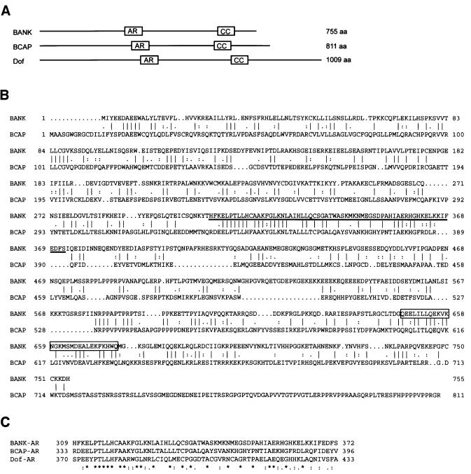
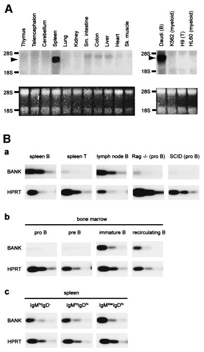
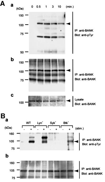
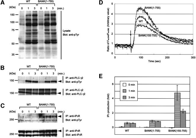
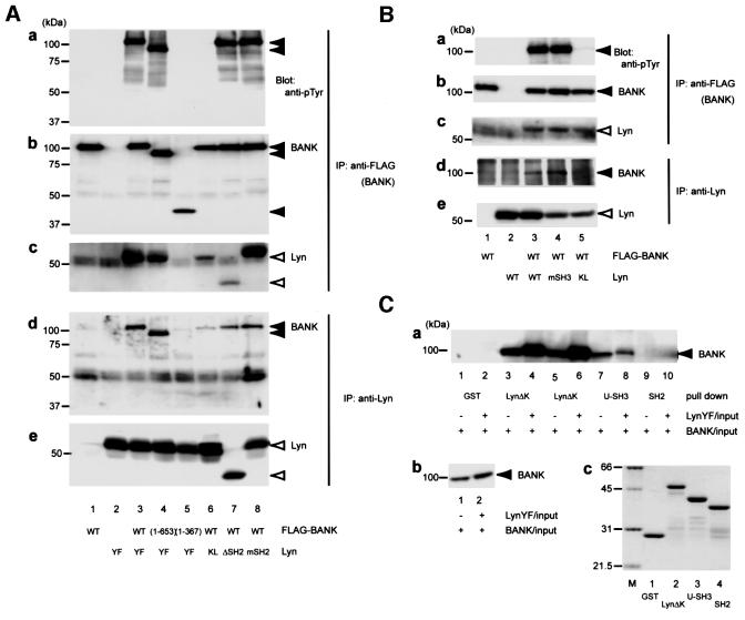
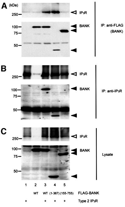
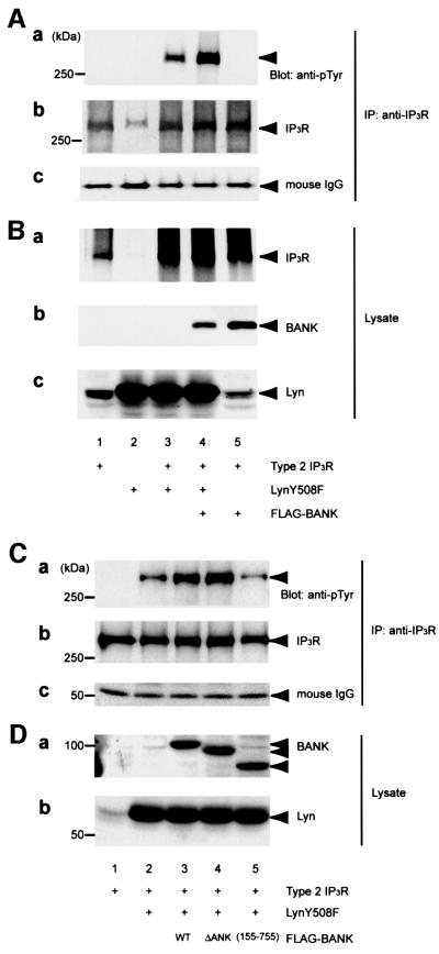
B-cell activation mediated through the antigen receptor is dependent on activation of protein tyrosine kinases (PTKs) such as Lyn and Syk and subsequent phosphorylation of various signaling proteins. Here we report on the identification and characterization of the B-cell scaffold protein with ankyrin repeats (BANK), a novel substrate of tyrosine kinases. BANK is expressed in B cells and is tyrosine phosphorylated upon B-cell antigen receptor (BCR) stimulation, which is mediated predominantly by Syk. Overexpres sion of BANK in B cells leads to enhancement of BCR-induced calcium mobilization. We found that both Lyn and inositol 1,4,5-trisphosphate receptor (IP(3)R) associate with the distinct regions of BANK and that BANK promotes Lyn-mediated tyrosine phosphorylation of IP(3)R. Given that IP(3)R channel activity is up-regulated by its tyrosine phosphorylation, BANK appears to be a novel scaffold protein regulating BCR-induced calcium mobilization by connecting PTKs to IP(3)R. Because BANK expression is confined to functional BCR-expressing B cells, BANK-mediated calcium mobilization may be specific to foreign antigen-induced immune responses rather than to signaling required for B-cell development.


Figures

None
Fig. 1. Identification of a novel protein BANK. (A) Schematic representation of predicted motifs and structures of human BANK, mouse BCAP and Drosophila Dof proteins. The ankyrin repeat-like motifs (amino acids 309–372 of BANK) and the presumptive coiled coils (647–675) are indicated by boxes AR and CC, respectively. (B) Alignment of the amino acid sequences of human BANK and mouse BCAP proteins was performed with the GAP program. The vertical bars indicate identical amino acids, while colons and dots indicate similar amino acids. The ankyrin repeat-like motif is underlined and the predicted coiled coil is boxed. (C) The sequences of the ankyrin repeat-like motifs of human BANK, mouse BCAP and Drosophila Dof proteins were aligned by the Clustal program.
None
Fig. 2. Cell type-specific expression of BANK mRNA. (A) Northern blot analysis of BANK expression in mouse tissues and human cell lines (upper parts). As a loading control, images of the ethidium bromide staining of RNA are shown in the lower parts. (B) Semi-quantitative RT–PCR analysis of BANK expression in B- and T-lineage cells. The RNA of described cells was isolated and reverse transcribed to cDNA. RT–PCR reactions were performed using three different template concentrations by 3.5-fold dilution. The amount of cDNA in the first dilution corresponds to 300 cells. (a) Cells indicated were isolated by MACS as described in Materials and methods. (b and c) Cells indicated were isolated by FACS as described in Materials and methods.
None
Fig. 3. BCR-induced tyrosine phosphorylation of BANK. (A) Tyrosine phosphorylation of BANK in Daudi cells. At the time points indicated after BCR stimulation (10 µg/ml anti-human IgM), whole-cell lysates and anti-BANK immunoprecipitates were analyzed by immunoblotting with anti-phosphotyrosine (anti-pTyr, a) or anti-BANK (b and c; immunoprecipitates and whole-cell lysates, respectively). The positions of BANK are indicated by arrowheads. (B) BANK tyrosine phos phorylation in DT40 cells deficient in Lyn, Syk or Btk, unstimulated (minus symbol) or stimulated with 4 µg/ml anti-chicken IgM (M4) for 2 min (plus symbol). All experiments were performed at least three times.
None
Fig. 4. Regulation of BCR-induced calcium mobilization by BANK. (A) BCR-induced tyrosine phosphorylation in wild-type (WT) and BANK-overexpressing (BANK 1–755) DT40 cells. (B) Tyrosine phosphorylation of PLC-γ2. (C) Tyrosine phosphorylation of type 2 IP3R. At the time points indicated after BCR stimulation (4 µg/ml M4), whole-cell lysates (A) and anti-PLC-γ2 (B) and anti-type 2 IP3R (C) immunoprecipitates were analyzed by immunoblotting with anti-pTyr. (D) Intracellular free calcium levels in Fura-2-loaded cells were monitored by spectrofluorometry after stimulation with M4 (4 µg/ml). The ratios of the fluorescence intensities at excitation wavelengths of 340 nm (Fex340) and 380 nm (Fex380) are shown. An arrow indicates the time point of M4 antibody addition. These data are representative of five independent experiments with two independent BANK-overexpressing DT40 clones (BANK 1–755), mutant BANK-overexpressing DT40 clones (BANK 155–755) and parental DT40 cells (WT). (E) BCR-induced IP3 production in BANK(1–755), BANK(155–755) and wild-type DT40 cells. Results are shown as the mean from three independent experiments. Error bars represent the SD from the mean.
None
Fig. 5. Association between BANK and Lyn. (A and B) HEK293T cells were transiently transfected with combinations of plasmids expressing various FLAG-tagged mutant BANK and Lyn mutants. Anti-FLAG (BANK) and anti-Lyn immunoprecipitates were subjected to immunoblotting with anti-pTyr, anti-BANK and anti-Lyn antibodies. (C) HEK293T cells were transiently transfected with plasmids expressing FLAG-tagged BANK, with (+) or without (–) expressing LynY508F. The lysates were incubated with 1 µg of the GST fusion proteins indicated immobilized on glutathione– Sepharose. The precipitates were washed and immunoblotted with anti-BANK antibody (a). The amount of BANK in HEK293T cell lysates (b). The amount and purity of GST fusion proteins were verified by Coomassie Blue staining (c; M, molecular weight standards). Abbreviations are explained in the text.
None
Fig. 6. In vivo association between BANK and IP3R. HEK293T cells were transiently transfected with combinations of plasmids expressing various FLAG-tagged mutant BANK and type 2 IP3R. Anti-FLAG (BANK, A) and anti-IP3R (B) immunoprecipitates and whole-cell lysates (C) were subjected to immunoblotting with anti-IP3R (top) or anti-FLAG (bottom) antibodies. These data are representative of four independent experiments with similar results.
None
Fig. 7. Promotion of Lyn-mediated tyrosine phosphorylation of IP3R by BANK. HEK293T cells were transiently transfected with various combinations of plasmids expressing FLAG-tagged BANK, LynY508F and type 2 IP3R. (A) IP3R was immunoprecipitated and subjected to immunoblotting with anti-pTyr antibody (a). The filter used in (a) was stripped and reprobed with anti-IP3R (b). The amounts of anti-IP3R antibody used in immunoprecipitation were equal (c). Expression of BANK, IP3R and Lyn in whole-cell lysates was confirmed by immunoblotting (B). The effects of BANK mutations were examined in similar experiments (C and D). All experiments were performed at least four times with similar results.

References

    1. Bourguignon L.Y., Jin,H., Iida,N., Brandt,N.R. and Zhang,S.H. (1993) The involvement of ankyrin in the regulation of inositol 1,4,5-trisphosphate receptor-mediated internal Ca2+ release from Ca2+ storage vesicles in mouse T-lymphoma cells. J. Biol. Chem., 268, 7290–7297. - PubMed
    1. Bourguignon L.Y., Chu,A., Jin,H. and Brandt,N.R. (1995) Ryanodine receptor–ankyrin interaction regulates internal Ca2+ release in mouse T-lymphoma cells. J. Biol. Chem., 270, 17917–17922. - PubMed
    1. Cameron A.M., Steiner,J.P., Roskams,A.J., Ali,S.M., Ronnett,G.V. and Snyder,S.H. (1995) Calcineurin associated with the inositol 1,4,5-trisphosphate receptor–FKBP12 complex modulates Ca2+ flux. Cell, 83, 463–472. - PubMed
    1. Fukunaga R. and Hunter,T. (1997) MNK1, a new MAP kinase-activated protein kinase, isolated by a novel expression screening method for identifying protein kinase substrates. EMBO J., 16, 1921–1933. - PMC - PubMed
    1. Ishiai M. et al. (1999) BLNK required for coupling Syk to PLCγ2 and Rac1-JNK in B cells. Immunity, 10, 117–125. - PubMed

Publication types

MeSH terms

Associated data