. 2002 Jan 1;16(1):127-37.
doi: 10.1101/gad.950902.

An ERK2 docking site in the Pointed domain distinguishes a subset of ETS transcription factors

Affiliations

An ERK2 docking site in the Pointed domain distinguishes a subset of ETS transcription factors

Jeffrey J Seidel et al. Genes Dev. .

Abstract

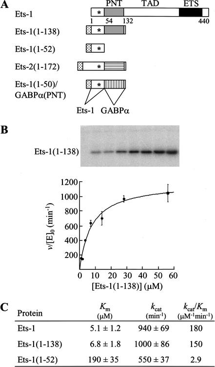
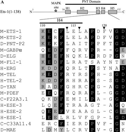
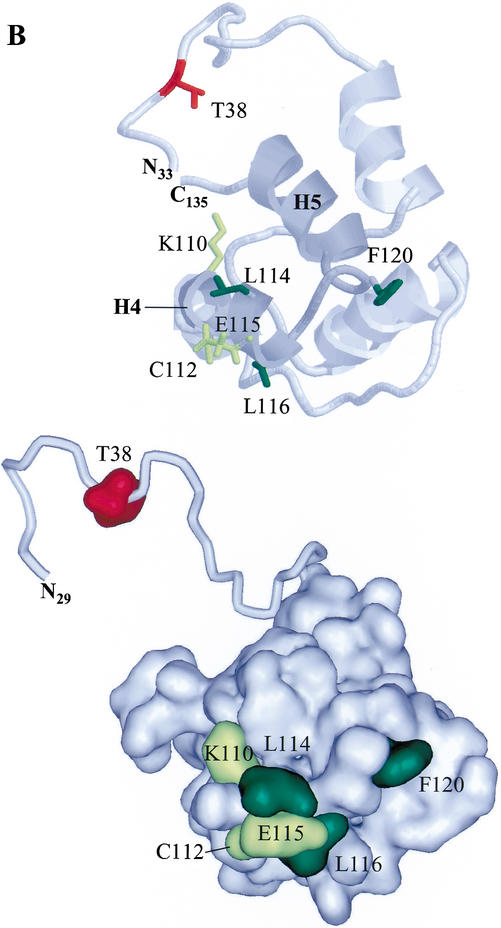
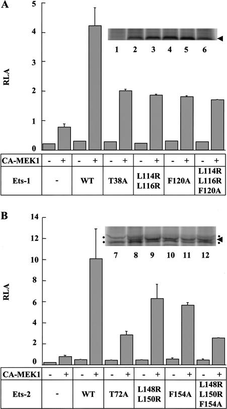
The ETS transcription factors perform distinct biological functions despite conserving a highly similar DNA-binding domain. One distinguishing property of a subset of ETS proteins is a conserved region of 80 amino acids termed the Pointed (PNT) domain. Using enzyme kinetics we determined that the Ets-1 PNT domain contains an ERK2 docking site. The docking site enhances the efficiency of phosphorylation of a mitogen-activated protein kinase (MAPK) site N-terminal to the PNT domain. The site enhances ERK2 binding rather than catalysis. Three hydrophobic residues are involved in docking, and the previously determined NMR structure indicates that these residues are clustered on the surface of the Ets-1 PNT domain. The docking site function is conserved in the PNT domain of the highly related Ets-2 but not in the ets family member GABPalpha. Ablation of the docking site in Ets-1 and Ets-2 prevented Ras pathway-mediated enhancement of the transactivation function of these proteins. This study provides structural insight into the function of a MAPK docking site and describes a unique activity for the PNT domain among a subset of ets family members.


Figures

Figure 1
Figure 1
An ERK2 docking site is localized to the PNT domain. (A) Schematic of Ets-1 and fragments of ETS proteins used in kinase assays. MAPK phosphorylation site (T38, asterisk) and histidine tag (stippled). (TAD) Transactivation domain. (B) Phosphorylation of Ets-1 by ERK2 follows Michaelis–Menten kinetics. Ets-1(1–138) was phosphorylated by ERK2 with [γ-32P]ATP under steady-state conditions as described in the Materials and Methods. PhosphorImage of a 15% SDS-polyacrylamide gel (top) displays reaction products. Initial reaction velocities, v, were divided by the ERK2 concentration, [E]0. Duplicate v/[E]0 values from a single experiment were averaged and are shown fit to the Michaelis–Menten equation v/[E]0 = kcat[S]/(Km + [S]), where [S] is the ETS protein concentration (bottom). Error bars indicate the range. (C) Kinetic parameters of Ets-1 and fragments phosphorylated by ERK2. Mean values for Km and kcat are reported ±S.E. and were determined as described in the Materials and Methods and B using data from two (Ets-1), five (Ets-1(1–138)), and three (Ets-1(1–52)) independent experiments. kcat/Km was determined by dividing individual values. A similar value for the 1–138 fragment (19 μM) was recently reported (Waas and Dalby 2001).
Figure 2
Figure 2
An ERK2 docking site lies on the surface of the Ets-1 PNT domain. (A) Position of docking residues in the secondary structure of Ets-1(1–138). Rectangles indicate α-helices, and the asterisk indicates the position of the MAPK phosphorylation site (T38) within the flexible N-terminal region of Ets-1 (top; Slupsky et al. 1998). Sequence lineup (bottom) of helix H4 and the loop between helices H4 and H5 of the PNT domains of ets family members as well as Drosophila Mae, which lacks an ETS domain. Residues conserved in >50% of proteins are shaded in black; conservative substitutions found in >50% of proteins are shaded in gray. (M) Murine; (H) human; (D) Drosophila; (C) C. elegans. Numbering is for murine Ets-1. Arrowheads mark residues involved in ERK2 docking in Ets-1 and Ets-2 (Tables 1, 2). (B) Positions of docking residues in the NMR solution structure of Ets-1(29–138) (Slupsky et al. 1998). Ribbon display (top) and surface rendering (bottom) show α-helices (H1–H5). Helices H4 and H5, and the connecting loop are shaded in dark gray (top). Mutagenized residues are highlighted: residues that gave negligible increases in Km when mutated (yellow); residues that gave fourfold or higher increases in Km when mutated (green). The disordered N-terminal region containing the phosphoacceptor (T38; red) is shown in ribbon display and in different orientations to highlight its flexibility (top and bottom). The PNT domain also shows strong structural similarity to the SAM domain in helices H2–H5 (Stapleton et al. 1999). The figure was generated using RasMol version 2.6 and WebLab ViewerPro 4.0 (Accelrys, San Diego, CA).
Figure 2
Figure 2
An ERK2 docking site lies on the surface of the Ets-1 PNT domain. (A) Position of docking residues in the secondary structure of Ets-1(1–138). Rectangles indicate α-helices, and the asterisk indicates the position of the MAPK phosphorylation site (T38) within the flexible N-terminal region of Ets-1 (top; Slupsky et al. 1998). Sequence lineup (bottom) of helix H4 and the loop between helices H4 and H5 of the PNT domains of ets family members as well as Drosophila Mae, which lacks an ETS domain. Residues conserved in >50% of proteins are shaded in black; conservative substitutions found in >50% of proteins are shaded in gray. (M) Murine; (H) human; (D) Drosophila; (C) C. elegans. Numbering is for murine Ets-1. Arrowheads mark residues involved in ERK2 docking in Ets-1 and Ets-2 (Tables 1, 2). (B) Positions of docking residues in the NMR solution structure of Ets-1(29–138) (Slupsky et al. 1998). Ribbon display (top) and surface rendering (bottom) show α-helices (H1–H5). Helices H4 and H5, and the connecting loop are shaded in dark gray (top). Mutagenized residues are highlighted: residues that gave negligible increases in Km when mutated (yellow); residues that gave fourfold or higher increases in Km when mutated (green). The disordered N-terminal region containing the phosphoacceptor (T38; red) is shown in ribbon display and in different orientations to highlight its flexibility (top and bottom). The PNT domain also shows strong structural similarity to the SAM domain in helices H2–H5 (Stapleton et al. 1999). The figure was generated using RasMol version 2.6 and WebLab ViewerPro 4.0 (Accelrys, San Diego, CA).
Figure 3
Figure 3
Trypsin sensitivity of Ets-1(1–138) fragments is unaffected by mutation of the docking site. (A) Schematic of Ets-1(1–138) depicting potential trypsin cleavage sites. Histidine tag (stippled), PNT domain with α-helices (gray with white boxes), potential trypsin cleavage sites (vertical lines). Ets-1(1–138;L114R;L116R;F120A) has two additional potential cleavage sites (L114R and L116R, longer lines). Arrowheads indicate residues, clustered as T1 and T2, that were cleaved by partial proteolysis of Ets-1(1–138) as determined by N-terminal sequencing. (B) Partial trypsin digest of Ets-1(1–138) (top) and Ets-1(1–138;L114R;L116R;F120A) (bottom). Proteins were incubated with trypsin for indicated times then electrophoresed on an 18% SDS-polyacrylamide gel. Digital image shows Coomassie Blue-stained gels with size standards noted (in kilodaltons). Clusters of cleavage sites (T1 and T2) in A are the proposed N termini of the indicated protein bands.
Figure 4
Figure 4
Docking site mutants of Ets-1 and Ets-2 affect Ras-pathway-induced transcription. (A) Reporter assay with Ets-1 docking site mutants. NIH3T3 cells were transfected with a firefly luciferase gene reporter under the control of a MMP-9 Ras-responsive element (RRE) (2.5 μg) and the pRL-null internal control plasmid (0.5 μg). As indicated, a plasmid encoding a constitutively active form of MEK1 (CA-MEK1; 0.1 μg) and a plasmid encoding wild-type or mutant Ets-1 (0.1 μg) were cotransfected. Firefly luciferase activity was normalized to Renilla luciferase activity (relative luciferase activity, RLA). Bars depict the mean ± S.D. of a representative experiment with each assay performed in duplicate (Ets-1 and mutants thereof transfected without CA-MEK1 were performed once). The experiment was repeated four times with similar results. Inset shows protein expression levels as detected by immunoprecipitation from metabolically labeled, transfected cells: (lane 1) mock-transfected; (lane 2) FLAG-Ets-1; (lane 3) FLAG-Ets-1(T38A); (lane 4) FLAG-Ets-1(L114R;L116R); (lane 5) FLAG-Ets-1(F120A); (lane 6) FLAG-Ets-1(L114R;L116R;F120A). (B) Reporter assay with Ets-2 docking site mutants. Transfection as in A with plasmids encoding wild-type or mutant Ets-2 (0.1 μg). Inset shows protein expression levels as determined above; dots indicate background bands; arrowhead, ETS protein. (Lane 7) Mock-transfected; (lane 8) FLAG-Ets-2; (lane 9) FLAG-Ets-2(T72A); (lane 10) FLAG-Ets-2(L148R;L150R); (lane 11) FLAG-Ets-2(F154A); (lane 12) FLAG-Ets-2(L148R;L150R;F154A).
Figure 5
Figure 5
Potential docking interfaces of ERK2. Potential docking interfaces of ERK2 as determined by molecular modeling (area within dashed line) on two views of the activated ERK2 structure (Canagarajah et al. 1997). Dual-phosphorylated ERK2 can dimerize with a Kd of 7.5 nM (Khokhlatchev et al. 1998); however, monomeric ERK2 was used for modeling because most ERK2 was estimated to be monomeric at the concentrations (0.1 nM) used in the kinase assays. Hydrophobic residues (Val, Ile, Leu, Trp, Phe, Ala, and Met) are shown in green. Surfaces involved in the catalytic site (1) and ERK2 dimer interface (2) are noted. The ED (TT) site (3), CD domain (4), and surface containing Y314 and Y315 (5) are implicated to dock with other proteins (Tanoue et al. 2000, 2001; Xu et al. 2001). The figure was generated using WebLab ViewerPro 4.0 (Accelrys, San Diego, CA).

References

    1. Baker DA, Mille-Baker B, Wainwright SM, Ish-Horowicz D, Dibb NJ. Mae mediates MAP kinase phosphorylation of Ets transcription factors in Drosophila. Nature. 2001;411:330–334. - PubMed
    1. Bartel FO, Higuchi T, Spyropoulos DD. Mouse models in the study of the Ets family of transcription factors. Oncogene. 2000;19:6443–6654. - PubMed
    1. Brunner D, Ducker K, Oellers N, Hafen E, Scholz H, Klambt C. The ETS domain protein pointed-P2 is a target of MAP kinase in the sevenless signal transduction pathway. Nature. 1994;370:386–389. - PubMed
    1. Canagarajah BJ, Khokhlatchev A, Cobb MH, Goldsmith EJ. Activation mechanism of the MAP kinase ERK2 by dual phosphorylation. Cell. 1997;90:859–869. - PubMed
    1. Chakrabarti SR, Sood R, Ganguly S, Bohlander S, Shen Z, Nucifora G. Modulation of TEL transcription activity by interaction with the ubiquitin-conjugating enzyme UBC9. Proc Natl Acad Sci. 1999;96:7467–7472. - PMC - PubMed

Publication types

Substances

LinkOut - more resources