. 2002 Feb 19;99(4):2344-9.
doi: 10.1073/pnas.022438099. Epub 2002 Jan 8.

Embryonic stem cells develop into functional dopaminergic neurons after transplantation in a Parkinson rat model

Affiliations

Embryonic stem cells develop into functional dopaminergic neurons after transplantation in a Parkinson rat model

Lars M Bjorklund et al. Proc Natl Acad Sci U S A. .

Abstract

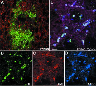
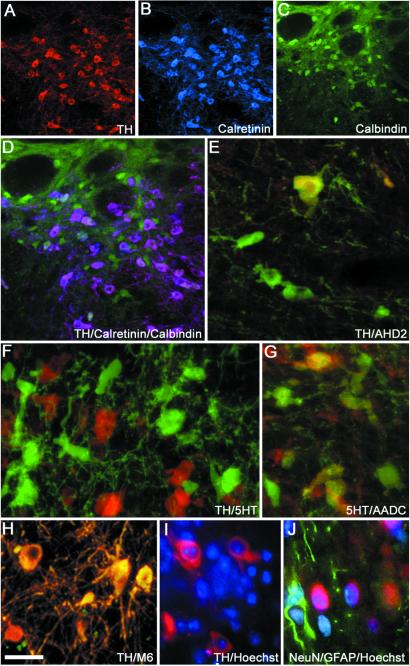
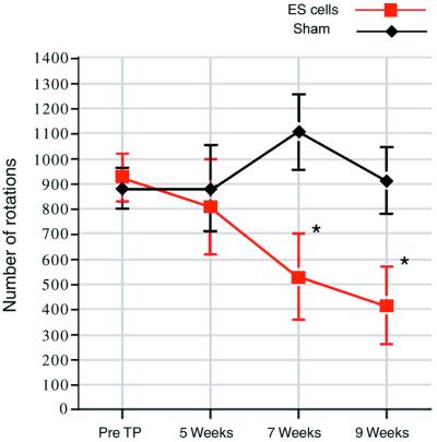
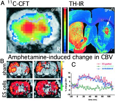
Although implantation of fetal dopamine (DA) neurons can reduce parkinsonism in patients, current methods are rudimentary, and a reliable donor cell source is lacking. We show that transplanting low doses of undifferentiated mouse embryonic stem (ES) cells into the rat striatum results in a proliferation of ES cells into fully differentiated DA neurons. ES cell-derived DA neurons caused gradual and sustained behavioral restoration of DA-mediated motor asymmetry. Behavioral recovery paralleled in vivo positron emission tomography and functional magnetic resonance imaging data demonstrating DA-mediated hemodynamic changes in the striatum and associated brain circuitry. These results demonstrate that transplanted ES cells can develop spontaneously into DA neurons. Such DA neurons can restore cerebral function and behavior in an animal model of Parkinson's disease.


Figures

Figure 1
Figure 1
Immunohistochemical staining of a graft 16 weeks after implantation of a low concentration (1,000–2,000 cells per μl) of D3 ES cells into adult 6-OHDA lesioned striatum. Numerous TH-positive neurons were found within the graft (A and B, green). All TH-positive profiles coexpressed the neuronal marker NeuN (A, red). TH (B) also was coexpressed with DAT (C, red) and AADC (D, blue), demonstrated by white triple labeling (E). (Scale bars: A, 150 μm; B–D, 50 μm; E, 25 μm.)
Figure 2
Figure 2
Photomicrograph showing TH-positive neurons (A and D) coexpressing (D) typical midbrain DA neuron markers such as calretinin (B and D) and calbindin (C and D). TH-positive neurons (E, green) also coexpressed the A9 marker aldehyde dehydrogenase 2 (E, yellow coexpression). Numerous 5HT neurons were found in grafts (F, green), and 5HT neurons also coexpressed AADC (G, green, yellow coexpression with 5HT). Grafted mouse ES cell-derived DA neurons (H, yellow, and I, red) were identified by colabeling with mouse-specific antibodies, M6 (H, yellow coexpression with TH), or by mouse-specific intranuclear fluorescent inclusions after Hoechst staining (I, blue). Astrocytes (glial fibrillary acidic protein-positive, J, green) and neurons (NeuN-positive, J, red) show mouse intranuclear fluorescent inclusions (Hoechst, J, blue). (Scale bars: A–C, 100 μm; D, 67 μm; E–J, 20 μm.)
Figure 3
Figure 3
Rotational behavior in response to amphetamine (4 mg/kg) was tested pretransplantation (pre TP) and at 5, 7, and 9 weeks postgrafting. A significant decrease in absolute numbers of amphetamine-induced turning was seen in animals with ES cell neural DA grafts in the striatum (n = 9) compared with control animals that received sham surgery (n = 13). Animals with sham surgery showed no change in rotational score over time (t = 1.51, P = 0.14). In contrast, animals with ES cell-derived neural grafts showed a significant reduction in rotations over time (t = −5.16, P < 0.001). We then examined at what time point rotational decrease was reduced significantly compared with pretransplantation scores. Because we performed post hoc comparisons, Bonferroni correction was applied to the significance criterion (adjusted criterion, P = 0.05/3 = 0.017). At 5 weeks postgrafting, ES cell-grafted animals showed no significant difference in rotations compared with pretransplantation scores (808 ± 188 vs. 924 ± 93 rotations, t = −0.62, P = 0.58). However, a clear and significant difference was evident at 7 weeks (530 ± 170 vs. 924 ± 93 rotations, t = −3.66, P = 0.0064) and further at 9 weeks (413 ± 154 vs. 924 ± 93 rotations, t = −4.30, P = 0.0026). *, P < 0.01.
Figure 4
Figure 4
(A) By using PET and the specific DAT ligand [11C]CFT, we identified specific binding in the right grafted striatum, as shown in this brain slice (A, Left) acquired 26 min after injection of the ligand into the tail vein (acquisition time was 15 sec). Color-coded (activity) PET images were overlaid with MRI images for anatomical localization. The increase in [11C]CFT binding in the right striatum was correlated with the postmortem presence of TH-immunoreactive (IR) neurons in the graft (A, Right). (B) Neuronal activation mediated by DA release in response to amphetamine (2 mg/kg) was restored in animals receiving ES grafts. Color-coded maps of the percentage of change in rCBV are shown at two striatal levels for control (Upper) and an ES cell-derived DA graft (Lower). A 6-OHDA lesion results in a complete absence of CBV response to amphetamine on striatum and cortex ipsilateral to the lesion (Upper). Recovery of signal change in motor and somatosensory cortex (arrows) and to a minor extent in the striatum was observed only in ES-grafted animals. (C) Graphic representation of signal changes over time in the same animal shown in B. The response on the grafted (red line) and normal (blue line) striata was similar in magnitude and time course, whereas no changes were observed in sham-grafted animals (green line). Baseline was collected for 10 min before and 10 min after monocrystalline iron oxide nanocolloid injection, and amphetamine was injected at time 0. cc, corpus callosum.

Comment in

References

    1. Olanow C W, Tatton W G. Annu Rev Neurosci. 2000;22:123–144. - PubMed
    1. Olanow C W, Obeso J A. Ann Neurol. 2000;47:167–178. - PubMed
    1. Freed C R, Greene P E, Breeze R E, Tsai W Y, DuMouchel W, Kao R, Dillon S, Winfield H, Culver S, Trojanowski J Q, Eidelberg D, Fahn S. N Engl J Med. 2001;344:710–719. - PubMed
    1. Hauser R A, Freeman T B, Snow B J, Nauert M, Gauger L, Kordower J H, Olanow C W. Arch Neurol (Chicago) 1999;56:179–187. - PubMed
    1. Piccini P, Brooks D J, Bjorklund A, Gunn R N, Grasby P M, Rimoldi O, Brundin P, Hagell P, Rehncrona S, Widner H, Lindvall O. Nat Neurosci. 1999;2:1137–1140. - PubMed

Publication types