. 2002 Jan 8;99(1):167-72.
doi: 10.1073/pnas.012607899.

The large GTPase dynamin regulates actin comet formation and movement in living cells

Affiliations

The large GTPase dynamin regulates actin comet formation and movement in living cells

James D Orth et al. Proc Natl Acad Sci U S A. .

Abstract

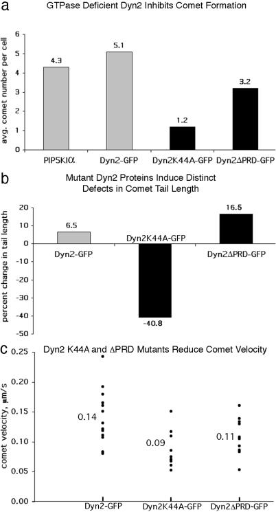
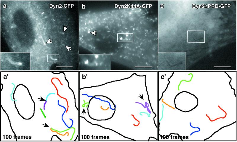
The large GTPase dynamin (Dyn2) has been demonstrated by us and others to interact with several different actin-binding proteins. To define how Dyn2 might participate in actin dynamics in livings cells we have expressed green fluorescent protein (GFP)-tagged Dyn2 in cultured cells and observed labeling of comet-like vesicles and macropinosomes. The comet structures progressed with a constant velocity and were reminiscent of actin comets associated with motile vesicles in cells expressing type I phosphatidylinositol phosphate 5-kinases. Based on these observations we sought to determine whether Dyn2 is an integral component of actin comets. Cells expressing type I phosphatidylinositol phosphate 5-kinase and Dyn2-GFP revealed a prominent colocalization of Dyn2 and actin in comet structures. Interestingly, comet formation and motility were normal in cells expressing wild-type Dyn2-GFP but altered markedly in Dyn2 mutant-expressing cells. Dyn2K44A-GFP mutant cells displayed a significant reduction in comet number, length, velocity, and efficiency of movement. In contrast, comets in cells expressing Dyn2DeltaPRD-GFP appeared dark and did not incorporate the mutant Dyn2 protein, indicating that the proline-rich domain (PRD) is required for Dyn2 recruitment. Further, these comets were significantly longer and slower than those in control cells. These findings demonstrate a role for Dyn2 in actin-based vesicle motility.


Figures

Figure 1
Figure 1
Dyn2-GFP incorporates into endogenous comet-like vesicles and macropinosomes in living cells. (a) Confocal time-lapse imaging of Clone 9 hepatocytes expressing Dyn2-GFP. Dynamic comet-like structures were observed. N, nucleus. (be) Higher magnification revealed that these motile structures consisted of a brightly stained head followed by a tail structure (arrows) that extends 180° from the direction of movement. (f) The velocity of the movement was uniform as revealed by the consistent distances between projected frames of the time-lapse images (arrows). (gk) Confocal time-lapse imaging of Dyn2-GFP in NIH 3T3 fibroblasts revealed large vesicular structures associated with small tails of Dyn2-GFP (arrows and boxes). (h′ and k′) High magnifications of individual macropinosomes from frames h and k, respectively. Consistent with the formation of macropinosomes, these structures formed from zones of active membrane ruffling at the cell periphery. (Bar, 10 μm.) See Movies 1 and 2, which are published as supporting information on the PNAS web site, www.pnas.org.
Figure 2
Figure 2
Dyn2 is an integral component of actin comets. Cultured rat fibroblasts and Clone 9 hepatocytes showing dynamin incorporated into PIP5KIα-induced actin comets. (a and a′) Comets in a rat fibroblast expressing Dyn2aa-GFP and colabeled with cortactin. The Dyn2 labeling is brightest at the head of the comet (arrows) but is prominent also in the tail (Inset, arrows). In contrast, cortactin staining is localized predominantly to the comet tail. RF, rat fibroblast; αCortY, anti-cortactin. (b and b′) Comets in a Clone 9 hepatocyte immunolabeled for dynamin (αDyn2) and actin (rhodamine-phalloidin). Note the Dyn2 enrichment in the head and staining in the tails (arrows and Inset), whereas actin stains the tail only. (a: bar, 3 μm; b: bar, 2 μm.)
Figure 3
Figure 3
Dyn2 regulates comet formation, actin tail length, and comet velocity. For quantitation of comet formation and actin tail length, rat fibroblasts coexpressing Myc-PIP5KIα and wild-type or mutant Dyn2-GFP were fixed and stained for actin (rhodamine-phalloidin). (a) Cells expressing the K44A mutant show on average only 1.2 comets per cell, a 72.1% reduction compared with cells expressing PIP5KIα alone (4.3), and Dyn2ΔPRD expression shows a reduction in comet formation of 25.6% (3.2 comets) compared with PIP5Kα control. (b) The average comet tail length in cells expressing PIP5KIα and Dyn2 was only 6.5% longer than PIP5KIα control (5.7 μm), whereas those in cells expressing Dyn2K44A were 40.8% shorter (3.2 μm). In contrast, comet tails in Dyn2ΔPRD-expressing cells were 16.5% longer (6.2 μm) than control. (c) Particle-tracking analysis showed that Dyn2K44A-GFP and Dyn2ΔPRD-GFP comets progressed with reduced velocities of 0.09 and 0.11 μm/s, respectively, whereas wild-type Dyn2-GFP comets progressed at 0.14 μm/s. See Movies 3–5 (which are published as supporting information on the PNAS web site) for comparison of the Dyn2 comets.
Figure 4
Figure 4
Mutant dynamin proteins induce abnormal comet movements. Rat fibroblasts coexpressing Myc-PIP5KIα and wild-type or mutant forms of Dyn2-GFP were imaged for 100 frames, and the movement of comet structures was followed. (a) Cells expressing wild-type Dyn2-GFP formed numerous comets (arrows and Inset) that actively incorporated the tagged Dyn2 (see Movie 3). (a′) One hundred frames from the time-lapse images were stacked to show the movement characteristics of individual comets. Each color represents a distinct comet path. Note the linear quality of comet movement. Arrows, multiple comets appeared to form from a single domain. (b) A K44A-GFP-expressing cell with comets (arrows). The comets are fewer and much smaller than those of wild type (see Movie 4). (b′) One hundred stacked frames from the K44A-GFP time-lapse. These comets often had curved and wandering paths (arrows) and were less efficient in their translocation. See the Dyn2K44a-GFP video (Movie 4, arrow on right) for an example of defective movement. (c) Comets in cells expressing the truncated Dyn2ΔPRD-GFP are dark and do not incorporate the mutant protein (see Movie 5). (c′) One hundred frames from the ΔPRD-GFP video revealed that these comets moved in a similar manner to those of wild-type Dyn2, with smooth curvilinear paths. (Bars, 10 μm.)
Figure 5
Figure 5
GTPase-defective Dyn2 comets move with decreased efficiency. Dyn2K44A-GFP comets show a dramatically reduced MI compared with actin-GFP, Dyn2-GFP, and Dyn2ΔPRD-GFP comets. One hundred frames from GFP time-lapse videos were stacked and superimposed over a circle with an outer radius of 5.0 μm (red) to provide a point of origin of individual comets. The distance traveled (yellow) to completely traverse the 5.0-μm radius was determined for at least 12 comets. The ratio of 5.0 μm and the distance traveled is the MI. (a and c) Actin-GFP, Dyn2-GFP, and Dyn2ΔPRD-GFP comets moved nearly linearly (1.0). (b and c) Dyn2K44A-GFP comets have an MI of only 0.54, indicating their efficiency of movement was impaired severely (decreased linearity). Note that Dyn2ΔPRD-GFP comets also had linear movement despite having a reduced velocity as shown in Fig. 3c. (Bar, 5.0 μm.)
Figure 6
Figure 6
Comets assemble on macropinosomes forming from membrane ruffles in PIP5KIα-expressing cells. Time-lapse confocal imaging of microinjected rat fibroblasts expressing PIP5KIα and actin-GFP. These cells often formed large macropinocytic structures from peripheral membrane ruffles. (a) Actin-GFP fluorescence in a cell undergoing active membrane ruffling (box). (bd) Enlarged image of the boxed area in a. Time-lapse frames at 10-s intervals revealed macropinosomes forming directly from the peripheral membrane ruffles (arrowhead). The large vesicle head and short, trailing, actin tail are resolved easily (arrows). Similar to the endogenous Dyn2-GFP macropinosomes (Fig. 1), the tail remained 180° opposed from the direction of movement. (Bar, 10 μm.) See Movie 6.

References

    1. McNiven M A, Cao H, Pitts K R, Yoon Y. Trends Biochem Sci. 2000;25:115–120. - PubMed
    1. Hinshaw J E. Annu Rev Cell Dev Biol. 2000;16:483–519. - PMC - PubMed
    1. Takei K, Mundigl O, Daniell L, De Camilli P. J Cell Biol. 1996;133:1237–1250. - PMC - PubMed
    1. Henley J R, McNiven M A. J Cell Biol. 1996;133:761–775. - PMC - PubMed
    1. McNiven M A, Kim L, Krueger E W, Orth J D, Cao H, Wong T W. J Cell Biol. 2000;151:187–198. - PMC - PubMed

Publication types

MeSH terms

LinkOut - more resources