. 2002 Jan 15;22(2):404-12.
doi: 10.1523/JNEUROSCI.22-02-00404.2002.

Urocortin, but not urocortin II, protects cultured hippocampal neurons from oxidative and excitotoxic cell death via corticotropin-releasing hormone receptor type I

Affiliations

Urocortin, but not urocortin II, protects cultured hippocampal neurons from oxidative and excitotoxic cell death via corticotropin-releasing hormone receptor type I

Ward A Pedersen et al. J Neurosci. .

Abstract

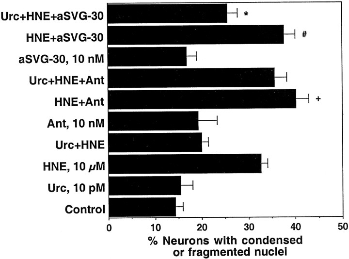
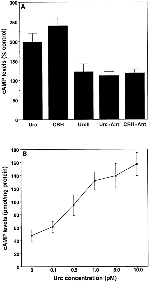
Urocortin and urocortin II are members of the corticotropin-releasing hormone (CRH) family of neuropeptides that function to regulate stress responses. Two high-affinity G-protein-coupled receptors have been identified that bind CRH and/or urocortin I and II, designated CRHR1 and CRHR2, both of which are present in hippocampal regions of mammalian brain. The hippocampus plays an important role in regulating stress responses and is a brain region in which neurons are vulnerable during disease and stress conditions, including cerebral ischemia, Alzheimer's disease, and anxiety disorders. Here we report that urocortin exerts a potent protective action in cultured rat hippocampal neurons with concentrations in the range of 0.5-5.0 pm, increasing the resistance of the cells to oxidative (amyloid beta-peptide, 4-hydroxynonenal, ferrous sulfate) and excitotoxic (glutamate) insults. We observed that urocortin is 10-fold more potent than CRH in protecting hippocampal neurons from insult, whereas urocortin II is ineffective. RT-PCR and sequencing analyses revealed the presence of both CRHR1 and CRHR2 in the hippocampal cultures, with CRHR1 being expressed at much higher levels than CRHR2. Using subtype-selective CRH receptor antagonists, we provide evidence that the neuroprotective effect of exogenously added urocortin is mediated by CRHR1. Furthermore, we provide evidence that the signaling pathway that mediates the neuroprotective effect of urocortin involves cAMP-dependent protein kinase, protein kinase C, and mitogen-activated protein kinase. This is the first demonstration of a biological activity of urocortin in hippocampal neurons, suggesting a role for the peptide in adaptive responses of hippocampal neurons to potentially lethal oxidative and excitotoxic insults.


Figures

Fig. 1.
Fig. 1.
Protective effects of Urc against Aβ-induced cell death in cultured rat hippocampal neurons. A, Cultures were exposed for 24 hr to Locke's solution alone (Control) or to Locke's solution containing Urc at 10 pm, Aβ25-35 at 1 μm, or the combination of Urc at 10 pm and Aβ25-35 at 1 μm; the cultures were photographed under phase-contrast optics with a 10× objective. Many of the neurons exposed to Aβ25-35 exhibited morphological signs of degeneration, i.e., cell body shrinkage and neurite fragmentation (arrows), whereas the majority of neurons treated with a combination of Urc and Aβ25-35 had neurites and cell bodies with normal appearance. B, The neuroprotective effect of Urc is concentration dependent. Cultures were exposed for 24 hr to Locke's solution alone (Control) or to Locke's solution containing Aβ25-35 at 1 μm, Urc at the indicated concentrations, or the combination of Aβ25-35 at 1 μm and Urc at the indicated concentrations. The cultures were photographed under phase-contrast optics with a 10× objective, and the percentage of remaining viable neurons was determined. Values are the mean and SD of determinations made in three cultures per treatment condition (*p = 0.0007 and #p < 0.0001 vs Aβ; one-way ANOVA and Fisher's PLSD).
Fig. 2.
Fig. 2.
Comparison of the potencies and effectiveness of Urc, CRH, and UrcII in protecting cultured rat hippocampal neurons from oxidative and excitotoxic insults. A, Cultures were exposed for 24 hr to Locke's solution alone (Control) or to Locke's solution containing Urc at 10 pm, Aβ25-35 at 1 μm, HNE at 1 μm, FeSO4 at 3 μm,l-glut at 20 μm, or the combinations as indicated. Values are the mean and SD of determinations made in four cultures per treatment condition. The extent of cell death caused by each insult alone was reduced in the presence of Urc (*p < 0.0001; one-way ANOVA and Fisher's PLSD).B, Cultures were exposed for 24 hr to Locke's solution alone (Control) or to Locke's solution containing HNE at 1 μm with or without the combinations as indicated. Values are the mean and SD of determinations made in four cultures per treatment condition. Submaximal and equally potent protective effects were observed with Urc alone at 0.5 pmand with CRH alone at 5 pm (*p < 0.0001 vs HNE; one-way ANOVA and Fisher's PLSD). Maximal protective effects were observed with Urc at 1 pm, CRH at 10 pm, or with the combination of Urc at 0.5 pm and CRH at 5 pm(*p < 0.0001 vs HNE;#p < 0.002 vs Urc, 0.5 pm+ HNE; +p = 0.0001 vs CRH, 5 pm + HNE; **p < 0.008 vs Urc, 0.5 pm + HNE; or CRH, 5 pm + HNE; one-way ANOVA and Fisher's PLSD). C, Cultures were exposed for 24 hr to Locke's solution alone (Control) or to Locke's solution containing HNE at 1 μm, UrcII at 1, 10, and 100 pm, or the combinations as indicated. Values are the mean and SD of determinations made in four cultures per treatment condition. UrcII was ineffective at protecting against HNE-induced cell death at any of the concentrations that were tested.
Fig. 3.
Fig. 3.
Expression of CRHR1 and CRHR2 in cultured rat hippocampal and cortical neurons. Top, Total RNA extracted from hippocampal and cortical cultures and from the midbrain, hippocampus, and cortex of E18 rat was subjected to first-strand reverse transcription, followed by PCR that used primers specific for either CRHR1 or CRHR2. The PCR products were separated by electrophoresis in a 2% agarose gel stained with ethidium bromide and were visualized with a FUJIFilm FLA-3000 fluorescent image analyzer and Image Gauge version 3.2 software. Equal loading of PCR products onto the gel was determined by using internal primers specific for GAPDH (data not shown). Similar results were obtained in three independent experiments. Bottom, Identity of the PCR products corresponding to the hippocampal cultures was confirmed by cloning and sequencing. Underlined sequences are those recognized by the reverse primers used for PCR amplification.
Fig. 4.
Fig. 4.
The neuroprotective effects of Urc and CRH are blocked by a CRHR1 antagonist. A, Cultures were exposed for 1 hr to fresh Neurobasal with or without antalarmin (Ant) at 10 nm and subsequently were exposed for 24 hr to Neurobasal alone (Control) or to Neurobasal containing Urc at 10 pm, HNE at 10 μm, Ant at 10 nm, or the combinations as indicated. Values are the mean and SD of determinations made in four cultures per treatment condition. The extent of cell death in cultures treated with HNE alone and in cultures treated with Ant followed by Urc and HNE was not statistically different. B, Cultures were exposed for 1 hr to fresh Neurobasal with or without Ant at 10 nm and subsequently were exposed for 24 hr to Neurobasal alone (Control) or to Neurobasal containing CRH at 10 pm, HNE at 10 μm, Ant at 10 nm, or the combinations as indicated. Values are the mean and SD of determinations made in four cultures per treatment condition. The extent of cell death in cultures treated with HNE alone and in cultures treated with Ant followed by CRH and HNE was not statistically different.
Fig. 5.
Fig. 5.
The neuroprotective effects of Urc are not blocked by a CRHR2 antagonist. Cultures were exposed for 1 hr to fresh Neurobasal with or without Ant or antisauvagine-30 (aSVG-30) at 10 nm and subsequently were exposed for 24 hr to Neurobasal alone (Control) or to Neurobasal containing Urc at 10 pm, HNE at 10 μm, Ant at 10 nm, aSVG-30 at 10 nm, or the combinations as indicated. Values are the mean and SD of determinations made in four cultures per treatment condition. Pretreatment with Ant or aSVG-30 caused a slight potentiation of HNE-induced cell death (+p = 0.0001 and#p = 0.0074 vs HNE; one-way ANOVA and Fisher's PLSD). The protective effect of Urc was blocked in cultures pretreated with Ant, but not in cultures pretreated with aSVG-30 (*p < 0.0001 vs HNE + aSVG-30; one-way ANOVA and Fisher's PLSD).
Fig. 6.
Fig. 6.
Elevation in cAMP concentrations by Urc and CRH in cultured rat hippocampal neurons. A, Cultures were exposed for 1 hr to Locke's solution with 50 μm IBMX and then were exposed for 30 min to Locke's solution alone (as the control) or to Locke's solution containing Urc at 1 pm, CRH at 10 pm, or UrcII at 10 pm in the continued presence of IBMX. Additional cultures were pretreated for 1 hr with IBMX at 50 μm and Ant at 10 nm and subsequently were treated for 30 min with Urc at 1 pm or CRH at 10 pm in the continued presence of both IBMX and Ant. Values are the mean and SD of determinations made in four cultures per treatment condition, expressed as a percentage of control cAMP levels (58.8 ± 8.7 pmol/mg protein). There was a statistically significant increase in the levels of cAMP in Urc- and CRH-treated groups (p < 0.0001 vs control; one-way ANOVA and Fisher's PLSD), but the levels of cAMP in control, UrcII, Urc + Ant, and CRH + Ant groups were not statistically different.B, Cultures were exposed for 1 hr to Locke's solution with 50 μm IBMX and then were exposed for 30 min to Locke's solution containing Urc at 0, 0.1, 0.5, 1.0, 5.0, or 10.0 pm in the continued presence of IBMX. Values are the mean and SD of determinations made in four cultures per treatment condition. There was a statistically significant increase in the levels of cAMP in cultures treated with Urc at concentrations ≥0.5 pm(p < 0.005 vs Urc at 0 or Urc at 0.1 pm; one-way ANOVA and Fisher's PLSD).
Fig. 7.
Fig. 7.
The neuroprotective effect of Urc requires G-protein-coupled receptor activation of PKA, PKC, and MAP kinase.A, Cultures were exposed for 1 hr to fresh Neurobasal with or without NF 449 at 200 nm, PD 98059 at 4 μm, BIM I at 10 nm, or H-89 at 100 nm; subsequently, the cultures were exposed for 24 hr to Neurobasal alone (Control) or to Neurobasal containing Urc at 10 pm, HNE at 10 μm, NF 449 at 200 nm, PD 98059 at 4 μm, BIM I at 10 nm, H-89 at 100 nm, or the combinations as indicated. Values are the mean and SD of determinations made in four cultures per treatment condition. Pretreatment with each of the inhibitors followed by treatment with Urc and HNE caused a statistically significant increase in the extent of cell death (p < 0.0001 vs Urc + HNE; one-way ANOVA and Fisher's PLSD). B, Immunoblot analysis demonstrating Urc activation of MAP kinases ERK1 and ERK2. Cultures were exposed for 1 hr to PD 98059 at 4 μm, BIM I at 10 nm, or H-89 at 100 nm and subsequently were exposed for 15 min to Urc with or without each of the inhibitors. The increased phosphorylation of ERK1 (p44) and ERK2 (p42) caused by Urc treatment is abolished in cultures pretreated with PD 98059 or BIM I and is attenuated in cultures pretreated with H-89. The levels of ERK1 and ERK2 in these samples are shown for comparison.C, Densitometric analysis of the immunoblots shown inB. The levels of phosphorylated ERK1 and ERK2 were normalized to the total levels of ERK1 and ERK2.

Similar articles

Cited by

References

    1. Berger AJ, Hart AC, Kaplan JM. Gαs-induced neurodegeneration in Caenorhabditis elegans. J Neurosci. 1998;18:2871–2880. - PMC - PubMed
    1. Bishop GA, Seelandt CM, King JS. Cellular localization of corticotropin-releasing factor receptors in the adult mouse cerebellum. Neuroscience. 2000;101:1083–1092. - PubMed
    1. Bittencourt JC, Vaughan J, Arias C, Rissman RA, Vale WW, Sawchenko PE. Urocortin expression in rat brain: evidence against a pervasive relationship of urocortin-containing projections with targets bearing type 2 CRF receptors. J Comp Neurol. 1999;415:285–312. - PubMed
    1. Brar BK, Stephanou A, Okosi A, Lawrence KM, Knight RA, Marber MS, Latchman DS. CRH-like peptides protect cardiac myocytes from lethal ischaemic injury. Mol Cell Endocrinol. 1999;158:55–63. - PubMed
    1. Brar BK, Jonassen AK, Stephanou A, Santilli G, Railson J, Knight RA, Yellon DM, Latchman DS. Urocortin protects against ischemic and reperfusion injury via a MAPK-dependent pathway. J Biol Chem. 2000;275:8508–8514. - PubMed

Publication types

MeSH terms

LinkOut - more resources