. 2002 Jan 15;22(2):515-22.
doi: 10.1523/JNEUROSCI.22-02-00515.2002.

Amyloid-associated neuron loss and gliogenesis in the neocortex of amyloid precursor protein transgenic mice

Affiliations

Amyloid-associated neuron loss and gliogenesis in the neocortex of amyloid precursor protein transgenic mice

Luca Bondolfi et al. J Neurosci. .

Abstract

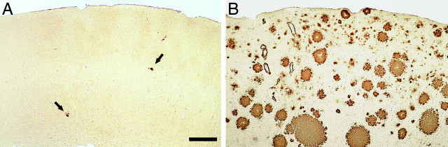
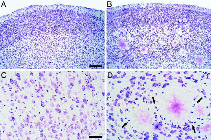
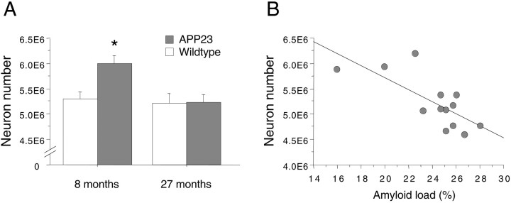
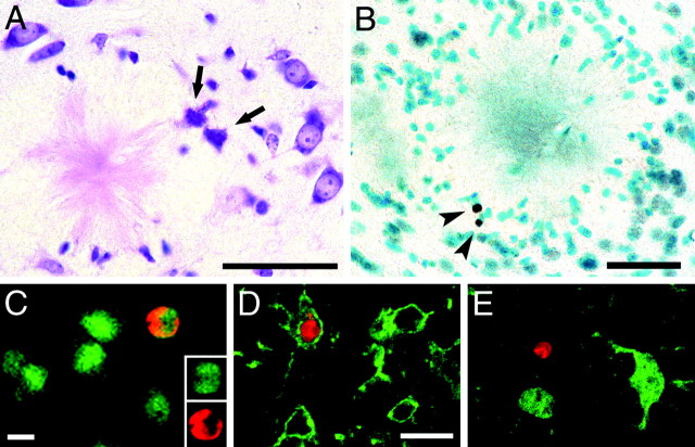
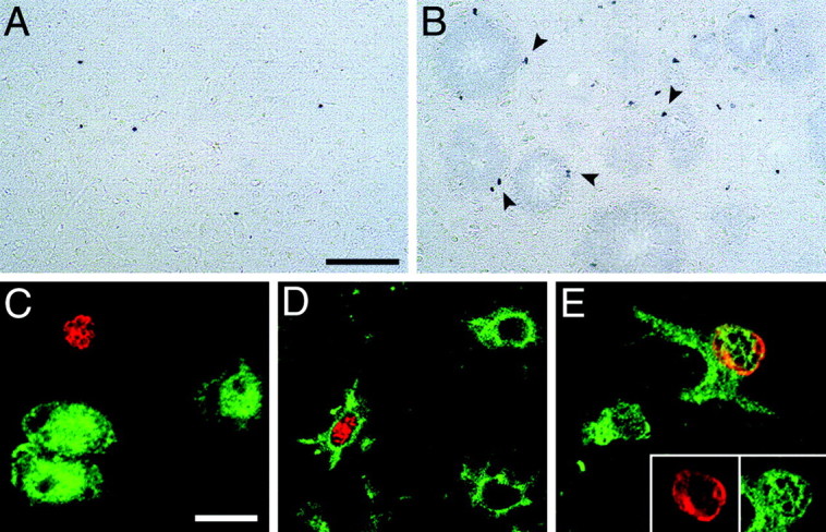
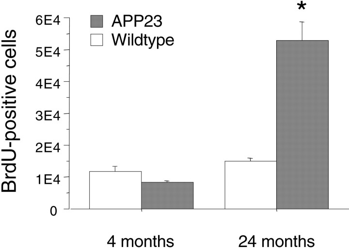
APP23 transgenic mice express mutant human amyloid precursor protein and develop amyloid plaques predominantly in neocortex and hippocampus progressively with age, similar to Alzheimer's disease. We have previously reported neuron loss in the hippocampal CA1 region of 14- to 18-month-old APP23 mice. In contrast, no neuron loss was found in neocortex. In the present study we have reinvestigated neocortical neuron numbers in adult and aged APP23 mice. Surprisingly, results revealed that 8-month-old APP23 mice have 13 and 14% more neocortical neurons compared with 8-month-old wild-type and 27-month-old APP23 mice, respectively. In 27-month-old APP23 mice we found an inverse correlation between amyloid load and neuron number. These results suggest that APP23 mice have more neurons until they develop amyloid plaques but then lose neurons in the process of cerebral amyloidogenesis. Supporting this notion, we found more neurons with a necrotic-apoptotic phenotype in the neocortex of 24-month-old APP23 mice compared with age-matched wild-type mice. Stimulated by recent reports that demonstrated neurogenesis after targeted neuron death in the mouse neocortex, we have also examined neurogenesis in APP23 mice. Strikingly, we found a fourfold to sixfold increase in newly produced cells in 24-month-old APP23 mice compared with both age-matched wild-type mice and young APP23 transgenic mice. However, subsequent cellular phenotyping revealed that none of the newly generated cells in neocortex had a neuronal phenotype. The majority were microglial and to a lesser extent astroglial cells. We conclude that cerebral amyloidosis in APP23 mice causes a modest neuron loss in neocortex and induces marked gliogenesis.


Figures

Fig. 1.
Fig. 1.
Cerebral amyloidosis in the neocortex of APP23 transgenic mice. A, In 8-month-old mice only few Aβ-immunostained amyloid deposits were found (arrows).B, In contrast, 27-month-old APP23 mice exhibited severe cerebral amyloidosis throughout the neocortex. The total amyloid load in the neocortex of the mouse shown was estimated to be 25.7%. Scale bar, 150 μm. A and B have the same magnification.
Fig. 2.
Fig. 2.
Amyloid plaques disrupt neocortical cytoarchitecture in aged APP23 mice. A, Cresyl violet staining in the neocortex of a 27-month-old wild-type mouse reveals the typical cortical cell layers. B, In contrast, in 27-month-old APP23 mice amyloid deposits disrupt the neurocytoarchitecture, and some of the layers are barely detectable.C, D, Higher magnifications of layer V neurons in a 27-month-old wild-type (C) and transgenic (D) mouse. Note the numerous glial cell nuclei (arrows in D) and the absence of neurons in the immediate vicinity of the amyloid plaques. Scale bars:A, B, 150 μm; C,D, 40 μm.
Fig. 3.
Fig. 3.
Stereological estimation of total neocortical neuron number. A, Total neuron number per neocortex per hemisphere in 8- and 27-month-old APP23 and wild-type control mice. For this graph, males and females were combined (n = 11–15 per group). Results revealed that 8-month-old APP23 mice have more neurons compared with the three other groups (*p < 0.01). B, For the 27-month-old APP23 mice, linear regression analysis revealed an inverse relation between neuron number and amyloid load.
Fig. 4.
Fig. 4.
Cell death with necrotic and apoptotic appearance in the vicinity of amyloid plaques. A, Neurons with a pyknotic appearance and an irregular membrane structure (arrows) were occasionally detected in 24-month-old APP23 mice. Such neurons were almost exclusively associated with amyloid deposits. B, TUNEL-positive cells (arrowheads) in the vicinity of an amyloid plaque. Using confocal microscopy TUNEL-positive cells (red) were labeled with markers for either neurons (C), microglia (D), or astrocytes (E) (all in green). Most of the cells could not be phenotyped. Only occasionally were TUNEL-positive cells labeled for NeuN or CD11b. No double labeling was observed with S100β (Table 1). The insert in Crepresents single confocal sections of both markers. Scale bars:A, B, 40 μm; C, D, 10 μm. D and E have same magnification.
Fig. 5.
Fig. 5.
BrdU-positive cells in the neocortex of aged APP23 mice. A, Only few labeled cells were observed in the neocortex of 24-month-old wild-type mice 4 weeks after BrdU injections.B, In contrast, there was an approximately fourfold increase in labeled cells in age-matched APP23 mice (for quantitative results, see Fig. 6). Most of these cells appeared to be associated with amyloid plaques (arrowheads). To study the cellular phenotype of these cells BrdU immunofluorescence (red) was combined with NeuN (C) CD11b (D) and S100β (E) (all ingreen), and colocalization was assessed with confocal microscopy. None of the BrdU-labeled cells were positive for NeuN (C). In contrast, the majority of the BrdU-labeled cells revealed colocalization with the microglia marker CD11b (D). Colocalization was also found with the astrocytic marker S100 β (E). Theinsert in E represents single confocal sections of both markers. Quantitative phenotyping is summarized in Table 1. Scale bars: A, B, 75 μm;C–E, 10 μm.
Fig. 6.
Fig. 6.
Increase in BrdU-positive cells in the neocortex of aged APP23 mice. Total BrdU-positive cells in the neocortex of young (4-month-old) and aged (24-month-old) wild-type and APP23 mice. Numbers are for one hemisphere only. Note the approximately fourfold increase in BrdU-labeled cells in the aged APP23 mice compared with all three other groups (*p values < 0.001).

References

    1. Biebl M, Cooper CM, Winkler J, Kuhn HG. Analysis of neurogenesis and programmed cell death reveals a self- renewing capacity in the adult rat brain. Neurosci Lett. 2000;291:17–20. - PubMed
    1. Bornemann KD, Wiederhold KH, Pauli C, Ermini F, Stalder M, Schnell L, Sommer B, Jucker M, Staufenbiel M. Abeta-induced inflammatory processes in microglia cells of APP23 transgenic mice. Am J Pathol. 2001;158:63–73. - PMC - PubMed
    1. Bundgaard MJ, Regeur L, Gundersen HJ, Pakkenberg B. Size of neocortical neurons in control subjects and in Alzheimer's disease. J Anat. 2001;198:481–489. - PMC - PubMed
    1. Calhoun ME, Kurth D, Phinney AL, Long JM, Hengemihle J, Mouton PR, Ingram DK, Jucker M. Hippocampal neuron and synaptophysin-positive bouton number in aging C57BL/6 mice. Neurobiol Aging. 1998a;19:599–606. - PubMed
    1. Calhoun ME, Wiederhold KH, Abramowski D, Phinney AL, Probst A, Sturchler-Pierrat C, Staufenbiel M, Sommer B, Jucker M. Neuron loss in APP transgenic mice. Nature. 1998b;395:755–756. - PubMed

Publication types

MeSH terms

Substances