Clinical Trial
. 2002 Jan 15;22(2):554-61.
doi: 10.1523/JNEUROSCI.22-02-00554.2002.

Functional connectivity of human premotor and motor cortex explored with repetitive transcranial magnetic stimulation

Affiliations
Clinical Trial

Functional connectivity of human premotor and motor cortex explored with repetitive transcranial magnetic stimulation

A Münchau et al. J Neurosci. .

Abstract

Connections between the premotor cortex and the primary motor cortex are dense and are important in the visual guidance of arm movements. We have shown previously that it is possible to engage these connections in humans and to measure the net amount of inhibition/facilitation from premotor to motor cortex using single-pulse transcranial magnetic stimulation (TMS). The aim of this study was to test whether premotor activation can affect the excitability of circuits within the primary motor cortex (M1) itself. Repetitive TMS (rTMS), which is known to produce effects that outlast the train at the site of stimulation, was given for 20 min at 1 Hz over premotor, primary motor, and sensory areas of cortex at an intensity of 80% of the active motor threshold for the motor hand area. The excitability of some corticocortical connections in M1 was probed by using paired-pulse testing of intracortical inhibition (ICI) and intracortical facilitation (ICF) with a coil placed over the motor cortex hand area. rTMS over the premotor cortex, but not other areas, changed the time course of the ICI/ICF for up to 1 hr afterward without affecting motor thresholds or motor-evoked potential recruitment. The cortical silent period was also shortened. The implication is that rTMS at a site distant from the motor cortex can change the excitability of circuits intrinsic to the motor cortex.


Figures

Fig. 1.
Fig. 1.
A, Design of the main experiment. RMTs and AMTs, the ICI/ICF, and the cortical SP were determined before and after rTMS. The ICI/ICF was tested in three different blocks, referred to as A–C. Each block consisted of four (5 for the 7 subjects in whom extra ISIs were studied) different conditions: the test stimulus alone and the test plus conditioning stimuli at three (or 4) different interstimulus intervals. The order of presentation of the different conditions within a block was changed randomly. B, Design of the control experiment, in which the time course of effects was studied. Before rTMS, RMT, and AMT, the ICI/ICF and the SP were determined. Testing of the ICI/ICF was performed in three blocks (A–C), as described in A. In addition, the amplitude of the MEP during slight voluntary contraction of the target muscle (Active MEP) was measured. RMT, AMT, SP, and active MEP were determined again immediately after rTMS. Then the ICI/ICF was retested. The active MEP was measured again between blocksA and B (5 min after rTMS), between blocks B and C (10 min after rTMS), and after block C (15 min after rTMS). The SP was also repeated after block C. Finally, the ICI/ICF (blocksA–C) was retested 1 and 2 hr after rTMS.Left, The coil position during TMS measurements and during rTMS. The positions of both the motor hot spot for the FDI muscle and the premotor area are indicated by a filledand an open circle, respectively.
Fig. 2.
Fig. 2.
A, RMTs and AMTs before and immediately after rTMS in the eight subjects who had received both motor and premotor rTMS. There was no significant change after motor or premotor rTMS. Error bars indicate SEM. B, MEP size of relaxed FDI muscle before and after motor rTMS and premotor rTMS in the same eight subjects. Measurements were repeated three times after rTMS, at 5, 10, and 15 min. Resting MEP amplitudes were slightly smaller 5 and 10 min after premotor rTMS, but this difference was not significant. C, MEP size during slight voluntary contraction of FDI muscle before and after premotor rTMS in four subjects. MEP size was determined 1, 5, 10, and 15 min after rTMS. There was no significant difference from baseline.
Fig. 3.
Fig. 3.
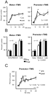
A, ICI/ ICF curves before and after rTMS at 80% of the AMT over motor and premotor areas. The mean (± SEM) time course of the conditioned test MEP after rTMS is superimposed on the time course at baseline. The size of the conditioned test response is expressed as a percentage of the unconditioned test size. Data from the eight subjects who had both motor and premotor rTMS are shown. Nine different ISIs were studied. After motor rTMS there was no significant change from baseline. In contrast, after premotor rTMS there was significantly increased facilitation at an ISI of 7 msec (t(7) = −2.5; p = 0.041; post hoc paired-samples t test).B, Comparison of the averaged size of the conditioned test response of adjacent time points, separated into early ISIs (2, 3, and 4 msec), medium ISIs (5, 6, and 7 msec), and later ISIs (10, 15, and 20 msec). rTMS had no effect on the ICI/ICF when applied over the motor cortex. In contrast, after premotor rTMS the conditioned MEP size was significantly increased compared with baseline at medium ISIs (p < 0.0001; post hocpaired-samples t tests) but not at the other intervals. C, ICI/ICF curves before and after rTMS at 80% of the AMT over the premotor area. Data from all 13 subjects who had premotor rTMS are shown. In this larger group there is increased facilitation at ISIs of 6 msec (t(12) = −2.3; p < 0.05) and 7 msec (t(12) = −3.5; p< 0.005). In 7 of these 13 subjects, additional ISIs (8, 9, and 12 msec) were studied. There was no significant difference from baseline at any of these ISIs.
Fig. 4.
Fig. 4.
Effects of rTMS on the duration of the SP. There was a significant reduction in the duration of the SP immediately after premotor rTMS (t(7) = 5.6;p = 0.01; paired-samples t test) but not after motor rTMS. The effect after premotor rTMS was still significant 15 min later (t(7) = 2.3;p < 0.05).
Fig. 5.
Fig. 5.
Time course of the increased ICF at ISIs of 6 and 7 msec after rTMS over the premotor area in four subjects. The conditioned MEPs at 6 and 7 msec were significantly larger immediately after rTMS (t(3) = −3.2;p < 0.05) and 1 hr later (t(3) = −13,2; p = 0.001; paired-samples t tests). They were still slightly increased compared with baseline at 2 hr, but this was no longer statistically significant.
Fig. 6.
Fig. 6.
A, ICI/ICF curves after premotor rTMS using different stimulus intensities in four subjects. There was no change after rTMS at 70 or 90% of the AMT. For comparison, the time course before and after rTMS at 80% of the AMT for the same four subjects is also shown. After rTMS at 80% of the AMT there was a significant facilitation at an ISI of 7 msec (t(3) = −3.1; p < 0.05; paired-samples t test). B, ICI/ICF curve after sensory rTMS in four subjects. There was no significant change from baseline. For comparison, the time course before and after rTMS at 80% of the AMT premotor cortex for the same four subjects is also shown. Significant facilitation is present at an ISI of 7 msec (t(3) = −3.8, p < 0.05; paired-samples t test).

References

    1. Ashby P, Reynolds C, Wennberg A, Lozano AM, Rothwell JC. On the focal nature of inhibition and facilitation in the human motor cortex. Clin Neurophysiol. 1999;110:550–555. - PubMed
    1. Brasil-Neto JP, Cohen LG, Panizza M, Nilsson J, Roth BJ, Hallett M. Optimal focal transcranial magnetic activation of the human motor cortex: effects of coil orientation, shape of the induced current pulse, and stimulus intensity. J Clin Neurophysiol. 1992;9:132–136. - PubMed
    1. Chen R, Classen J, Gerloff C, Celnik P, Wassermann EM, Hallett M, Cohen LG. Depression of motor cortex excitability by low-frequency transcranial magnetic stimulation. Neurology. 1997;48:1398–1403. - PubMed
    1. Civardi C, Cantello R, Asselman P, Rothwell JC. Transcranial magnetic stimulation can be used to test connections to primary motor areas from frontal and medial cortex in humans. NeuroImage. 2001;14:1444–1453. - PubMed
    1. Ferbert A, Priori A, Rothwell JC, Day BL, Colebatch JG, Marsden CD. Interhemispheric inhibition of the human motor cortex. J Physiol (Lond) 1992;453:525–546. - PMC - PubMed

Publication types

LinkOut - more resources